

Human Antibody Responses to Emerging Mayaro Virus and Cocirculating Alphavirus Infections Examined by Using Structural Proteins from Nine New and Old World Lineages
- PMID: 29577083
- PMCID: PMC5863033
- DOI: 10.1128/mSphere.00003-18
Human Antibody Responses to Emerging Mayaro Virus and Cocirculating Alphavirus Infections Examined by Using Structural Proteins from Nine New and Old World Lineages
Abstract
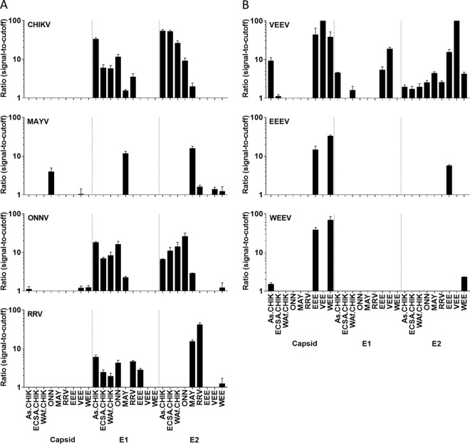
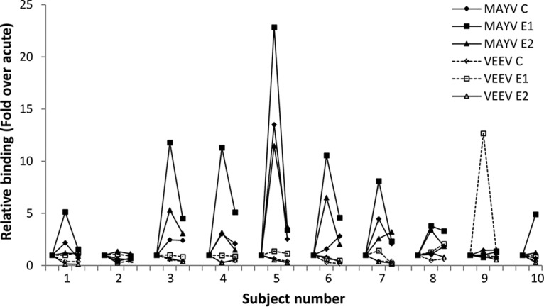
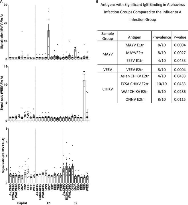
Mayaro virus (MAYV), Venezuelan equine encephalitis virus (VEEV), and chikungunya virus (CHIKV) are vector-borne alphaviruses that cocirculate in South America. Human infections by these viruses are frequently underdiagnosed or misdiagnosed, especially in areas with high dengue virus endemicity. Disease may progress to debilitating arthralgia (MAYV, CHIKV), encephalitis (VEEV), and death. Few standardized serological assays exist for specific human alphavirus infection detection, and antigen cross-reactivity can be problematic. Therefore, serological platforms that aid in the specific detection of multiple alphavirus infections will greatly expand disease surveillance for these emerging infections. In this study, serum samples from South American patients with PCR- and/or isolation-confirmed infections caused by MAYV, VEEV, and CHIKV were examined by using a protein microarray assembled with recombinant capsid, envelope protein 1 (E1), and E2 from nine New and Old World alphaviruses. Notably, specific antibody recognition of E1 was observed only with MAYV infections, whereas E2 was specifically targeted by antibodies from all of the alphavirus infections investigated, with evidence of cross-reactivity to E2 of o'nyong-nyong virus only in CHIKV-infected patient serum samples. Our findings suggest that alphavirus structural protein microarrays can distinguish infections caused by MAYV, VEEV, and CHIKV and that this multiplexed serological platform could be useful for high-throughput disease surveillance. IMPORTANCE Mayaro, chikungunya, and Venezuelan equine encephalitis viruses are closely related alphaviruses that are spread by mosquitos, causing diseases that produce similar influenza-like symptoms or more severe illnesses. Moreover, alphavirus infection symptoms can be similar to those of dengue or Zika disease, leading to underreporting of cases and potential misdiagnoses. New methods that can be used to detect antibody responses to multiple alphaviruses within the same assay would greatly aid disease surveillance efforts. However, possible antibody cross-reactivity between viruses can reduce the quality of laboratory results. Our results demonstrate that antibody responses to multiple alphaviruses can be specifically quantified within the same assay by using selected recombinant protein antigens and further show that Mayaro virus infections result in unique responses to viral envelope proteins.
Keywords: alphavirus; humoral immunity; protein microarray; viral antigen.
Figures
Similar articles
-
Chikungunya Virus Exposure Partially Cross-Protects against Mayaro Virus Infection in Mice.J Virol. 2021 Nov 9;95(23):e0112221. doi: 10.1128/JVI.01122-21. Epub 2021 Sep 22. J Virol. 2021. PMID: 34549980 Free PMC article.
-
Robustness of Serologic Investigations for Chikungunya and Mayaro Viruses following Coemergence.mSphere. 2020 Feb 5;5(1):e00915-19. doi: 10.1128/mSphere.00915-19. mSphere. 2020. PMID: 32024703 Free PMC article.
-
Adenoviral-Vectored Mayaro and Chikungunya Virus Vaccine Candidates Afford Partial Cross-Protection From Lethal Challenge in A129 Mouse Model.Front Immunol. 2020 Nov 4;11:591885. doi: 10.3389/fimmu.2020.591885. eCollection 2020. Front Immunol. 2020. PMID: 33224148 Free PMC article.
-
GloPID-R report on Chikungunya, O'nyong-nyong and Mayaro virus, part I: Biological diagnostics.Antiviral Res. 2019 Jun;166:66-81. doi: 10.1016/j.antiviral.2019.03.009. Epub 2019 Mar 21. Antiviral Res. 2019. PMID: 30905821 Review.
-
Antiviral Strategies against Arthritogenic Alphaviruses.Microorganisms. 2020 Sep 7;8(9):1365. doi: 10.3390/microorganisms8091365. Microorganisms. 2020. PMID: 32906603 Free PMC article. Review.
Cited by
-
Non-replicating adenovirus based Mayaro virus vaccine elicits protective immune responses and cross protects against other alphaviruses.PLoS Negl Trop Dis. 2021 Apr 1;15(4):e0009308. doi: 10.1371/journal.pntd.0009308. eCollection 2021 Apr. PLoS Negl Trop Dis. 2021. PMID: 33793555 Free PMC article.
-
Neutralizing antibodies against Mayaro virus require Fc effector functions for protective activity.J Exp Med. 2019 Oct 7;216(10):2282-2301. doi: 10.1084/jem.20190736. Epub 2019 Jul 23. J Exp Med. 2019. PMID: 31337735 Free PMC article.
-
Prediction of MAYV peptide antigens for immunodiagnostic tests by immunoinformatics and molecular dynamics simulations.Sci Rep. 2019 Sep 16;9(1):13339. doi: 10.1038/s41598-019-50008-3. Sci Rep. 2019. PMID: 31527652 Free PMC article.
-
Mayaro Virus: An Emerging Alphavirus in the Americas.Viruses. 2024 Aug 14;16(8):1297. doi: 10.3390/v16081297. Viruses. 2024. PMID: 39205271 Free PMC article. Review.
-
Developing a Prototype Pathogen Plan and Research Priorities for the Alphaviruses.J Infect Dis. 2023 Oct 18;228(Suppl 6):S414-S426. doi: 10.1093/infdis/jiac326. J Infect Dis. 2023. PMID: 37849399 Free PMC article.
References
-
- Pan American Health Organization 2017. Chikungunya: data, maps and statistics. Pan American Health Organization, Washington, DC: http://www.paho.org/hq/index.php?option=com_topics&view=readall&cid=5927.... Accessed: 5 September 2017.
-
- Lumsden WH. 1955. An epidemic of virus disease in Southern Province, Tanganyika Territory, in 1952–53. II. General description and epidemiology. Trans R Soc Trop Med Hyg 49:33–57. - PubMed
-
- Robinson MC. 1955. An epidemic of virus disease in Southern Province, Tanganyika Territory, in 1952–53. I. Clinical features. Trans R Soc Trop Med Hyg 49:28–32. - PubMed
-
- Centers for Disease Control and Prevention 2016. Chikungunya virus: geographic distribution. Centers for Disease Control and Prevention, Atlanta, GA. https://www.cdc.gov/chikungunya/geo/index.html. Accessed: 5 September 2017.
Grants and funding
LinkOut - more resources
Full Text Sources
Other Literature Sources
Research Materials
