Lymph node metastases can invade local blood vessels, exit the node, and colonize distant organs in mice
- PMID: 29567713
- PMCID: PMC6002772
- DOI: 10.1126/science.aal3622
Lymph node metastases can invade local blood vessels, exit the node, and colonize distant organs in mice
Abstract
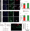
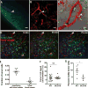
Lymph node metastases in cancer patients are associated with tumor aggressiveness, poorer prognoses, and the recommendation for systemic therapy. Whether cancer cells in lymph nodes can seed distant metastases has been a subject of considerable debate. We studied mice implanted with cancer cells (mammary carcinoma, squamous cell carcinoma, or melanoma) expressing the photoconvertible protein Dendra2. This technology allowed us to selectively photoconvert metastatic cells in the lymph node and trace their fate. We found that a fraction of these cells invaded lymph node blood vessels, entered the blood circulation, and colonized the lung. Thus, in mouse models, lymph node metastases can be a source of cancer cells for distant metastases. Whether this mode of dissemination occurs in cancer patients remains to be determined.
Copyright © 2018 The Authors, some rights reserved; exclusive licensee American Association for the Advancement of Science. No claim to original U.S. Government Works.
Conflict of interest statement
The authors declare no competing financial interests.
Figures


Comment in
-
Tumor Cells Metastasize from Lymph Nodes.Cancer Discov. 2018 Jun;8(6):OF10. doi: 10.1158/2159-8290.CD-NB2018-041. Epub 2018 Apr 12. Cancer Discov. 2018. PMID: 29650532
-
Take a left here.Nat Rev Cancer. 2018 Jun;18(6):337. doi: 10.1038/s41568-018-0011-x. Nat Rev Cancer. 2018. PMID: 29662237 No abstract available.
-
Exit Stage Left: A Tumor Cell's Journey from Lymph Node to Beyond.Trends Cancer. 2018 Aug;4(8):519-522. doi: 10.1016/j.trecan.2018.05.007. Epub 2018 Jun 6. Trends Cancer. 2018. PMID: 30064660
Similar articles
-
Lymph node blood vessels provide exit routes for metastatic tumor cell dissemination in mice.Science. 2018 Mar 23;359(6382):1408-1411. doi: 10.1126/science.aal3662. Science. 2018. PMID: 29567714
-
The Lymph Node and the Metastasis.N Engl J Med. 2018 May 24;378(21):2045-2046. doi: 10.1056/NEJMcibr1803854. N Engl J Med. 2018. PMID: 29791831 No abstract available.
-
Fate Mapping of Cancer Cells in Metastatic Lymph Nodes Using Photoconvertible Proteins.Methods Mol Biol. 2021;2265:363-376. doi: 10.1007/978-1-0716-1205-7_26. Methods Mol Biol. 2021. PMID: 33704727 Free PMC article.
-
Lymph node metastases. Indicators, but not governors of survival.Arch Surg. 1984 Sep;119(9):1067-72. doi: 10.1001/archsurg.1984.01390210063014. Arch Surg. 1984. PMID: 6383272 Review.
-
Model for mediastinal lymph node metastasis produced by orthotopic intrapulmonary implantation of lung cancer cells in mice.Hum Cell. 1999 Dec;12(4):197-204. Hum Cell. 1999. PMID: 10834106 Review.
Cited by
-
Phenotypic Heterogeneity, Bidirectionality, Universal Cues, Plasticity, Mechanics, and the Tumor Microenvironment Drive Cancer Metastasis.Biomolecules. 2024 Feb 3;14(2):184. doi: 10.3390/biom14020184. Biomolecules. 2024. PMID: 38397421 Free PMC article. Review.
-
Drug Combination Nanoparticles Containing Gemcitabine and Paclitaxel Enable Orthotopic 4T1 Breast Tumor Regression.Cancers (Basel). 2024 Aug 8;16(16):2792. doi: 10.3390/cancers16162792. Cancers (Basel). 2024. PMID: 39199565 Free PMC article.
-
The hybrid nanosystem for the identification and magnetic hyperthermia immunotherapy of metastatic sentinel lymph nodes as a multifunctional theranostic agent.Front Bioeng Biotechnol. 2024 Jul 29;12:1445829. doi: 10.3389/fbioe.2024.1445829. eCollection 2024. Front Bioeng Biotechnol. 2024. PMID: 39135950 Free PMC article.
-
Metastasis-Initiating Cells and Ecosystems.Cancer Discov. 2021 Apr;11(4):971-994. doi: 10.1158/2159-8290.CD-21-0010. Cancer Discov. 2021. PMID: 33811127 Free PMC article. Review.
-
Intranodal pressure of a metastatic lymph node reflects the response to lymphatic drug delivery system.Cancer Sci. 2020 Nov;111(11):4232-4241. doi: 10.1111/cas.14640. Epub 2020 Sep 18. Cancer Sci. 2020. PMID: 32882076 Free PMC article.
References
-
- Jatoi I, Hilsenbeck SG, Clark GM, Osborne CK. Significance of axillary lymph node metastasis in primary breast cancer. J Clin Oncol. 1999;17:2334–2340. - PubMed
-
- Kawada K, Taketo MM. Significance and mechanism of lymph node metastasis in cancer progression. Cancer research. 2011;71:1214–1218. - PubMed
-
- Saksena MA, Saokar A, Harisinghani MG. Lymphotropic nanoparticle enhanced MR imaging (LNMRI) technique for lymph node imaging. Eur J Radiol. 2006;58:367–374. - PubMed
-
- Starz H, Balda BR, Kramer KU, Buchels H, Wang H. A micromorphometry-based concept for routine classification of sentinel lymph node metastases and its clinical relevance for patients with melanoma. Cancer. 2001;91:2110–2121. - PubMed
Publication types
MeSH terms
Substances
Grants and funding
LinkOut - more resources
Full Text Sources
Other Literature Sources

