Fusion Inhibitory Lipopeptides Engineered for Prophylaxis of Nipah Virus in Primates
- PMID: 29566184
- PMCID: PMC6009590
- DOI: 10.1093/infdis/jiy152
Fusion Inhibitory Lipopeptides Engineered for Prophylaxis of Nipah Virus in Primates
Abstract
Background: The emerging zoonotic paramyxovirus Nipah virus (NiV) causes severe respiratory and neurological disease in humans, with high fatality rates. Nipah virus can be transmitted via person-to-person contact, posing a high risk for epidemic outbreaks. However, a broadly applicable approach for human NiV outbreaks in field settings is lacking.
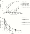
Methods: We engineered new antiviral lipopeptides and analyzed in vitro fusion inhibition to identify an optimal candidate for prophylaxis of NiV infection in the lower respiratory tract, and we assessed antiviral efficiency in 2 different animal models.
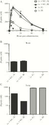
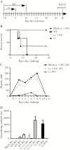
Results: We show that lethal NiV infection can be prevented with lipopeptides delivered via the respiratory route in both hamsters and nonhuman primates. By targeting retention of peptides for NiV prophylaxis in the respiratory tract, we avoid its systemic delivery in individuals who need only prevention, and thus we increase the safety of treatment and enhance utility of the intervention.
Conclusions: The experiments provide a proof of concept for the use of antifusion lipopeptides for prophylaxis of lethal NiV. These results advance the goal of rational development of potent lipopeptide inhibitors with desirable pharmacokinetic and biodistribution properties and a safe effective delivery method to target NiV and other pathogenic viruses.
Figures


Similar articles
-
Single-dose live-attenuated Nipah virus vaccines confer complete protection by eliciting antibodies directed against surface glycoproteins.Vaccine. 2014 May 7;32(22):2637-44. doi: 10.1016/j.vaccine.2014.02.087. Epub 2014 Mar 12. Vaccine. 2014. PMID: 24631094 Free PMC article.
-
Efficient reverse genetics reveals genetic determinants of budding and fusogenic differences between Nipah and Hendra viruses and enables real-time monitoring of viral spread in small animal models of henipavirus infection.J Virol. 2015 Jan 15;89(2):1242-53. doi: 10.1128/JVI.02583-14. Epub 2014 Nov 12. J Virol. 2015. PMID: 25392218 Free PMC article.
-
Griffithsin Inhibits Nipah Virus Entry and Fusion and Can Protect Syrian Golden Hamsters From Lethal Nipah Virus Challenge.J Infect Dis. 2020 May 11;221(Supplement_4):S480-S492. doi: 10.1093/infdis/jiz630. J Infect Dis. 2020. PMID: 32037447 Free PMC article.
-
Envelope-receptor interactions in Nipah virus pathobiology.Ann N Y Acad Sci. 2007 Apr;1102(1):51-65. doi: 10.1196/annals.1408.004. Ann N Y Acad Sci. 2007. PMID: 17470911 Free PMC article. Review.
-
Molecular characteristics of the Nipah virus glycoproteins.Ann N Y Acad Sci. 2007 Apr;1102:39-50. doi: 10.1196/annals.1408.003. Ann N Y Acad Sci. 2007. PMID: 17470910 Review.
Cited by
-
Dual Inhibition of Human Parainfluenza Type 3 and Respiratory Syncytial Virus Infectivity with a Single Agent.J Am Chem Soc. 2019 Aug 14;141(32):12648-12656. doi: 10.1021/jacs.9b04615. Epub 2019 Aug 5. J Am Chem Soc. 2019. PMID: 31268705 Free PMC article.
-
Nipah virus W protein harnesses nuclear 14-3-3 to inhibit NF-κB-induced proinflammatory response.Commun Biol. 2021 Nov 16;4(1):1292. doi: 10.1038/s42003-021-02797-5. Commun Biol. 2021. PMID: 34785771 Free PMC article.
-
Structure of SARS-CoV-2 Spike Glycoprotein for Therapeutic and Preventive Target.Immune Netw. 2021 Feb 19;21(1):e8. doi: 10.4110/in.2021.21.e8. eCollection 2021 Feb. Immune Netw. 2021. PMID: 33728101 Free PMC article. Review.
-
Structural and Functional Characterization of Membrane Fusion Inhibitors with Extremely Potent Activity against Human Immunodeficiency Virus Type 1 (HIV-1), HIV-2, and Simian Immunodeficiency Virus.J Virol. 2018 Sep 26;92(20):e01088-18. doi: 10.1128/JVI.01088-18. Print 2018 Oct 15. J Virol. 2018. PMID: 30089693 Free PMC article.
-
Nebulized fusion inhibitory peptide protects cynomolgus macaques from measles virus infection.Res Sq [Preprint]. 2022 Jun 1:rs.3.rs-1700877. doi: 10.21203/rs.3.rs-1700877/v1. Res Sq. 2022. Update in: Nat Commun. 2022 Oct 28;13(1):6439. doi: 10.1038/s41467-022-33832-6. PMID: 35677066 Free PMC article. Updated. Preprint.
References
Publication types
MeSH terms
Substances
Grants and funding
LinkOut - more resources
Full Text Sources
Other Literature Sources

