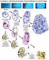
Granulosa Cell Apoptosis in the Ovarian Follicle-A Changing View
- PMID: 29551992
- PMCID: PMC5840209
- DOI: 10.3389/fendo.2018.00061
Granulosa Cell Apoptosis in the Ovarian Follicle-A Changing View
Abstract
Recent studies challenge the previous view that apoptosis within the granulosa cells of the maturing ovarian follicle is a reflection of aging and consequently a marker for poor quality of the contained oocyte. On the contrary, apoptosis within the granulosa cells is an integral part of normal development and has limited predictive capability regarding oocyte quality or the ensuing pregnancy rate in in vitro fertilization programs. This review article covers our revised understanding of the process of apoptosis within the ovarian follicle, its three phenotypes, the major signaling pathways underlying apoptosis as well as the associated mitochondrial pathways.
Keywords: aging effects; apoptosis signaling; bone morphogenetic proteins; fertility preservation; mitogenic growth; ovarian reserve; receptor of follicle stimulating hormone.
Figures


Similar articles
-
The effect of ovarian reserve and receptor signalling on granulosa cell apoptosis during human follicle development.Mol Cell Endocrinol. 2018 Jul 15;470:219-227. doi: 10.1016/j.mce.2017.11.002. Epub 2017 Nov 4. Mol Cell Endocrinol. 2018. PMID: 29113831
-
Mitochondrial Function in Modulating Human Granulosa Cell Steroidogenesis and Female Fertility.Int J Mol Sci. 2020 May 19;21(10):3592. doi: 10.3390/ijms21103592. Int J Mol Sci. 2020. PMID: 32438750 Free PMC article.
-
Beyond apoptosis: evidence of other regulated cell death pathways in the ovary throughout development and life.Hum Reprod Update. 2023 Jul 5;29(4):434-456. doi: 10.1093/humupd/dmad005. Hum Reprod Update. 2023. PMID: 36857094 Free PMC article. Review.
-
Growth hormone during in vitro fertilization in older women modulates the density of receptors in granulosa cells, with improved pregnancy outcomes.Fertil Steril. 2018 Dec;110(7):1298-1310. doi: 10.1016/j.fertnstert.2018.08.018. Fertil Steril. 2018. PMID: 30503129 Clinical Trial.
-
Oocyte-somatic cell interactions in the human ovary-novel role of bone morphogenetic proteins and growth differentiation factors.Hum Reprod Update. 2016 Dec;23(1):1-18. doi: 10.1093/humupd/dmw039. Epub 2016 Oct 26. Hum Reprod Update. 2016. PMID: 27797914 Free PMC article. Review.
Cited by
-
In pre-clinical study fetal hypoxia caused autophagy and mitochondrial impairment in ovary granulosa cells mitigated by melatonin supplement.J Adv Res. 2024 Oct;64:15-30. doi: 10.1016/j.jare.2023.11.008. Epub 2023 Nov 11. J Adv Res. 2024. PMID: 37956860 Free PMC article.
-
High Fat-High Fructose Diet Elicits Hypogonadotropism Culminating in Autophagy-Mediated Defective Differentiation of Ovarian Follicles.Cells. 2022 Oct 31;11(21):3447. doi: 10.3390/cells11213447. Cells. 2022. PMID: 36359843 Free PMC article.
-
Genome wide identification and characterization of fertility associated novel CircRNAs as ceRNA reveal their regulatory roles in sheep fecundity.J Ovarian Res. 2023 Jun 20;16(1):115. doi: 10.1186/s13048-023-01178-2. J Ovarian Res. 2023. PMID: 37340323 Free PMC article.
-
miR-128-3p Regulates Follicular Granulosa Cell Proliferation and Apoptosis by Targeting the Growth Hormone Secretagogue Receptor.Int J Mol Sci. 2024 Feb 27;25(5):2720. doi: 10.3390/ijms25052720. Int J Mol Sci. 2024. PMID: 38473968 Free PMC article.
-
Static magnetic field assisted thawing improves cryopreservation of mouse whole ovaries.Bioeng Transl Med. 2023 Oct 18;9(1):e10613. doi: 10.1002/btm2.10613. eCollection 2024 Jan. Bioeng Transl Med. 2023. PMID: 38193129 Free PMC article.
References
-
- Regan SLP, Knight PG, Yovich J, Stanger J, Leung Y, Arfuso F, et al. Dysregulation of granulosal bone morphogenetic protein receptor 1B density is associated with reduced ovarian reserve and the age-related decline in human fertility. Mol Cell Endocrinol (2016) 425:84–93.10.1016/j.mce.2016.01.016 - DOI - PubMed
Publication types
LinkOut - more resources
Full Text Sources
Other Literature Sources

