. 2018 Mar 1;32(5-6):389-401.
doi: 10.1101/gad.307132.117. Epub 2018 Mar 13.

Cooperative recruitment of Yan via a high-affinity ETS supersite organizes repression to confer specificity and robustness to cardiac cell fate specification

Affiliations

Cooperative recruitment of Yan via a high-affinity ETS supersite organizes repression to confer specificity and robustness to cardiac cell fate specification

Jean-François Boisclair Lachance et al. Genes Dev. .

Abstract

Cis-regulatory modules (CRMs) are defined by unique combinations of transcription factor-binding sites. Emerging evidence suggests that the number, affinity, and organization of sites play important roles in regulating enhancer output and, ultimately, gene expression. Here, we investigate how the cis-regulatory logic of a tissue-specific CRM responsible for even-skipped (eve) induction during cardiogenesis organizes the competing inputs of two E-twenty-six (ETS) members: the activator Pointed (Pnt) and the repressor Yan. Using a combination of reporter gene assays and CRISPR-Cas9 gene editing, we suggest that Yan and Pnt have distinct syntax preferences. Not only does Yan prefer high-affinity sites, but an overlapping pair of such sites is necessary and sufficient for Yan to tune Eve expression levels in newly specified cardioblasts and block ectopic Eve induction and cell fate specification in surrounding progenitors. Mechanistically, the efficient Yan recruitment promoted by this high-affinity ETS supersite not only biases Yan-Pnt competition at the specific CRM but also organizes Yan-repressive complexes in three dimensions across the eve locus. Taken together, our results uncover a novel mechanism by which differential interpretation of CRM syntax by a competing repressor-activator pair can confer both specificity and robustness to developmental transitions.

Keywords: Drosophila embryogenesis; ETS transcription factor; cis-regulatory syntax; even-skipped; heart development; receptor tyrosine kinase.


Figures

Figure 1.
Figure 1.
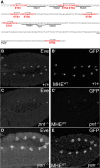
The MHE reliably reports the pattern of mesodermal Eve expression. (A) MHE sequence with putative ETS sites in red. Black arrows indicate site orientation. Previously characterized Mad and Twi sites are underlined. (BE) Lateral views, oriented with anterior to the left and ventral down, of the thoracic and abdominal segments of representative stage 11 embryos expressing two copies of the MHEWT-GFP reporter in w1118 (B), pntΔ88 (C), or yanER443 mutants (D,E). Costaining with anti-Eve (B,C) and anti-GFP (B′,C′) shows that the reporter-driven pattern matches closely that of endogenous Eve. (D,E) Loss of yan expands these Eve expressing and GFP-expressing cell clusters.
Figure 2.
Figure 2.
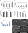
An overlapping pair of high-affinity ETS sites organizes MHE repression, while lower-affinity sites contribute predominantly to reporter activation. (A) Competitive gel shift assay testing the ability of MHE ETS sites 1–8 to outcompete a 32P-labeled ETS site probe bound to a YanA86D monomer. ETS sites 2, 3, and 8 competed effectively, whereas the remaining sites did not. (B) The impact of individual ETS site mutations (mut1–mut8) on the average cluster intensity of the MHE-GFP reporter suggests a role for strong-affinity sites in repression and lower-affinity sites in activation. In this and all subsequent diagrams, ETS sites and their sense/antisense orientations are depicted with arrowheads, with lower-affinity sites in gray, strong sites in black, and mutated sites in white. Error bars show SEM. The statistical significance of single mutants relative to MHEWT after Bonferroni correction is indicated. (***) P < 0.001. (C) The impact of double and triple mutants on MHE-GFP reporter expression. The statistical significance relative to each of the relevant single-site mutations after Bonferroni correction is provided in numerical order. (*) P < 0.05; (***) P < 0.001. The mut2,3 combination was unique in its nonadditive increase in reporter expression; although measurements were less linear at this higher range of GFP expression, all three quantification strategies tested revealed a similar trend (Supplemental Fig. S2C–E). Mutation of all three strong sites reduced reporter expression, showing that these sites also mediate activating inputs and that effective activation requires multiple sites. (D,D′) Representative stage 11 embryos showing the increased and ectopic expression driven by one copy of MHEmut2,3 in and outside Eve+ clusters. (E,F) Single and double mutations in MHE ETS sites 2 and 3 reduce the fold repression by twi-GAL4-driven YanACT (E) and fold activation by twi-GAL4-driven PntP1 (F) relative to MHEWT. All measurements were normalized to MHEWT, but, in F, the intensity of the confocal laser was set on MHEmu2,3/twi-GAL4,UAS-PntP1 embryos instead of MHEWT as in E. All differences are statistically different, with P < 0.001. Error bars show SEM.
Figure 3.
Figure 3.
Evolutionary conservation and sufficiency of an ETS2,3-like pair for MHE repression. (A) Phylogenetic tree (adapted from Gramates et al. 2017) and schematic representation of MHE ETS sites in eight different Drosophila species; light gray indicates species whose sequences were not analyzed. Full MHE sequences, as identified by Hare et al. (2008), are in Supplemental Figure S2L. (B) The introduction of ectopic paired high-affinity ETS sites can restore effective repression to the MHEmut2,3 reporter. Error bars show SEM. Statistical significance after Bonferroni correction from either MHEWT, MHEmut2,3, or MHEmut2,3 strong9,8 (where relevant) is shown. (*) P < 0.05; (***) P < 0.001.
Figure 4.
Figure 4.
The overlapping anti-parallel configuration of the ETS2,3 pair may preclude classic cooperative recruitment of Yan dimers. (AC) Gel shifts using 32P-labeled probes and recombinant Yan monomers (YanA86D) or Yan dimers (1:1 YanA86D:YanV105R). Increasing concentrations of total Yan protein are indicated above each gel. (A) SAM-mediated dimerization induces classic cooperative binding to probe with two consensus ETS sites (2× ETS). Single-bound (Y) and double-bound (Y–Y) species are indicated. (B) Cooperative binding requires two ETS sites. (C) The MHE ETS2,3 sites do not support simultaneous occupancy by two Yan molecules. The slower-migrating species (Y–Y) observed with Yan dimers occurs even when one ETS site is mutated, suggesting that it reflects association of the second Yan molecule to an adjacent nonspecific sequence. (D) A structural model of a Yan ETS DNA-binding domain (DBD) dimer in complex with the MHE ETS2,3 sequence, viewed from the side (D) and along the axis of the DNA (D′), predicts a strong steric clash that should preclude simultaneous occupancy of both ETS sites. The two Yan DBDs are colored in green and purple.
Figure 5.
Figure 5.
Loss of robustness in evemut2,3 embryos highlights the importance of ETS2,3-mediated regulation in cardiac cell fate specification. (A) Quantification of average Eve levels per cluster shows that the eveMHEmut2,3 background is sensitized to genetic perturbations that are well buffered against in eveMHEWT embryos. Error bars show SEM. Statistical significance after Bonferroni correction is depicted. (*) P < 0.05; (**) P < 0.01; (***) P < 0.001. (B) The average number of Eve+ cells per 10 clusters in the same genetic backgrounds as in A shows that specification of ectopic Eve+ cells occurs in the eveMHEmut2,3 background, but not in the eveMHEWT control, when the Pnt:Yan ratio is increased. Error bars show SEM. (C) The reduced survival of eveMHEmut2,3 embryos to adulthood was enhanced by temperature stress. (D) Increased pnt dose also decreased eveMHEmut2,3 survival. (E) ChIP-qPCR (chromatin immunoprecipitation [ChIP combined with quantitative PCR [qPCR]) from stage 11 embryos. Signals were normalized to a negative control (see Supplemental Methods). Mutation of the ETS2,3 site reduced occupancy at all three eve CRMs but not at argos (aos). (FI) Eve expression in representative stage 11 embryos. Examples of clusters with four or more Eve+ cells are indicated with white arrows. Error bars show SEM.
Figure 6.
Figure 6.
Mutation of the ETS2,3 site increases sensitivity to variation in upstream signaling. Quantification of average Eve levels per cluster (A) and average number of Eve+ cells (B) shows that aos heterozygosity increases the average number of Eve+ cells specified in eveMHEmut2,3 embryos. Error bars show SEM. (C,D) Representative stage 11 embryos, with clusters with extra Eve+ cells marked by a white arrow.

Similar articles

Cited by

References

    1. Arnold K, Bordoli L, Kopp J, Schwede T. 2006. The SWISS-MODEL workspace: a Web-based environment for protein structure homology modelling. Bioinformatics 22: 195–201. - PubMed
    1. Biasini M, Bienert S, Waterhouse A, Arnold K, Studer G, Schmidt T, Kiefer F, Cassarino TG, Bertoni M, Bordoli L, et al. 2014. SWISS-MODEL: modelling protein tertiary and quaternary structure using evolutionary information. Nucleic Acids Res 42: W252–W258. - PMC - PubMed
    1. Bischof J, Maeda RK, Hediger M, Karch F, Basler K. 2007. An optimized transgenesis system for Drosophila using germ-line-specific phiC31 integrases. Proc Natl Acad Sci 104: 3312–3317. - PMC - PubMed
    1. Boisclair Lachance JF, Peláez N, Cassidy JJ, Webber JL, Rebay I, Carthew RW. 2014. A comparative study of pointed and yan expression reveals new complexity to the transcriptional networks downstream of receptor tyrosine kinase signaling. Dev Biol 385: 263–278. - PMC - PubMed
    1. Carmena A, Gisselbrecht S, Harrison J, Jiménez F, Michelson AM. 1998. Combinatorial signaling codes for the progressive determination of cell fates in the Drosophila embryonic mesoderm. Genes Dev 12: 3910–3922. - PMC - PubMed

Publication types

MeSH terms

LinkOut - more resources