. 2018 Apr 4;26(4):1066-1081.
doi: 10.1016/j.ymthe.2018.01.023. Epub 2018 Feb 3.

miR-296-3p Negatively Regulated by Nicotine Stimulates Cytoplasmic Translocation of c-Myc via MK2 to Suppress Chemotherapy Resistance

Affiliations

miR-296-3p Negatively Regulated by Nicotine Stimulates Cytoplasmic Translocation of c-Myc via MK2 to Suppress Chemotherapy Resistance

Xiaojie Deng et al. Mol Ther. .

Abstract

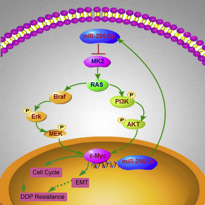
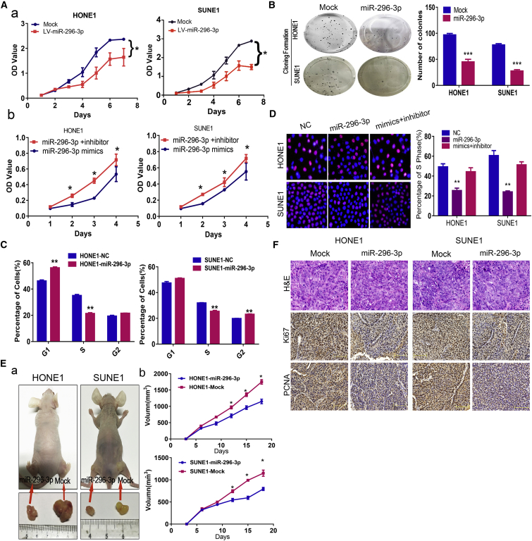
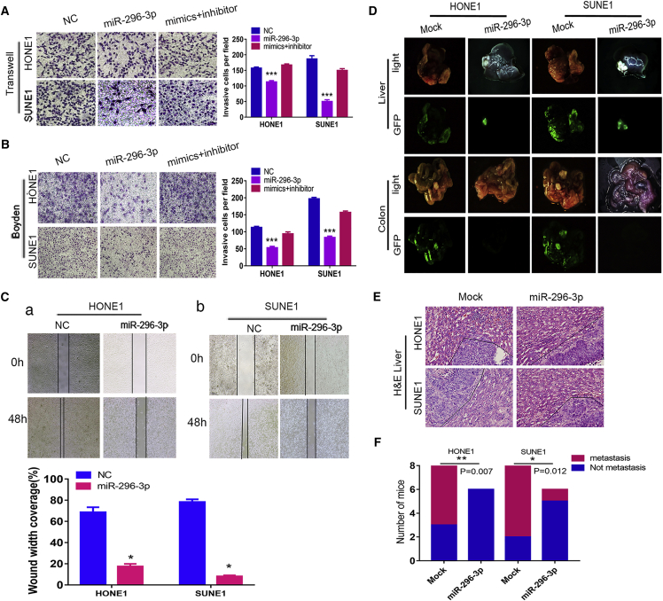
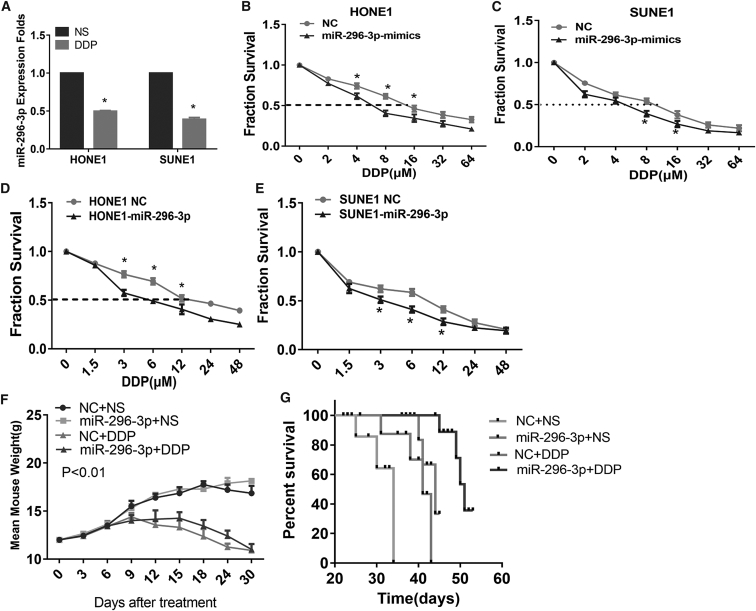
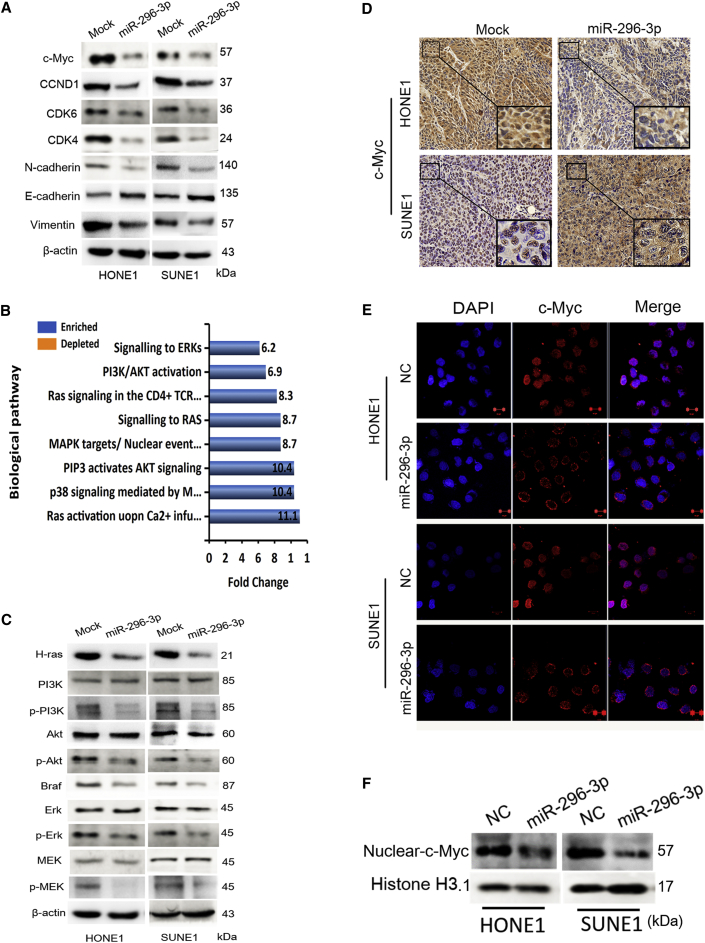
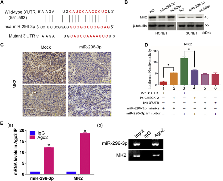
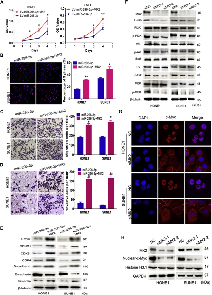
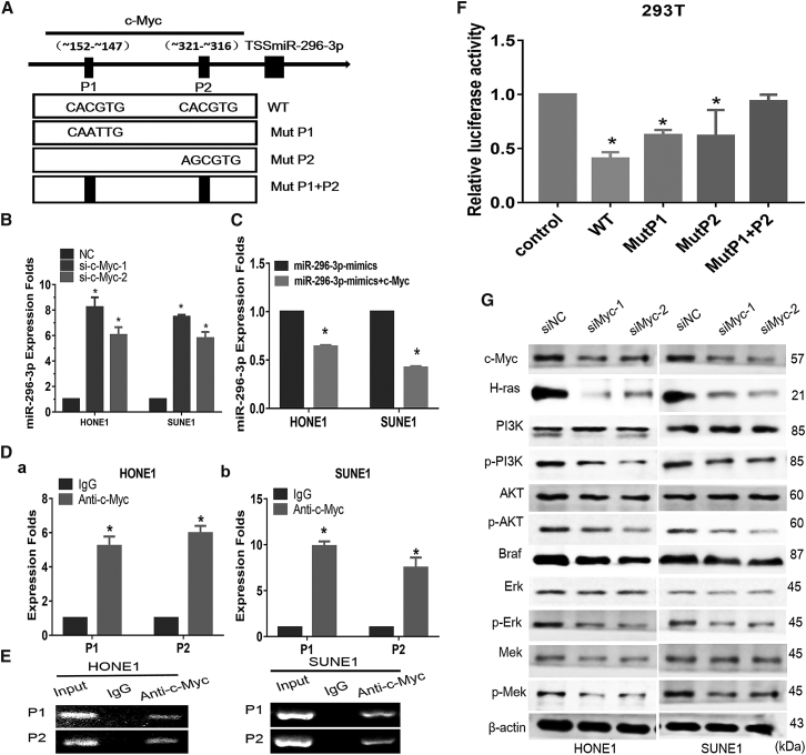
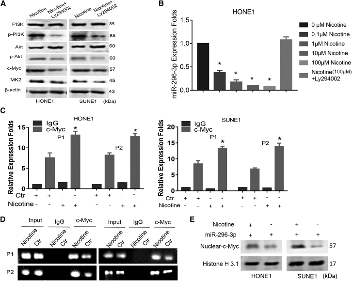
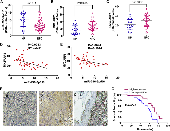
This study aimed to identify mechanisms by which microRNA 296-3p (miR-296-3p) functions as a tumor suppressor to restrain nasopharyngeal carcinoma (NPC) cell growth, metastasis, and chemoresistance. Mechanistic studies revealed that miR-296-3p negatively regulated by nicotine directly targets the oncogenic protein mitogen-activated protein kinase-activated protein kinase-2 (Mapkapk2) (MK2). Suppression of MK2 downregulated Ras/Braf/Erk/Mek/c-Myc and phosphoinositide-3-kinase (PI3K)/Akt/c-Myc signaling and promoted cytoplasmic translocation of c-Myc, which activated miR-296-3p expression by a feedback loop. This ultimately inhibited cell cycle progression, epithelial-to-mesenchymal transition (EMT), and chemoresistance of NPC. In addition, nicotine as a key component of tobacco was observed to suppress miR-296-3p and thus elevate MK2 expression by inducing PI3K/Akt/c-Myc signaling. In clinical samples, reduced miR-296-3p as an unfavorable factor was inversely correlated with MK2 and c-Myc expression. These results reveal a novel mechanism by which miR-296-3p negatively regulated by nicotine directly targets MK2-induced Ras/Braf/Erk/Mek/c-Myc or PI3K/AKT/c-Myc signaling to stimulate its own expression and suppress NPC cell proliferation and metastasis. miR-296-3p may thus serve as a therapeutic target to reverse chemotherapy resistance of NPC.

Keywords: MK2; miR-296-3p; nasopharyngeal carcinoma; nicotine.


Figures

None
Graphical abstract
Figure 1
Figure 1
miR-296-3p Suppresses NPC Cell Growth In Vitro and In Vivo (A) miR-296-3p lentivirus markedly inhibited cell proliferation (a) and inhibitor restored cell proliferation in SUNE1 and HONE1 cells by MTT assay (b). Student’s t test, one-way ANOVA, and Dunnett’s multiple comparison test. Mean ± SD, *p < 0.05; **p < 0.01; ***p < 0.001. (B) Colony formation assay and of HONE1 and SUNE1 cells was performed after transfection with NC, miR-296-3p lentivirus, and inhibitor, as indicated. Scale bar, 15 mm. Parametric generalized linear model with random effects. (C and D) FACS assays (C) and EdU incorporation assays (D) showed miR-296-3p mimics markedly inhibited G1 to S and G2 cell cycle transition in vitro. Student’s t test, one-way ANOVA, and Dunnett’s multiple comparison test. Mean ± SD, *p < 0.05; **p < 0.01; ***p < 0.001. (E) The in vivo effect of miR-296-3p was evaluated in xenograft mouse models bearing tumors originating from HONE1 and SUNE1 cells, N = 6/group (a); tumor volume was periodically measured for each mouse (b). Parametric generalized linear model with random effects, *p < 0.05. (F) Representative H&E as well as Ki67 and PCNA IHC staining of primary tumor tissues are shown. Scale bar, 30 mm.
Figure 2
Figure 2
miR-296-3p Significantly Inhibits NPC Cell Metastasis In Vitro and In Vivo (A–C) Transwell assay (A), Boyden assay (B), and wound-healing assays (C) evaluated the migration and invasion of NPC cell treatment with NC, miR-296-3p mimics, and inhibitor, respectively. Scale bar, 30 or 60 μm. Student’s t test, mean ± SD, ***p < 0.001. (D) HONE1 and SUNE1 cells with stable miR-296-3p expression or their respective control cells were inoculated under the liver capsules of nude mice. Fluorescent images are shown for four representative mouse models. Intrahepatic dissemination and intestine metastasis predominantly appeared in the control mice compared with miR-296-3p cell-injected mouse models. Data are presented as mean ± SD for three independent experiments (*p < 0.05). Ctr, negative control cells. (E) H&E staining of NPC metastasis tumor from mouse liver. Original magnification, × 400; scale bar, 25 μm. Data are presented as mean ± SD for three independent experiments. (F) Graph described the numbers of mice, in which metastasis occurred or not; χ2 test, *p < 0.05, **p < 0.01.
Figure 3
Figure 3
miR-296-3p Enhances Cisplatin Chemosensitivity In Vitro and In Vivo (A) miR-296-3p expression was examined by RT-PCR after treatment with DDP for 18 hr. (B and C) miR-296-3p mimics significantly elevated NPC cell sensitivity to DDP in HONE1(B) and SUNE1 (C) cells. (D and E) miR-296-3p lentivirus remarkably elevated NPC cell sensitivity to DDP in HONE1 (D) and SUNE1 (E) cells. (F) Bodyweight of mice was measured after 30 days of treatment with DDP. (G) Animals were divided into four groups: control cells (NC) + NS, miR-296-3p+NS, NC+DDP, or miR-296-3p+DDP (each group: N = 10). The rank of overall survival time was as follows: NC+NS < miR-296-3p+NS < NC+DDP < miR-296-3p+DDP.
Figure 4
Figure 4
miR-296-3p Suppresses Ras/Braf/Erk/Mek and PI3K/AKT Pathways, Reducing c-Myc Transcriptional Activity (A) Expression levels of c-Myc, CCND1, CDK6, CDK4, N-cadherin, E-cadherin, and Vimentin were detected following transfection with miR-296-3p mimics. β-actin was used as a loading control. (B) Schematic diagram of the potential pathway was modulated by miR-296-3p. (C) Expression levels of H-ras, Braf, Erk, Mek, PI3K, and AKT were detected after transfection of miR-296-3p mimics. β-actin served as a loading control. (D) c-Myc IHC staining of primary tumor tissues is shown. Scale bar, 30 mm. (E) Immunofluorescence of c-Myc in HONE1 and SUNE1 cells is shown after transfection of miR-296-3p and NC. (F) Nuclear c-Myc was detected after transfection of miR-296-3p and NC. Histone H3.1 served as a loading control.
Figure 5
Figure 5
miR-296-3p Directly Targets MK2 (A) Bioinformatics analysis revealed the 3′ UTR region of MK2 was similar to the seed sequence of miR-296-3p. Mutants were generated in the binding region of MK2 3′ UTR. (B) MK2 protein levels were examined in miR-296-3p-overexpressing/suppressing HONE1 cells and SUNE1 cells by western blot. (C) MK2 IHC staining of primary tumor tissues is shown. Scale bar, 30 mm. (D) miR-296-3p directly targets MK2 as confirmed by dual-luciferase reporter assay. One-way ANOVA and Dunnett’s multiple comparison test; *p < 0.05. (E) RT-qPCR (a) and PCR (b) showed that miR-296-3p and MK2 bind the AGO2 protein; *p < 0.05.
Figure 6
Figure 6
Ectopic Expression of MK2 Mitigates miR-296-3p Suppression of NPC Proliferation (A and B) MTT assay (A) and EdU incorporation assay (B) were performed after transfection with NC, ectopic MK2, or miR-296-3p, as indicated. Scale bar, 15 mm. Parametric generalized linear model with random effects, Student’s t test, one-way ANOVA, and Dunnett’s multiple comparison test. Mean ± SD, *p < 0.05; **p < 0.01. (C and D) Transwell assay (C) and Boyden assay (D) of miR-296-3p-overexpressing cells or treated with MK2 plasmid and mimics. Scale bar, 50 μm. Student’s t test, one-way ANOVA, *p < 0.05; **p < 0.01. (E) MK2 elevated the expression levels of N-cadherin, Vimentin, CCND1, CDK6, and CDK4, but reduced E-cadherin in miR-296-3p-overexpressing HONE1 and SUNE1 cells. β-tubulin served as a loading control. (F) Expression levels of H-ras, Braf, Erk, Mek, PI3K, and AKT were detected after MK2 plasmid transfection in miR-296-3p-overexpressing cells. β-tubulin served as a loading control. (G) Immunofluorescence of c-Myc was examined in HONE1 and SUNE1 cells after transfection of MK2 siRNA and NC. (H) Nuclear c-Myc detection was examined after transfection of MK2 siRNA and NC. Histone H3.1 served as a loading control.
Figure 7
Figure 7
Endogenous c-Myc Negatively Modulates miR-296-3p Expression by Directly Binding to the Promoter Region (A) Schematic of the promoter regions of miR-296-3p with potential c-Myc binding sites (P1 and P2) and the structure of the wild-type (WT) and mutant binding sites (Mut P1, Mut P2, and Mut P1+P2). (B) miR-296-3p expression in c-Myc-suppressed HONE1 and SUNE1 cells was examined by RT-PCR, normalized to U6. One-way ANOVA and Dunnett’s multiple comparison test; *p < 0.05. (C) miR-296-3p expression was detected after co-transfection of mimics and c-Myc plasmid compared to the mimics group by RT-PCR, normalized to U6. One-way ANOVA and Dunnett’s multiple comparison test; *p < 0.05. (D and E) RT-PCR (D) (a and b) and PCR gel (E) showing amplification of c-Myc-binding sites P1+P2, P1, and P2 after ChIP using antibody against c-Myc. IgG antibody was used as a negative control. Gel figures were accompanied by the locations of molecular weight markers. (F) Dual-luciferase reporter assay identified the luciferase activities of the wild-type and Mut P1, Mut P2, and Mut P1+P2 of the miR-296-3p promoter in HONE1 and SUNE1 cells transfected with c-Myc plasmid. One-way ANOVA and Dunnett’s multiple comparison test; *p < 0.05. (G) Expression levels of H-ras, Braf, Erk, Mek, PI3K, and AKT were detected after c-Myc siRNA transfection. β-actin served as a loading control.
Figure 8
Figure 8
Nicotine Decreases miR-296-3p via PI3K/Akt/c-Myc Signaling (A) Ly294002 decreased p-PI3K, p-Akt, c-Myc, and MK2 in NPC cells treated with nicotine. (B) RT-qPCR showed miR-296-3p expression was decreased after treatment with different concentrations of nicotine and Ly294002 restored the effect of nicotine. Student’s t test, *p < 0.05. (C and D) Nicotine enhanced binding of c-Myc, with the promoter region of miR-296-3p based on RT-qPCR (C) and gel electrophoresis assays (D). Student’s t test, *p < 0.05. (E) Nicotine stimulated c-Myc nuclear translocation in miR-296-3p-overexpressing cells.
Figure 9
Figure 9
Pathoclinical Features of miR-296-3p Expression and Their Correlation with MK2 and c-Myc Expression (A–C) mRNA expression of miR-296-3p (A), MK2 (B), and c-Myc (C) in fresh NPC and NP samples was detected by RT-PCR, normalized to U6 and ARF5, respectively. Student’s t test, mean ± SD, p = 0.011; p = 0.0023; p = 0.0087. (D and E) miR-296-3p expression was negatively correlated with expression of MK2 mRNA (D) and c-Myc mRNA (E) in NPC tissues. (F) miR-296-3p expression was examined in paraffin NPC and NP samples by in situ hybridization (ISH). Weak expression level of miR-296-3p was shown in NPC samples (a); strong staining of miR-296-3p was shown in NPC samples (b); and positive expression of miR-296-3p was shown in NP samples (c) (original magnification × 400). Scale bar, 30 mm. (G) Kaplan-Meier survival analysis showing overall survival of 110 NPC patients on the basis of miR-296-3p expression. (Log-rank test, p = 0.0042)

Similar articles

Cited by

References

    1. Di Leva G., Garofalo M., Croce C.M. MicroRNAs in cancer. Annu. Rev. Pathol. 2014;9:287–314. - PMC - PubMed
    1. Kohlhapp F.J., Mitra A.K., Lengyel E., Peter M.E. MicroRNAs as mediators and communicators between cancer cells and the tumor microenvironment. Oncogene. 2015;34:5857–5868. - PMC - PubMed
    1. Bonci D., Coppola V., Patrizii M., Addario A., Cannistraci A., Francescangeli F., Pecci R., Muto G., Collura D., Bedini R. A microRNA code for prostate cancer metastasis. Oncogene. 2016;35:1180–1192. - PMC - PubMed
    1. Wang S.N., Luo S., Liu C., Piao Z., Gou W., Wang Y., Guan W., Li Q., Zou H., Yang Z.Z. miR-491 inhibits osteosarcoma lung metastasis and chemoresistance by targeting αB-crystallin. Mol. Ther. 2017;25:2140–2149. - PMC - PubMed
    1. Liu W., Zhang B., Chen G., Wu W., Zhou L., Shi Y., Zeng Q., Li Y., Sun Y., Deng X. Targeting miR-21 with sophocarpine inhibits tumor progression and reverses epithelial-mesenchymal transition in head and neck cancer. Mol. Ther. 2017;25:2129–2139. - PMC - PubMed

Publication types

MeSH terms