

Interpretation of P53 Immunohistochemistry in Endometrial Carcinomas: Toward Increased Reproducibility
- PMID: 29517499
- PMCID: PMC6127005
- DOI: 10.1097/PGP.0000000000000488
Interpretation of P53 Immunohistochemistry in Endometrial Carcinomas: Toward Increased Reproducibility
Abstract
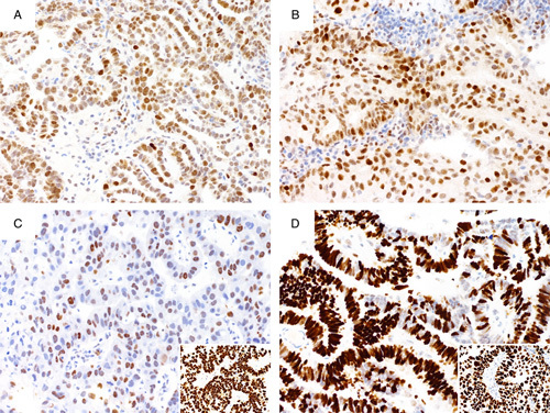
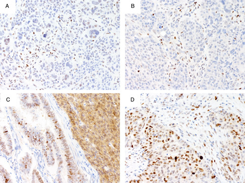
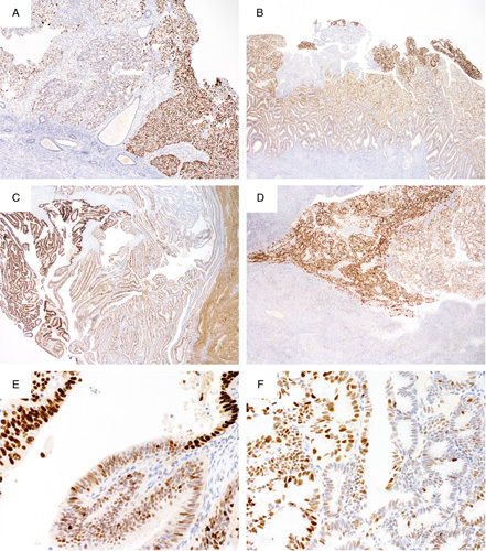
P53 immunohistochemistry has evolved into an accurate surrogate reflecting the underlying TP53 mutation status of a tumor, and has utility in the diagnostic workup of endometrial carcinomas. Recent work predominantly carried out in tubo-ovarian high-grade serous carcinoma has revealed 4 main patterns of p53 staining (normal/wild-type, complete absence, overexpression, and cytoplasmic); the latter 3 patterns are variably termed abnormal/aberrant/mutation-type and are strongly predictive of an underlying TP53 mutation. The aim of this review is to provide practical advice to pathologists regarding various aspects of p53 immunohistochemical staining. These include laboratory methods to optimize staining, a description of the different patterns of staining, advice regarding the interpretation, and reporting of p53 staining and practical uses of p53 staining in endometrial carcinoma diagnosis. Illustrations are provided to aid in the interpretational problems.
Conflict of interest statement
The authors declare no conflict of interest.
Figures


Similar articles
-
p53 immunohistochemistry is an accurate surrogate for TP53 mutational analysis in endometrial carcinoma biopsies.J Pathol. 2020 Mar;250(3):336-345. doi: 10.1002/path.5375. Epub 2020 Jan 29. J Pathol. 2020. PMID: 31829441
-
Cytoplasmic Pattern p53 Immunoexpression in Pelvic and Endometrial Carcinomas With TP53 Mutation Involving Nuclear Localization Domains: An Uncommon But Potential Diagnostic Pitfall With Clinical Implications.Am J Surg Pathol. 2021 Nov 1;45(11):1441-1451. doi: 10.1097/PAS.0000000000001713. Am J Surg Pathol. 2021. PMID: 33899789
-
The Many Uses of p53 Immunohistochemistry in Gynecological Pathology: Proceedings of the ISGyP Companion Society Session at the 2020 USCAP Annual9 Meeting.Int J Gynecol Pathol. 2021 Jan;40(1):32-40. doi: 10.1097/PGP.0000000000000725. Int J Gynecol Pathol. 2021. PMID: 33290354 Review.
-
Immunohistochemical staining patterns of p53 can serve as a surrogate marker for TP53 mutations in ovarian carcinoma: an immunohistochemical and nucleotide sequencing analysis.Mod Pathol. 2011 Sep;24(9):1248-53. doi: 10.1038/modpathol.2011.85. Epub 2011 May 6. Mod Pathol. 2011. PMID: 21552211
-
Does a p53 "Wild-type" Immunophenotype Exclude a Diagnosis of Endometrial Serous Carcinoma?Adv Anat Pathol. 2018 Jan;25(1):61-70. doi: 10.1097/PAP.0000000000000171. Adv Anat Pathol. 2018. PMID: 28945609 Review.
Cited by
-
Correlation of PD-L1 immunohistochemical expression with microsatellite instability and p53 status in endometrial carcinoma.Eur J Obstet Gynecol Reprod Biol X. 2022 Nov 21;16:100172. doi: 10.1016/j.eurox.2022.100172. eCollection 2022 Dec. Eur J Obstet Gynecol Reprod Biol X. 2022. PMID: 36440057 Free PMC article.
-
Implementing an integrated molecular classification for gastric cancer from endoscopic biopsies using on-slide tests.Rom J Morphol Embryol. 2024 Apr-Jun;65(2):257-265. doi: 10.47162/RJME.65.2.12. Rom J Morphol Embryol. 2024. PMID: 39020540 Free PMC article.
-
p53 immunohistochemistry in endometrial cancer: clinical and molecular correlates in the PORTEC-3 trial.Mod Pathol. 2022 Oct;35(10):1475-1483. doi: 10.1038/s41379-022-01102-x. Epub 2022 Jun 25. Mod Pathol. 2022. PMID: 35752743 Free PMC article. Clinical Trial.
-
Molecular classification grade 3 endometrial endometrioid carcinoma using a next-generation sequencing-based gene panel.Front Oncol. 2022 Aug 8;12:935694. doi: 10.3389/fonc.2022.935694. eCollection 2022. Front Oncol. 2022. PMID: 36003784 Free PMC article.
-
Potential Markers to Differentiate Uterine Leiomyosarcomas from Leiomyomas.Int J Med Sci. 2024 May 13;21(7):1227-1240. doi: 10.7150/ijms.93464. eCollection 2024. Int J Med Sci. 2024. PMID: 38818470 Free PMC article. Review.
References
-
- Stelloo E, Nout RA, Osse EM, et al. Improved risk assessment by integrating molecular and clinicopathological factors in early-stage endometrial cancer-combined analysis of the PORTEC cohorts. Clin Cancer Res 2016;22:4215–24. - PubMed
-
- Talhouk A, McConechy MK, Leung S, et al. Confirmation of ProMisE: a simple, genomics-based clinical classifier for endometrial cancer. Cancer 2017;123:802–13. - PubMed
-
- Chen W, Husain A, Nelson GS, et al. Immunohistochemical profiling of endometrial serous carcinoma. Int J Gynecol Pathol 2016;36:128–39. - PubMed
-
- Altman AD, Ferguson SE, Atenafu EG, et al. Canadian high risk endometrial cancer (CHREC) consortium: analyzing the clinical behavior of high risk endometrial cancers. Gynecol Oncol 2015;139:268–74. - PubMed
Publication types
MeSH terms
Substances
Grants and funding
LinkOut - more resources
Full Text Sources
Other Literature Sources
Research Materials
Miscellaneous
