

Claudin-3 Inhibits Lung Squamous Cell Carcinoma Cell Epithelial-mesenchymal Transition and Invasion via Suppression of the Wnt/β-catenin Signaling Pathway
- PMID: 29511369
- PMCID: PMC5835704
- DOI: 10.7150/ijms.22927
Claudin-3 Inhibits Lung Squamous Cell Carcinoma Cell Epithelial-mesenchymal Transition and Invasion via Suppression of the Wnt/β-catenin Signaling Pathway
Abstract
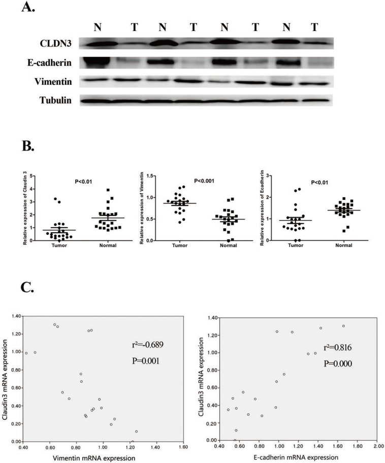
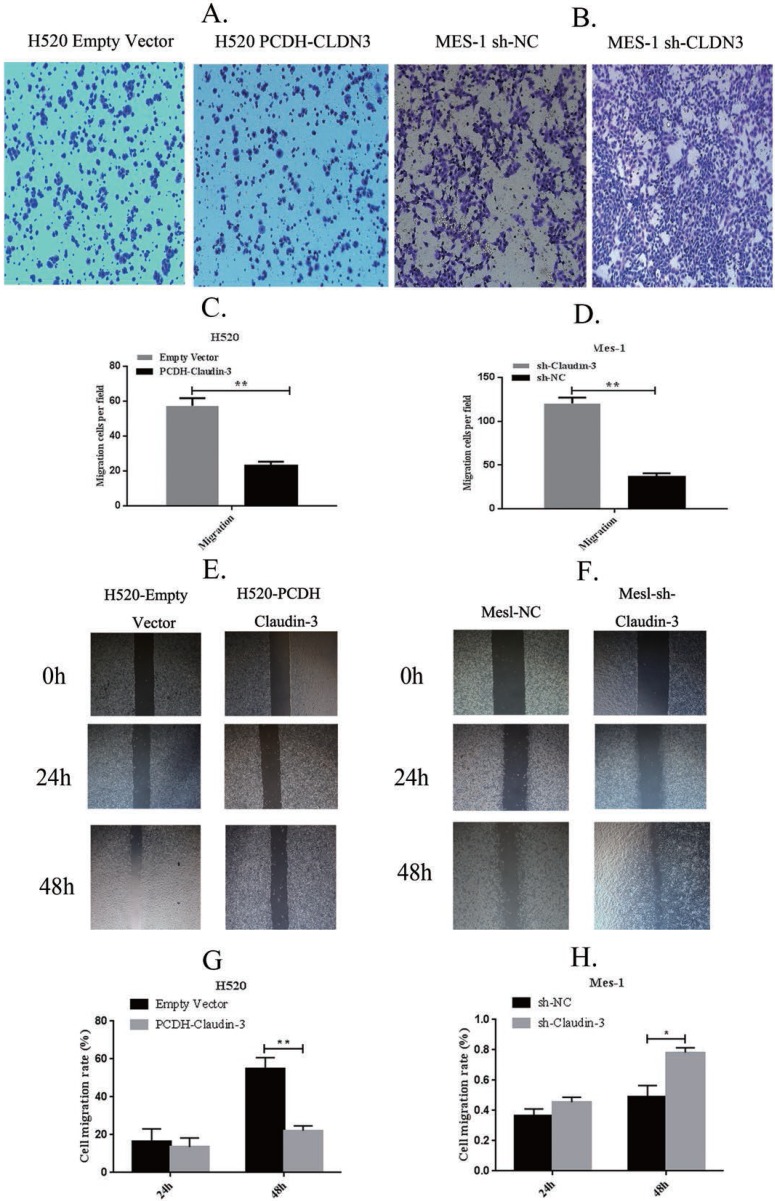
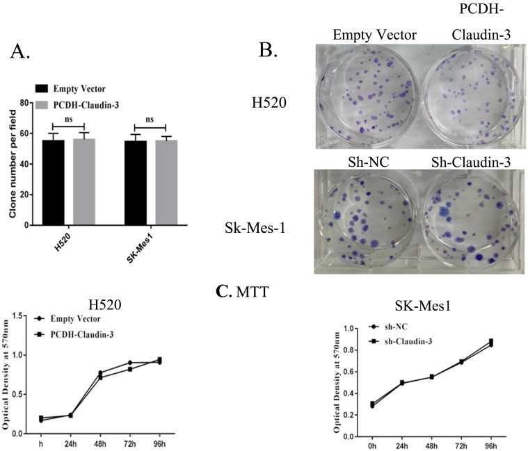
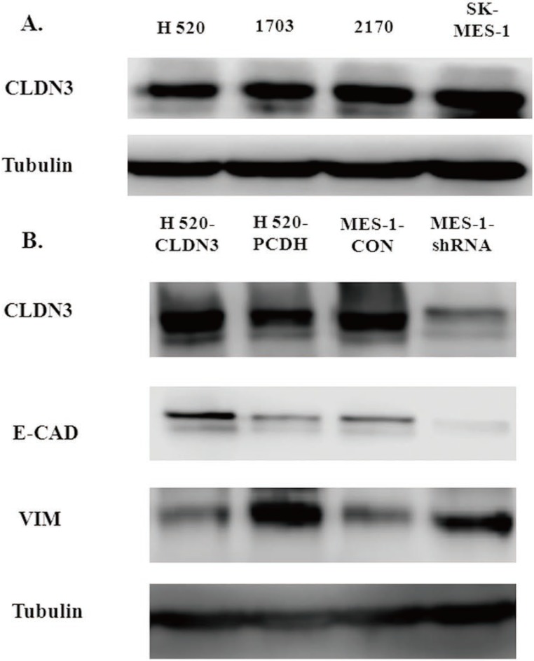
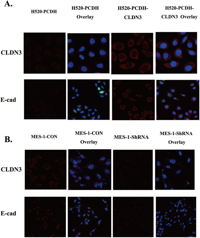
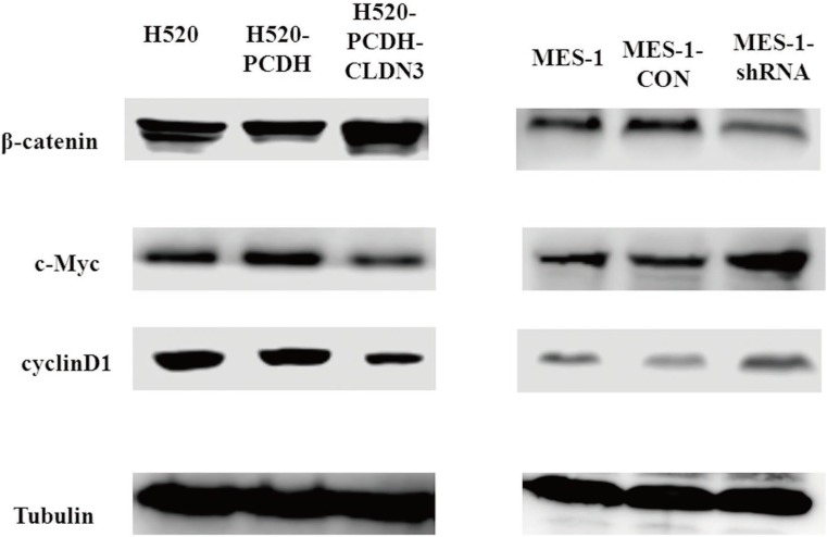
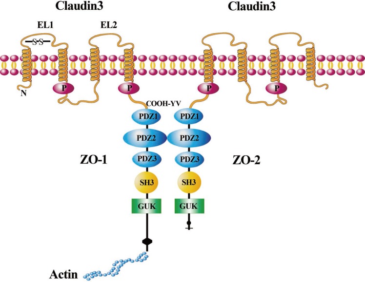
Altered expression of claudin-3 (CLDN3), a key cytoskeletal structural protein of the tight junctions in the epithelium, is associated with the development and metastasis of various human cancers. CLDN3 expression has been shown to be significantly associated with the prognosis of lung squamous cell carcinoma (SqCC). This study investigated the role of CLDN3 in inhibiting lung SqCC cell migration and invasion as well as the underlying molecular mechanisms. The CLDN3 levels were assessed between 20 paired lung SqCC tissues and adjacent normal tissues using quantitative real-time polymerase chain reaction (qRT-PCR) and western blot. The ectopic CLDN3 overexpression or knockdown was generated by using a plasmid carrying CLDN3 cDNA or shRNA, respectively. CLDN3 expression was significantly reduced in lung SqCC tissues vs. the adjacent normal tissues. The ectopic CLDN3 overexpression markedly inhibited the migration, invasion, and epithelial-mesenchymal transition (EMT) of lung cancer H520 cells, whereas CLDN3 knockdown had an inverse effect on SK-MES-1 cells. However, cell viability and plate colony formation assays showed that both CLDN3 knockdown and overexpression did not affect SqCC cell proliferation. Both tissue and cell data revealed that CLDN3 expression was significantly associated with the expression of the EMT biomarkers E-cadherin and Vimentin. Furthermore, CLDN3-modulated EMT and expression of the EMT markers were through regulation of the Wnt/β-catenin signaling pathway. In conclusion, this study identified reduced CLDN3 expression in lung SqCC tissues, which was associated with the progression and metastasis of lung SqCC and was attributed to EMT by activation of the Wnt pathway. Thus, CLDN3 could be further evaluated as a novel biomarker for predicting the prognosis of lung SqCC and as a target for the treatment of lung SqCC in the future.
Keywords: EMT; Lung squamous cell carcinoma; Wnt; claudin-3; metastasis.
Conflict of interest statement
Competing Interests: The authors have declared that no competing interest exists.
Figures
Similar articles
-
Decreased expression of claudin-3 is associated with a poor prognosis and EMT in completely resected squamous cell lung carcinoma.Tumour Biol. 2015 Aug;36(8):6559-68. doi: 10.1007/s13277-015-3350-1. Epub 2015 Mar 28. Tumour Biol. 2015. PMID: 25820701
-
Angiogenin elevates the invasive potential of squamous cell lung carcinoma cells through epithelial‑mesenchymal transition.Oncol Rep. 2016 Nov;36(5):2836-2842. doi: 10.3892/or.2016.5107. Epub 2016 Sep 19. Oncol Rep. 2016. PMID: 27667357
-
CLDN3 inhibits cancer aggressiveness via Wnt-EMT signaling and is a potential prognostic biomarker for hepatocellular carcinoma.Oncotarget. 2014 Sep 15;5(17):7663-76. doi: 10.18632/oncotarget.2288. Oncotarget. 2014. PMID: 25277196 Free PMC article.
-
Long non-coding RNAs as the pivotal regulators of epithelial mesenchymal transition through WNT/β-catenin signaling pathway in tumor cells.Pathol Res Pract. 2024 Nov;263:155683. doi: 10.1016/j.prp.2024.155683. Epub 2024 Oct 28. Pathol Res Pract. 2024. PMID: 39471528 Review.
-
Biomarkers of epithelial-mesenchymal transition in squamous cell carcinoma.J Dent Res. 2013 Feb;92(2):114-21. doi: 10.1177/0022034512467352. Epub 2012 Nov 5. J Dent Res. 2013. PMID: 23128109 Free PMC article. Review.
Cited by
-
The role and mechanism of claudins in cancer.Front Oncol. 2022 Dec 22;12:1051497. doi: 10.3389/fonc.2022.1051497. eCollection 2022. Front Oncol. 2022. PMID: 36620607 Free PMC article. Review.
-
Clinical Implications of HSC70 Expression in Clear Cell Renal Cell Carcinoma.Int J Med Sci. 2021 Jan 1;18(1):239-244. doi: 10.7150/ijms.43100. eCollection 2021. Int J Med Sci. 2021. PMID: 33390792 Free PMC article.
-
Pre-exposure to hydrogen sulfide modulates the innate inflammatory response to organic dust.Cell Tissue Res. 2021 Apr;384(1):129-148. doi: 10.1007/s00441-020-03333-3. Epub 2021 Jan 6. Cell Tissue Res. 2021. PMID: 33409657 Free PMC article.
-
Expression patterns of claudins in cancer.Heliyon. 2023 Oct 29;9(11):e21338. doi: 10.1016/j.heliyon.2023.e21338. eCollection 2023 Nov. Heliyon. 2023. PMID: 37954388 Free PMC article. Review.
-
Increased expression of claudin-12 promotes the metastatic phenotype of human bronchial epithelial cells and is associated with poor prognosis in lung squamous cell carcinoma.Exp Ther Med. 2019 Jan;17(1):165-174. doi: 10.3892/etm.2018.6964. Epub 2018 Nov 13. Exp Ther Med. 2019. PMID: 30651778 Free PMC article.
References
-
- Isla D, Majem M, Viñolas N, Artal A, Blasco A, Felip E. et al. A consensus statement on the gender perspective in lung cancer. Clin Transl Oncol. 2017;19:527–35. - PubMed
-
- Auerbach O, Hammond EC, Garfinkel L. Changes in bronchial epithelium in relation to cigarette smoking, 1955-1960 vs. 1970-1977. N Engl J Med. 1979;300:381–5. - PubMed
-
- Halpern MT, Gillespie BW, Warner KE. Patterns of absolute risk of lung cancer mortality in former smokers. J Natl Cancer Inst. 1993;85:457–64. - PubMed
-
- Lam S, Mac Aulay C, Hung J, LeRiche J, Profio AE, Palcic B. Detection of dysplasia and carcinoma in situ with a lung imaging fluorescence endoscopedevice. J Thorac Cardiovasc Surg. 1993;105:1035–40. - PubMed
MeSH terms
Substances
LinkOut - more resources
Full Text Sources
Other Literature Sources
Medical
Miscellaneous
