. 2018 Jun;17(6):528-534.
doi: 10.1038/s41563-018-0028-2. Epub 2018 Mar 5.

A facile approach to enhance antigen response for personalized cancer vaccination

Affiliations

A facile approach to enhance antigen response for personalized cancer vaccination

Aileen Weiwei Li et al. Nat Mater. 2018 Jun.

Abstract

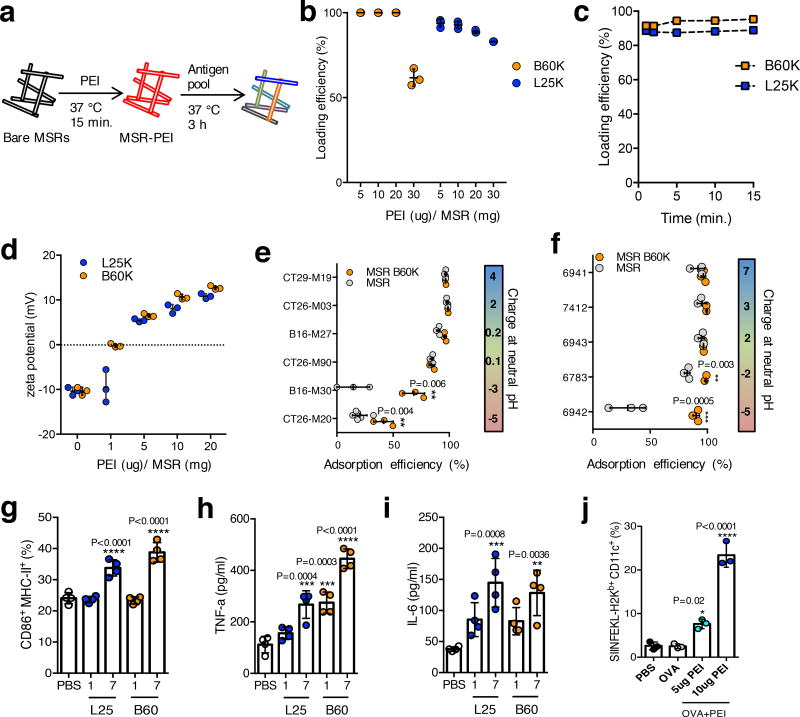
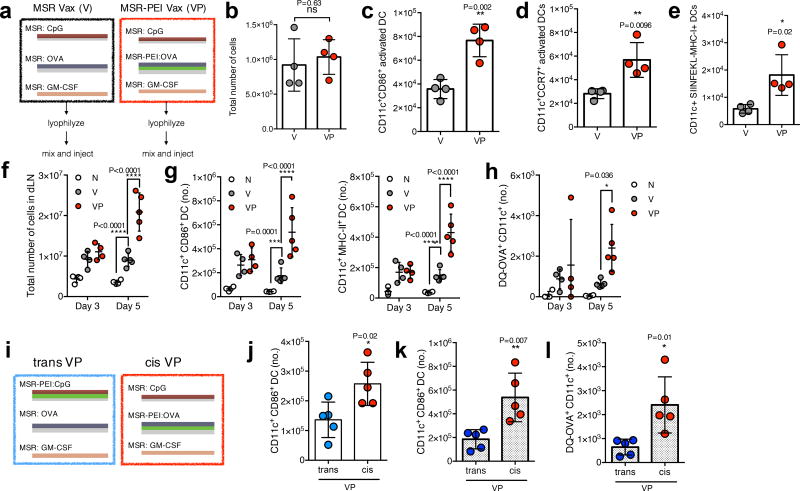
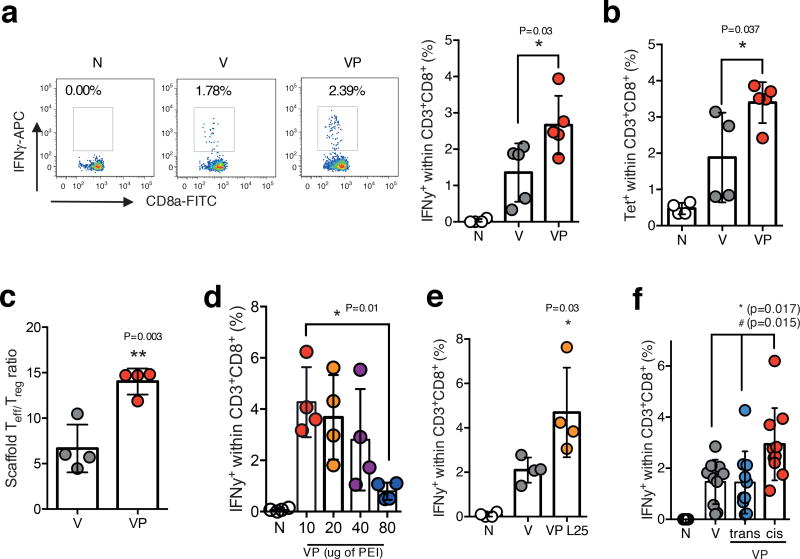
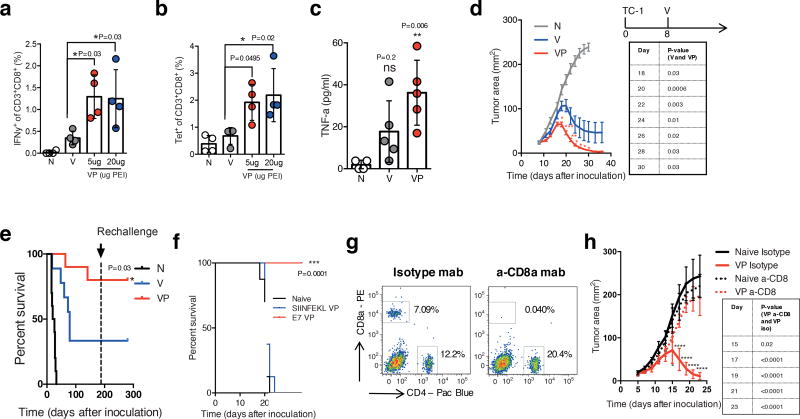
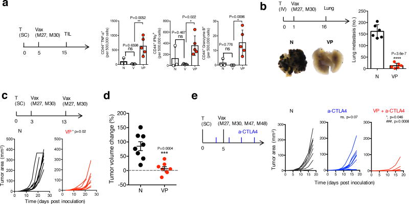
Existing strategies to enhance peptide immunogenicity for cancer vaccination generally require direct peptide alteration, which, beyond practical issues, may impact peptide presentation and result in vaccine variability. Here, we report a simple adsorption approach using polyethyleneimine (PEI) in a mesoporous silica microrod (MSR) vaccine to enhance antigen immunogenicity. The MSR-PEI vaccine significantly enhanced host dendritic cell activation and T-cell response over the existing MSR vaccine and bolus vaccine formulations. Impressively, a single injection of the MSR-PEI vaccine using an E7 peptide completely eradicated large, established TC-1 tumours in about 80% of mice and generated immunological memory. When immunized with a pool of B16F10 or CT26 neoantigens, the MSR-PEI vaccine eradicated established lung metastases, controlled tumour growth and synergized with anti-CTLA4 therapy. Our findings from three independent tumour models suggest that the MSR-PEI vaccine approach may serve as a facile and powerful multi-antigen platform to enable robust personalized cancer vaccination.


Conflict of interest statement

Competing Financial Interests:

The authors declare no competing financial interests.

Figures

Figure 1
Figure 1. PEI can be rapidly incorporated onto MSRs and leads to murine and human DC activation
(a) Schematics of PEI and subsequent antigen adsorption onto bare MSRs. (b) Incorporation efficiency of various doses of soluble B60K and L25K PEI into MSRs (n=3). (c) Incorporation kinetics of soluble B60K and L25K PEI into MSRs (representative data, repeated at least 3 times). (d) Zeta potential of MSR-PEI particles using various doses of soluble B60K and L25K PEI (n=3). Incorporation efficiency of (e) murine and (f) human neoantigen peptides (sorted according to the net charge at neutral pH) onto bare MSR or MSR-PEI particles using B60K PEI (n=3, two-tailed T test). (g) Flow cytometry analysis of CD86 and MHC-II expression on murine BMDCs after 24 hours of stimulation with 1ug or 7ug of soluble PEI or PBS (n=4, compared to PBS by one-way ANOVA). ELISA analysis of (h) TNFα and (i) IL-6 concentration in murine BMDC supernatant after 24 hours of stimulation with 1ug or 7ug of soluble B60K and L25K PEI or PBS (n=4, compared to PBS by one-way ANOVA). (j) Flow cytometry analysis of SIINFEKL presenting murine BMDCs after stimulation with PBS, OVA and OVA with 5ug or 10ug of soluble B60K PEI (n=3, compared to OVA by one-way ANOVA). Data depicts mean +/− sd.
Figure 2
Figure 2. MSR-PEI vaccine enhances DC activation and trafficking in situ
(a) Schematics of the MSR vaccine (V) and MSR-PEI vaccine (VP). (b) Total cell number at the vaccine site explanted on day 3 post immunization with the MSR vaccine (V), the MSR-PEI vaccine (VP) using B60K PEI (n=4, two-tailed T test). Total number of (c) CD11c+ CD86+ activated DCs (n=4, two-tailed T test), (d) CD11c+ CCR7+ LN homing DCs (n=4, two-tailed T test) and (e) SIINFEKL presenting DC (n=4, two-tailed T test) recruited to the vaccine site on day 3 post immunization with the MSR vaccine (V), the MSR-PEI vaccine (VP) using B60K PEI. (f) Total number of cells (n=4 for d3 and n=5 for d5, two-way ANOVA), of (g) CD11c+ CD86+ or CD11c+ MHC-II+ activated DCs (n=4 for d3 and n=5 for d5, two-way ANOVA), and (h) OVA+ DC (n=4 for d3 and n=5 for d5, two-way ANOVA) in the dLN on days 3 and day 5 post immunization with the MSR vaccine (V), the MSR-PEI vaccine (VP) using B60K PEI or left unimmunized (N). (i) Schematics of the MSR-PEI trans vaccine (trans VP) and the MSR-PEI cis vaccine (cis VP). (j) Total number of CD11c+ CD86+ activated DCs at the vaccine site on day 3 post immunization with the trans VP vaccine or the cis VP vaccine (n=5, two-tailed T test). Total number of (k) CD11c+ CD86+ activated DCs and (l) CD11c+ OVA+ DCs in the dLN on day 5 post immunization with the trans VP vaccine or the cis VP vaccine (n=5, two-tailed T test). Data depicts mean +/− sd
Figure 3
Figure 3. MSR-PEI vaccine enhances CD8 cytotoxic T cell response against OVA
(a) Percentage of IFNγ+ CD8+ T cells isolated from peripheral blood on day 7 after immunization with the MSR vaccine (V), the MSR-PEI vaccine (VP) using B60K PEI, or left unimmunized (N), and stimulated with SIINFEKL (primary FACS plots on the left, quantifications from the FACS plots on the right) (n=5, one-way ANOVA). (b) Percentage of SIINFEKL-tetramer+ CD8+ T cells isolated from peripheral blood on day 7 after immunization with the MSR vaccine (V), the MSR-PEI vaccine (VP) using B60K PEI or left unimmunized (N) (n=5 for VP, n=4 for N and V, one-way ANOVA). (c) Ratio of CD8+ effector T cells (Teff) to CD4+ Foxp3+ regulatory T cells (Treg) at the MSR vaccine site on day 11 after immunization with the MSR vaccine (V) or the MSR-PEI vaccine (VP) using B60K PEI (n=5, two-tailed T test). (d) Percentage of IFNγ+CD8+ T cells isolated from peripheral blood on day 7 after immunization with the MSR-PEI vaccine (VP) containing various doses of B60K PEI or left unimmunized (N) (n=4, one-way ANOVA). (e) Percentage of IFNγ+CD8+ T cells isolated from peripheral blood at day 7 after immunization with the MSR vaccine (V), the MSR-PEI vaccine using L25K PEI (VP L25) or left unimmunized (N) (n=4, one-way ANOVA). (f) Percentage of IFNγ+CD8+ T cells isolated from peripheral blood at day 7 after immunization with the MSR vaccine (V), the MSR-PEI trans vaccine (trans, VP) using B60K PEI, and the MSR-PEI cis vaccine (cis, VP) using B60K PEI, or left unimmunized (N) and stimulated with SIINFEKL (n=9, * between cis VP and trans VP, # between cis VP and V by one-way ANOVA). Data depicts mean +/− sd.
Figure 4
Figure 4. MSR-PEI vaccine enhances CD8 cytotoxic T cell response against E7 and regresses established tumors
(a) Percentage of IFNγ+CD8+ T cells in response to RAHYNIVTF stimulation and (b) percentage of tetramer+CD8+ T cells in peripheral blood on day 7 after immunization with the MSR E7 vaccine (V), the MSR-PEI E7 vaccine (VP) using 5ug or 20ug of B60K PEI, or left unimmunized (N) (n=4, one-way ANOVA). ELISA analysis of (c) TNFa level in serum 24 hours post vaccination with the MSR vaccine (V), the MSR-PEI (B60K) vaccine (VP), or left unimmunized (N) (n=4, compared to N by one-way ANOVA). (d) Tumor growth and (e) overall survival of mice bearing established E7 expressing TC-1 tumors (allowed to develop for 8 days) and treated with the MSR vaccine (V) or the MSR-PEI vaccine (VP) using L25K PEI, or left untreated (N), and subsequently rechallenged with TC-1 cells at 6 months post first inoculation (n=10, compared to V by two-way ANOVA for d, by Log-rank Test for e). (f) Overall survival of mice bearing established E7 expressing TC-1 tumors and treated with the MSR-PEI vaccine containing E7 (E7 VP) or the MSR-PEI vaccine containing SIINFEKL (SIINFEKL VP), or left untreated (Naive) (n=8, compared to SIINFEKL VP by Log-rank Test). (g) Flow cytometry analysis of blood T cells 3 days after treatment with a-CD8a mab or an isotype mab (representative data, repeated 3 times). (h) Tumor growth of mice bearing established E7 expressing TC-1 tumors and treated with the MSR-PEI vaccine with either a-CD8a mab or an isotype mab (n=8, compared to VP a-CD8 by two-way ANOVA). In (a–c), data depicts mean +/− sd and in (d,h), data depicts mean +/− sem
Figure 5
Figure 5. MSR-PEI vaccine enhances melanoma TIL effector function and induces tumor control and synergy with anti-CTLA4 therapy using combined B16 neoantigens
(a) Number of CD44+IFNγ+, CD44+TNFα+ and CD44+Granzyme B+ TILs per 500,000 tumor cells on day 15 post inoculation. Mice bearing established B16F10 tumors (allowed to develop for 5 days) and treated with the MSR vaccine (V) or the MSR-PEI vaccine (VP) using L25K PEI and 50ug of the B16 neoantigens, or left untreated (N) (n=5 for VP, n=3 for N and V, one-way ANOVA). (b) Number of lung metastases formed on day 16 post inoculation in mice that received IV inoculation of B16F10 melanoma cells (allowed to develop for 1 day) and treated with the MSR-PEI vaccine (VP) using L25K PEI and 50ug of the B16 neoantigens, or left untreated (N). Primary representative photographs of excised lungs are shown in the figure (n=6, two-tailed T test). (c) Tumor growth in mice bearing established B16F10 tumors (allowed to develop for 3 days) and treated with two injections of the MSR-PEI vaccine (VP) using L25K PEI and 50ug of the B16 neoantigens on days 3 and 13, or left untreated (N) (n=8, two-tailed T test). (d) Tumor volume change between days 13 and 17 after tumor inoculation (n=8, two-tailed T test). (e) Tumor growth of mice bearing established B16F10 tumors (inoculated with 1×10⁵ cells) and treated with anti-CTLA4 antibody (a-CTLA4), anti-CTLA4 antibody in combination with the MSR-PEI vaccine (VP+a-CTLA4) using L25K PEI and 50ug of the B16 neoantigens on days 5, or left untreated (N) (n=8, * between VP+a-CTLA4 and a-CTLA4, ### between VP+a-CTLA4, ns between a-CTLA4 and N by one-way ANOVA). In (a, b, d), data depicts mean +/− sd, in (c and e) data depicts individual tumor growth.

Comment in

  • Smart delivery of vaccines.
    Melief CJM. Melief CJM. Nat Mater. 2018 Jun;17(6):482-483. doi: 10.1038/s41563-018-0085-6. Nat Mater. 2018. PMID: 29795220 No abstract available.

Similar articles

Cited by

References

    1. Gubin MM, Artyomov MN, Mardis ER, Schreiber RD. Tumor neoantigens: building a framework for personalized cancer immunotherapy. The Journal of clinical investigation. 2015;125:3413–3421. - PMC - PubMed
    1. Schumacher TN, Schreiber RD. Neoantigens in cancer immunotherapy. Science. 2015;348:69–74. - PubMed
    1. Hacohen N, Fritsch EF, Carter TA, Lander ES, Wu CJ. Getting personal with neoantigen-based therapeutic cancer vaccines. Cancer immunology research. 2013;1:11–15. - PMC - PubMed
    1. Linnemann C, et al. High-throughput epitope discovery reveals frequent recognition of neo-antigens by CD4+ T cells in human melanoma. Nature medicine. 2015;21:81–85. - PubMed
    1. Kreiter S, et al. Mutant MHC class II epitopes drive therapeutic immune responses to cancer. Nature. 2015;520:692–696. - PMC - PubMed

Publication types