. 2018 Apr 20;293(16):5878-5894.
doi: 10.1074/jbc.M117.816298. Epub 2018 Feb 15.

Biochemical characterization and essentiality of Plasmodium fumarate hydratase

Affiliations

Biochemical characterization and essentiality of Plasmodium fumarate hydratase

Vijay Jayaraman et al. J Biol Chem. .

Abstract

Plasmodium falciparum (Pf), the causative agent of malaria, has an iron-sulfur cluster-containing class I fumarate hydratase (FH) that catalyzes the interconversion of fumarate to malate, a well-known reaction in the tricarboxylic acid cycle. In humans, the same reaction is catalyzed by class II FH that has no sequence or structural homology with the class I enzyme from Plasmodium Fumarate is generated in large quantities in the parasite as a by-product of AMP synthesis and is converted to malate by FH and then used in the generation of the key metabolites oxaloacetate, aspartate, and pyruvate. Previous studies have identified the FH reaction as being essential to P. falciparum, but biochemical characterization of PfFH that may provide leads for the development of specific inhibitors is lacking. Here, we report on the kinetic characterization of purified recombinant PfFH, functional complementation of fh deficiency in Escherichia coli, and mitochondrial localization in the parasite. We found that the substrate analog mercaptosuccinic acid is a potent PfFH inhibitor, with a Ki value in the nanomolar range. The fh gene could not be knocked out in Plasmodium berghei when transfectants were introduced into BALB/c mice; however, fh knockout was successful when C57BL/6 mice were used as host, suggesting that the essentiality of the fh gene to the parasite was mouse strain-dependent.

Keywords: Plasmodium; class I fumarate hydratase; enzyme inhibitor; essentiality of fumarate hydratase; gene knockout; mercaptosuccinic acid; parasitology; tricarboxylic acid cycle (TCA cycle) (Krebs cycle).


Conflict of interest statement

The authors declare that they have no conflicts of interest with the contents of this article

Figures

Figure 1.
Figure 1.
Generation of PfFH-GFP strain encoding FH-GFP and localization of PfFH. a, scheme showing the integration locus with the regulatable fluorescent affinity (RFA) (GFP + EcDHFR degradation domain + HA) tag in tandem with the fh gene in the strain PfFH-GFP. Oligonucleotides P1 and P2 and P3 and P4 were used for checking the 5′ and 3′ integration, respectively (Table S1). P1 and P4 are beyond the sites of integration in the genome. b, left panel, genotyping by PCR for validating 5′ integration. The templates used in different lanes are as follows: L1, genomic DNA from PfFH-GFP; L3, P. falciparum PM1KO genomic DNA. A band size of 2483 bp validates 5′ integration. Right panel, genotyping by PCR for validating 3′ integration. The templates used in different lanes are as follows: L2, genomic DNA from PfFH-GFP; L3, P. falciparum PM1KO genomic DNA. A band size of 2467 bp validates 3′ integration. Molecular weight markers are in lanes L2 and L1 in left and right panels, respectively. c, upper panel shows a trophozoite, and the lower panel shows a schizont. As evident from the merge, PfFH localizes to the mitochondrion. The Pearson correlation coefficients for the images are 0.8972 (upper panel) and 0.8954 (lower panel).
Figure 2.
Figure 2.
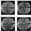
Phenotyping of the E. coli strain ΔfumACB and functional complementation by P. falciparum FH. Growth phenotype of the E. coli strains with at least one copy of fumarate hydratase gene deleted (ΔA, ΔB, and ΔC) and with all three genes deleted (ΔACB) on malate- (MAL) (a) and fumarate (FUM) (b)-containing minimal medium. As evident from the phenotype, ΔfumACB strainACB) is not able to grow on fumarate-containing minimal medium. c and d, the growth of ΔfumACB strains expressing either PfFHFL (FL) or PfFHΔ40 (Δ40) or PfFHΔ120 (Δ120) of P. falciparum fumarate hydratase on malate- (c) and fumarate (d)-containing minimal medium. The plates were scored after 48 h of incubation at 37 °C. ΔfumACB strain containing just pQE30 (E) was used as a control. The experiment was repeated three times, and the images shown correspond to one of the replicates.
Figure 3.
Figure 3.
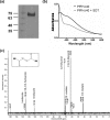
Purification and activity of PfFHΔ40. a, 1st lane, protein molecular mass markers (numbers indicated are in kDa); 2nd lane, Ni-NTA-purified PfFHΔ40. b, UV-visible absorption spectrum of purified and reconstituted PfFH shows characteristic peaks at 360 and at 405 nm that indicate the presence of a 4Fe-4S cluster. The spectrum of the protein with oxidized iron–sulfur cluster is shown as a solid line and that with reduced iron–sulfur cluster upon addition of 1 mm sodium dithionite (SDT) is shown as a dashed line. c, validation of malic acid formation by 13C NMR. The NMR spectrum of assay mixture consisting of 50 μm 2,3-[13C]fumaric acid in 100 mm potassium phosphate, pH 7.4, incubated with 100 μg of purified PfFHΔ40 enzyme, shows the presence of peaks corresponding to [13C]malic acid. Unreacted [13C]fumaric acid is also present. The inset shows the chemical structure of [13C]fumaric acid. The spectrum is an average of 3000 scans acquired using Bruker 400-MHz NMR spectrometer. The peaks corresponding to imidazole and glycerol are from the protein solution.
Figure 4.
Figure 4.
Growth of E. coli strain ΔfumACB expressing PfFH on different carbon sources. a–f, the growth of ΔfumACB strains expressing either PfFHFL (FL) or PfFHΔ40 (Δ40) or PfFHΔ120 (Δ120) were tested on meso-tartrate (a), mesaconate (b), d-tartrate (c), l-tartrate (d), itaconate (e), and glucose-containing minimal medium (f). The plates were scored after 48 h of incubation at 37 °C. ΔfumACB strain containing just pQE30 (E) was used as a control. The experiment was repeated three times, and the images correspond to one of the replicates. The glucose-containing plate served as a control for the number of cells plated across the different constructs.
Figure 5.
Figure 5.
Specificity of dl-MSA for class I FH. a, structures of l-malic acid and mercaptosuccinic acid. b, Lineweaver-Burk plot of initial velocity at varied malate and different fixed MSA concentrations. c, Lineweaver-Burk plot of the initial velocity at varied fumarate and different fixed MSA concentrations. d, inhibition of the growth of the ΔfumACB_pPfFHΔ40 strain of E. coli by MSA. e, rescue of MSA-mediated growth inhibition of ΔfumACB_pPfFHΔ40 upon addition of malate. f, inhibition of the in vitro growth of intra-erythrocytic asexual stages of P. falciparum by MSA.
Figure 6.
Figure 6.
Genotyping of P. berghei clones of knockout of fumarate hydratase. a, schematic representation of the selectable marker cassette inserted into the fh gene locus of P. berghei genome. Primers (P1–P8) used for diagnostic PCRs are indicated. b, schematic representation of the fh gene (PBANKA_0828100) flanked by 5′ UTR and 3′ UTR showing the location of primers P9 and P10. Shown is agarose gel electrophoresis of PCRs with genomic DNA from clones A–C (c, left panel) and clones H–M (c, right panel) for detection of 5′ integration; d, clones J and M for detection of 3′ integration (other clones did not answer for this PCR); e, clone A for the detection of fh gene; f, clones A–C for the presence of selectable marker cassette; g, clones C and O using primers P3 and P8. Shown in this figure is the genotyping of representative P. berghei clones, and the data on all 17 clones that were characterized are provided in Fig. S5. Clones C and M and O did not answer for 5′ integration, and only clones J, M, and Q answered for 3′ integration (c and d here and Fig. S5, c and d). All clones answered for the presence of the fh gene (e here and Fig. S5e). All clones except C and O answered by PCR with primers P2 and P6 indicating the integration of the entire selectable marker cassette into the genome (f here and Fig. S5f). Clones C and O answered for a shorter fragment of the selectable marker cassette covered by primers P3 and P8 (g here and Fig. S5g). hDHFR-yFCU, human DHFR-yeast cytosine and uridyl phosphoribosyltransferase; mr, molecular weight marker; numbers to the left of c–g are the sizes of the marker DNA fragments in kbp.
Figure 7.
Figure 7.
Genotyping of transfectants grown and drug-selected in different mouse strains. a, presence/absence of fumarate hydratase gene was validated by PCR using oligonucleotides P9 and P10 and genomic DNA isolated from respective parasites as template (lanes bracketed as fh). As a positive control for the presence of genomic DNA, PCR was performed with oligonucleotides corresponding to a segment of mqo gene loci (lanes bracketed as mqo). b, validation of 5′ and 3′ integration using primer pairs P1 and P4 and P5 and P7, respectively. Primer locations are as in Fig. 6. NC, negative control lacking template DNA; WT, P. berghei ANKA wildtype genomic DNA; BALB/c, genomic DNA from transfectants grown in BALB/c mouse; C57BL/6, genomic DNA from transfectants grown in C57BL/6 mouse; M, marker. Numbers to the right of figure are the sizes of the marker DNA fragments in kbp. The sequences of the oligonucleotides used are provided in Table S1.
Figure 8.
Figure 8.
Metabolic consequences of fumarate hydratase gene deletion in Plasmodium. Dashed arrows indicate the flow of metabolites into a pathway, and the dotted arrows indicate transport across compartments. Gray arrows show possible metabolic consequences of fh gene deletion (see text for explanation). AAT, aspartate aminotransferase; HGPRT, hypoxanthine-guanine phosphoribosyltransferase; MDH, malate dehydrogenase, MQO, malate-quinone oxidoreductase; PEPCK, phosphoenolpyruvate carboxykinase; α-Kg, α-ketoglutarate; Hyp, hypoxanthine; PEP, phosphoenolpyruvate; PRPP, phosphoribosyl 5′-pyrophosphate, SAMP, succinyl-AMP, UQ, ubiquinone, UQH, ubiquinol.

Similar articles

Cited by

References

    1. Roth E. (1990) Plasmodium falciparum carbohydrate metabolism: a connection between host cell and parasite. Blood Cells 16, 453–460 - PubMed
    1. MacRae J. I., Dixon M. W., Dearnley M. K., Chua H. H., Chambers J. M., Kenny S., Bottova I., Tilley L., and McConville M. J. (2013) Mitochondrial metabolism of sexual and asexual blood stages of the malaria parasite Plasmodium falciparum. BMC Biol. 11, 67 10.1186/1741-7007-11-67 - DOI - PMC - PubMed
    1. Bulusu V., Jayaraman V., and Balaram H. (2011) Metabolic fate of fumarate, a side product of the purine salvage pathway in the intra-erythrocytic stages of Plasmodium falciparum. J. Biol. Chem. 286, 9236–9245 10.1074/jbc.M110.173328 - DOI - PMC - PubMed
    1. Teipel J. W., Hass G. M., and Hill R. L. (1968) The substrate specificity of fumarase. J. Biol. Chem. 243, 5684–5694 - PubMed
    1. Chen B.-S., Otten L. G., and Hanefeld U. (2015) Stereochemistry of enzymatic water addition to C=C bonds. Biotechnol. Adv. 33, 526–546 10.1016/j.biotechadv.2015.01.007 - DOI - PubMed

Publication types

MeSH terms