. 2018 Feb 13;15(1):42.
doi: 10.1186/s12974-018-1069-9.

Trovafloxacin attenuates neuroinflammation and improves outcome after traumatic brain injury in mice

Affiliations

Trovafloxacin attenuates neuroinflammation and improves outcome after traumatic brain injury in mice

Charu Garg et al. J Neuroinflammation. .

Abstract

Background: Trovafloxacin is a broad-spectrum antibiotic, recently identified as an inhibitor of pannexin-1 (Panx1) channels. Panx1 channels are important conduits for the adenosine triphosphate (ATP) release from live and dying cells that enhances the inflammatory response of immune cells. Elevated extracellular levels ATP released upon injury activate purinergic pathways in inflammatory cells that promote migration, proliferation, phagocytosis, and apoptotic signals. Here, we tested whether trovafloxacin administration attenuates the neuroinflammatory response and improves outcomes after brain trauma.

Methods: The murine controlled cortical impact (CCI) model was used to determine whether in vivo delivery of trovafloxacin has anti-inflammatory and neuroprotective actions after brain trauma. Locomotor deficit was assessed using the rotarod test. Levels of tissue damage markers and inflammation were measured using western blot, qPCR, and immunofluorescence. In vitro assays were used to evaluate whether trovafloxacin blocks ATP release and cell migration in a chemotactic-stimulated microglia cell line.

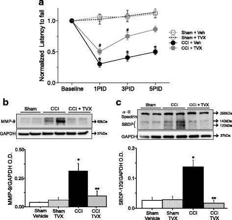
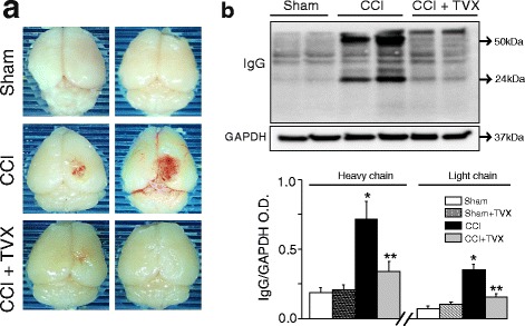
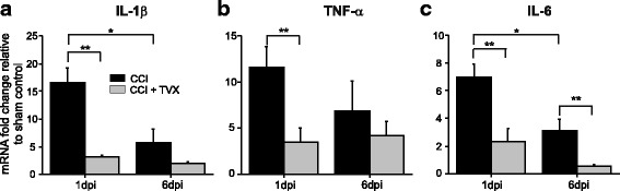
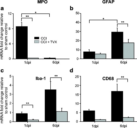
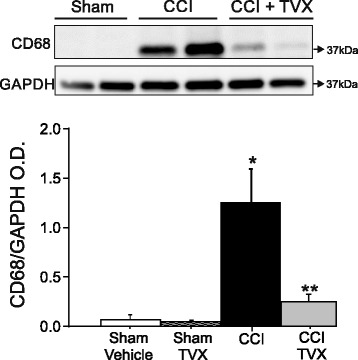
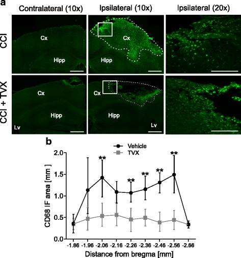
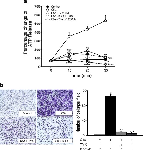
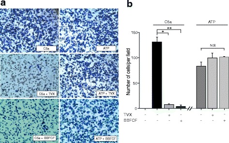
Results: Trovafloxacin treatment of CCI-injured mice significantly reduced tissue damage markers and improved locomotor deficits. In addition, trovafloxacin treatment significantly reduced mRNA levels of several pro-inflammatory cytokines (IL-1β, IL-6, and TNF-α), which correlates with an overall reduction in the accumulation of inflammatory cell types (neutrophils, microglia/macrophages, and astroglia) at the injury zone. To determine whether trovafloxacin exerted these effects by direct action on immune cells, we evaluated its effect on ATP release and cell migration using a chemotactic-stimulated microglial cell line. We found that trovafloxacin significantly inhibited both ATP release and migration of these cells.

Conclusion: Our results show that trovafloxacin administration has pronounced anti-inflammatory and neuroprotective effects following brain injury. These findings lay the foundation for future studies to directly test a role for Panx1 channels in pathological inflammation following brain trauma.

Keywords: Brain injury; Hemichannel; Microglia; Neuroinflammation; Pannexin.


Conflict of interest statement

Ethics approval and consent to participate

Not applicable.

Consent for publication

Not applicable.

Competing interests

The authors declare that they have no competing interests.

Publisher’s Note

Springer Nature remains neutral with regard to jurisdictional claims in published maps and institutional affiliations.

Figures

Fig. 1
Fig. 1
Trovafloxacin treatment improves locomotor behavior and attenuates brain damage. a The rotarod test was used to evaluate latency (time) to fall in sham treated with vehicle (gray open circles), sham treated with trovafloxacin (TVX, black open circles), CCI-injured mice treated with vehicle (gray closed circles), or trovafloxacin (TVX, black closed circles) for up to 5 days post-CCI. Latency to fall was normalized to the baseline behavior for each group. Values are represented as mean ± SEM (n = 8 per group). Linear regression analysis was used to determine statistical difference between different groups at each time point (p values calculated for treatment and treatment*time). *p < 0.05, sham vehicle vs. CCI + vehicle; #p < 0.05 sham vehicle vs. CCI + TVX. Identical p values at their respective PDI were found for sham + TVX vs. CCI vehicle or CCI + TVX (not symbols display). b Representative western blots images showing MMP-9 protein levels at 6 days post-injury in sham treated with vehicle and CCI-injured mice treated with vehicle or trovafloxacin. Bottom western blot corresponds to the GAPDH, which was used as housekeeping gene. Note that sham mice treated with TVX are not display in the western blots. Graph shows to densitometric quantification of MMP-9 normalized to GAPDH values. Values are represented as mean ± SEM (n = 5 per group). c α–ΙΙ spectrin and SPDBs (140 and 120 kDa) expression levels at 6 days post-injury in sham and CCI-injured mice treated with vehicle or trovafloxacin. Bottom western blot shows GAPDH. Note that sham mice treated with TVX are not displayed in the western blots. Graph shows densitometric quantification of SPDB 120 kDa normalized to GAPDH expression. Values are represented as mean ± SEM. (n = 5 per group). Two-way ANOVA followed by Tukey’s HSD test was used to determine statistical significance. *p < 0.01, sham vehicle vs. CCI group; **p < 0.01, CCI vehicle vs. CCI + TVX. No differences were found between sham vehicle and sham TVX (see also Additional file 1: Figure S1)
Fig. 2
Fig. 2
Trovafloxacin treatment attenuates blood brain barrier leakage in CCI-injured mice. a Representative images of perfused and fixed mice brains from sham and CCI-injured mice treated with vehicle or trovafloxacin (TVX) after 6 days post-CCI. b Representative western blot showing IgG protein levels from injury of sham and CCI-injured mice treated with vehicle or TVX 6 days post-injury. Bottom western blot corresponds to GAPDH. b Densitometric quantification of IgG levels for each group (n ≥ 5 per group) relative to their corresponding GAPDH expression levels. Note that sham mice treated with TVX are not displayed in the western blots. Values are represented as mean ± SEM. Two-way ANOVA followed by Tukey’s HSD test was used to determine statistical significance. *p < 0.01, sham vs. CCI group; **p < 0.01, CCI vehicle vs. CCI + TVX. No differences were found between sham vehicle and sham TVX (see also Additional file 1: Figure S1)
Fig. 3
Fig. 3
Pro-inflammatory cytokines mRNA levels are decreased in trovafloxacin-treated CCI mice. RNA was isolated from ipsilateral cortex from sham and CCI-injured mice treated with vehicle or trovafloxacin (TVX). Gene expression levels for a IL-1β, b TNF-α, and c IL-6 were measured at 1 and 6 days post-injury by qPCR. Values are expressed as mean fold change (± SEM) relative to sham (n = 5), and GAPDH was used as endogenous control. Linear regression analyses were used to determine statistical significance. (p values calculated for dpi and treatment, for IL-1β, TNF-α, and IL-6 separately). *p < 0.01, CCI 1 dpi vs. CCI 6 dpi, **p < 0.05, CCI vehicle vs. CCI + TVX
Fig. 4
Fig. 4
Trovafloxacin treatment attenuates the expression levels of markers for leukocytes and glia cells in the injured brain. RNA was isolated from ipsilateral cortex from each group, and gene expression levels were determined by qPCR at 1 and 6 days post-injury: a myeloperoxidase (MPO), marker of neutrophils; b glial fibrillary protein (GFAP), marker of astrocytes; c ionized calcium-binding adapter molecule 1 (Iba-1), marker of microglia; and d cluster of differentiation 68 (CD68), marker of activated microglia. Values are expressed as mean fold change (± SEM) relative to sham, and GAPDH was used as endogenous control. Linear regression analyses were used to determine statistical significance. (p values calculated for dpi and treatment, for MPO, GFAP, Iba-1, and CD68 separately). *p < 0.01, CCI 1 dpi vs. CCI 6 dpi, **p < 0.05, CCI vehicle vs. CCI + TVX
Fig. 5
Fig. 5
Trovafloxacin treatment reduces CD68 protein expression levels at 6 days post-injury. Representative western blots showing CD68 protein levels expressions at 6 days post-injury in sham or CCI-injured mice treated with vehicle or trovafloxacin (TVX). Graph shows densitometric quantification of CD68 is represented in each group (n = 5 per group). Values are represented as mean ± SEM. Two-way ANOVA followed by Tukey’s HSD test was used to determine statistical significance. *p < 0.01, sham vs. CCI group; **p < 0.01, CCI vehicle vs. CCI + TVX
Fig. 6
Fig. 6
Microglia/macrophage accumulation is reduced in trovafloxacin-treated CCI mice. Mice that received CCI injury where treated with trovafloxacin (TVX) or vehicle and were sacrificed at 6 dpi for histology analysis. a Representative images of CD68 immunoreactivity of brain sections from non-treated or TVX-treated CCI-injured mice at the core of the injury (AP − 1.96 to AP − 2.56). These areas were examined in the same anatomical location for each animal. Scale bar = 0.5 mm. Dotted lines demarcated high-density areas of CD68+ staining. b Correspond to the quantification of high-density areas of CD68+ cells (n = 5 per group). Error bars indicate SE of the mean. Linear regression analysis was performed to analyze statistical differences. (p values calculated for distance from Bregma and treatment). **p < 0.05 CCI vehicle vs. CCI + TVX
Fig. 7
Fig. 7
Trovafloxacin inhibits ATP release and migration in C5a-stimulated BV-2 cells. a BV-2 cells were stimulated with 10 nM of C5a in the presence or absence of Panx1 channel blockers, trovafloxacin (TVX, 1 μM), Brilliant Blue FCF (BBFCF, 5 μM), or 10Pnx1 (200 μM) for 30 min. Aliquots were collected from the medium every 10 min for ATP measurement. ATP was measured using the luciferin–luciferase assay. Values are represented as mean ± SEM. Percentage values were estimated with respect to basal ATP release before simulation at each condition. Each experiment was repeated at least three times in triplicates. Statistical difference was determined using linear regression with log(ATP) as the response. (p values calculated for treatment and treatment*time). *p < 0.05, C5a vs. control; **p < 0.05, C5a vs. TVX; ***p < 0.05, C5a vs. BBFCF, ****p < 0.05, C5a vs. 10Panx1. b Left panel shows representative images of BV-2 cells that were seeded in the upper compartment of the transwells for transmigration assay and treated with C5a in the absence or presence of TVX (1 μM) or BBFCF (5 μM). After 4 h, cells that had migrated to the bottom of the transwell were fixed, stained with crystal violet, and counted. The images are representative of the control treated with vehicle solutions, C5a, C5a + TVX, and C5a + BBFCF-treated cells. Graph corresponds to the quantification of transmigrated BV-2 cells after 4 h stimulation from images taken in five different fields at 20× per condition. Each experiment was repeated at least three times. One-way ANOVA with Tukey’s HSD test was used to measure statistical differences among groups. Values are expressed as mean ± SEM, *p < 0.05, control vs. C5a; **p < 0.05 C5a vs. C5a + TVX, or ***p < 0.05, C5a vs. C5a + BBFCF
Fig. 8
Fig. 8
Trovafloxacin does not affect downstream of ATP signaling. a Representative images of BV-2 cells that were seeded for transmigration assay and stimulated with C5a (10 nM) or ATP (200 μM) for 24 h in the presence or absence of trovafloxacin (TVX, 1 μM) or Brilliant Blue FCF (BBFCF, 5 μM). Cells were fixed and stained with crystal violet. b Quantification of transmigrated BV-2 cells at 24 h from five different fields at 20× per condition. Each experiment was repeated at least three times. One-way ANOVA with Tukey’s HSD test was used to measure statistical differences among groups. Values are expressed as mean ± SEM. *p < 0.05, C5a vs. C5a + trovafloxacin. **p < 0.05, C5a vs. C5a + BBFCF. No significant changes in the number of migrated cells were observed between ATP, ATP + TVX, and ATP + BBFCF (p > 0.05)

Similar articles

Cited by

References

    1. Gootz TD, Zaniewski R, Haskell S, Schmieder B, Tankovic J, Girard D, Courvalin P, Polzer RJ. Activity of the new fluoroquinolone trovafloxacin (CP-99,219) against DNA gyrase and topoisomerase IV mutants of Streptococcus pneumoniae selected in vitro. Antimicrob Agents Chemother. 1996;40:2691–2697. - PMC - PubMed
    1. Poon IK, Chiu YH, Armstrong AJ, Kinchen JM, Juncadella IJ, Bayliss DA, Ravichandran KS. Unexpected link between an antibiotic, pannexin channels and apoptosis. Nature. 2014;507:329–334. doi: 10.1038/nature13147. - DOI - PMC - PubMed
    1. Chiu YH, Ravichandran KS, Bayliss DA. Intrinsic properties and regulation of pannexin 1 channel. Channels (Austin) 2014;8:103–109. doi: 10.4161/chan.27545. - DOI - PMC - PubMed
    1. Dahl G. ATP release through pannexon channels. Philos Trans R Soc Lond Ser B Biol Sci. 2015;370 - PMC - PubMed
    1. Brough D, Pelegrin P, Rothwell NJ. Pannexin-1-dependent caspase-1 activation and secretion of IL-1beta is regulated by zinc. Eur J Immunol. 2009;39:352–358. doi: 10.1002/eji.200838843. - DOI - PMC - PubMed

MeSH terms