. 2018 Feb 13;18(1):182.
doi: 10.1186/s12885-018-4097-z.

A novel miR-375-HOXB3-CDCA3/DNMT3B regulatory circuitry contributes to leukemogenesis in acute myeloid leukemia

Affiliations

A novel miR-375-HOXB3-CDCA3/DNMT3B regulatory circuitry contributes to leukemogenesis in acute myeloid leukemia

Laixi Bi et al. BMC Cancer. .

Abstract

Background: Acute myeloid leukemia (AML) is a heterogeneous group of hematopoietic malignancies due to sophisticated genetic mutations and epigenetic dysregulation. MicroRNAs (miRNAs), a class of small non-coding RNAs, are important regulators of gene expression in all biological processes, including leukemogenesis. Recently, miR-375 has been reported to be a suppressive miRNA in multiple types of cancers, but its underlying anti-leukemia activity in AML is largely unknown.

Methods: Quantitative reverse transcriptase PCR (qRT-PCR) was used to measure the expression of miR-375 and HOXB3 in leukemic cells and normal controls. Targets of miR-375 were confirmed by western blot and luciferase assay. Phenotypic effects of miR-375 overexpression and HOXB3 knockdown were assessed using viability (trypan blue exclusion assay), colony formation/replating, as well as tumor xenograft assays in vivo.

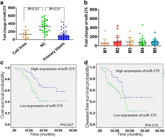
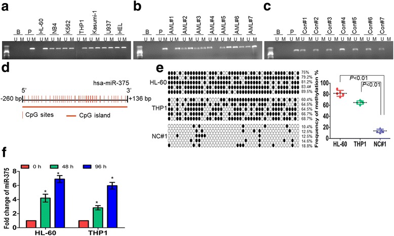
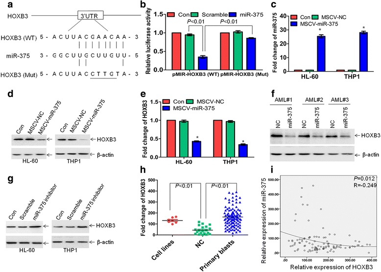
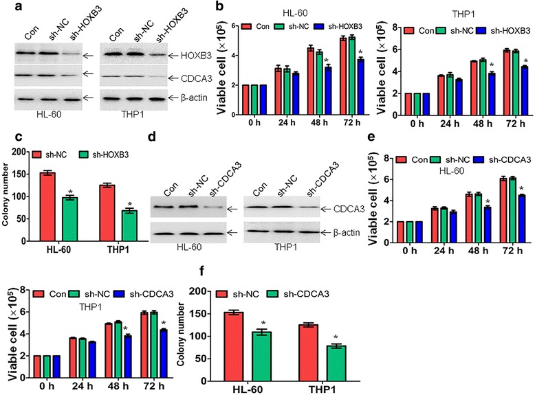
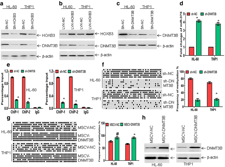
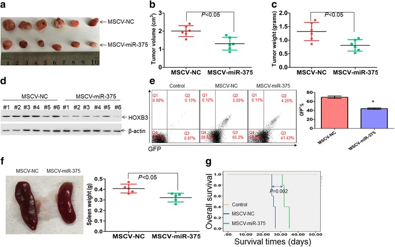
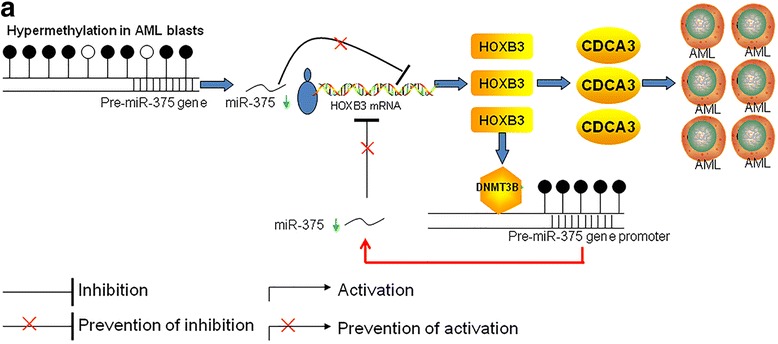
Results: The expression of miR-375 was substantially decreased in leukemic cell lines and primary AML blasts compared with normal controls, because DNA hypermethylation of precursor-miR-375 (pre-miR-375) promoter was discovered in leukemic cells but not in normal controls. Lower expression of miR-375 predicted poor outcome in AML patients. Furthermore, forced expression of miR-375 not only decreased proliferation and colony formation in leukemic cells but also reduced xenograft tumor size and prolonged the survival time in a leukemia xenograft mouse model. Mechanistically, overexpression of miR-375 reduced HOXB3 expression and repressed the activity of a luciferase reporter through binding 3'-untranslated regions (3'-UTR) of HOXB3 mRNA. Overexpression of HOXB3 partially blocked miR-375-induced arrest of proliferation and reduction of colony number, suggesting that HOXB3 plays an important role in miR-375-induced anti-leukemia activity. Knockdown of HOXB3 by short hairpin RNAs reduced the expression of cell division cycle associated 3 (CDCA3), which decreased cell proliferation. Furthermore, HOXB3 induced DNA methyltransferase 3B (DNMT3B) expression to bind in the pre-miR-375 promoter and enhanced DNA hypermethylation of pre-miR-375, leading to the lower expression of miR-375.

Conclusions: Collectively, we have identified a miR-375-HOXB3-CDCA3/DNMT3B regulatory circuitry which contributes to leukemogenesis and suggests a therapeutic strategy of restoring miR-375 expression in AML.

Keywords: AML; DNA hypermethylation; DNMT3B; HOXB3; miR-375.


Conflict of interest statement

Ethics approval and consent to participate

All procedures performed in studies involving human participants were in accordance with the ethical standards of the Ethics Committee of the First Affiliated Hospital of Wenzhou Medical University (2012025). All patients signed an informed consent for participation. All animal procedures and care were conducted in accordance with institutional guidelines and in compliance with national and international laws and policies.

Consent for publication

Not applicable.

Competing interests

The authors declare that they have no competing interests.

Publisher’s Note

Springer Nature remains neutral with regard to jurisdictional claims in published maps and institutional affiliations.

Figures

Fig. 1
Fig. 1
Decreased expression of miR-375 in AML patients predicts poor clinical outcome. a MiR-375 expressions were detected by qRT-PCR in several leukemic cell lines including NB4, HL-60, Kasumi-1, HEL, THP1, U937, and K562, 102 primary blasts from AML patients, and 20 normal controls (CD34+ cells, NC) by qRT-PCR. The lowest expression of miR-375 in one AML blast was set to 1.0 and then all other specimens were normalized by this lowest specimen. Housekeeping gene U6 is used as a reference. b MiR-375 expressions were detected in primary AML blasts according to FAB subtype (M1–M5). (c and d) The median expression of miR-375 was used as the cutoff. Kaplan-Meier overall survival curve (c) and disease-free survival curve (d) were indicated according to miR-375 expression level
Fig. 2
Fig. 2
DNA hypermethylation results in the low expression of miR-375. ac The methylation status of miR-375 was analyzed by MSP in 7 leukemic cell lines (a), 40 primary AML blasts (b), and 20 normal controls (c). B: Blank; P: positive control of methylated DNA. Bands of ‘M’ or ‘U’ are PCR products amplified by methylation-specific or unmethylation-specific primers, respectively. Shown are the representative figures for primary AML blasts and normal controls. d A CpG map of the sequenced region was analyzed by MethPrimer software. e Bisulfite genomic sequencing was performed to detect the methylation status of the DNA sequences at − 260 bp − + 136 bp in the pre-miR-375 gene upstream region in HL-60, THP1, and one normal control (NC#1). Five PCR products were shown for each sample. Each row of circles represents the sequence of an individual clone. Black circles and empty circles represent methylated and unmethylated CpG dinucleotides, respectively (Left). Shown was the summary of frequencies of methylated CpG dinucleotides detected in HL-60, THP1, and one NC by bisulfite genomic sequencing (Right). f The expression of miR-375 was detected in HL-60 and THP1 cells treated with 5 μM AZA for 2 and 4 days. *P < 0.01 versus untreated cells
Fig. 3
Fig. 3
MiR-375 targets HOXB3 via binding 3′-UTR of HOXB3. a Schematic of putative binding sites for miR-375 in 3′-UTR of HOXB3. b 293 T cells were transfected with wide-type pMIR-HOXB3UTR (WT), pMIR-HOXB3UTR (Mut), pMIR-NC, and pRL-SV40 containing Renilla luciferase gene for 24 h, followed by the transfection with miR-375 mimic or Scramble for another 24 h. Firefly and Renilla luciferase activities were both detected and histograms showed that the Firefly luciferase activities were normalized to Renilla luciferase activities. c The expression of miR-375 was detected in HL-60 and THP1 cells transduced with MSCV-miR-375 or MSCV-NC. *P < 0.01 versus MSCV-NC. (d and e) HOXB3 protein and mRNA expressions were detected by western blot and qRT-PCR in HL-60 and THP1 cells following transduction with MSCV-miR-375 or MSCV-NC, respectively. *P < 0.01 versus MSCV-NC. f HOXB3 protein expression was measured in three primary AML blasts, which were transduced with MSCV-miR-375 or MSCV-NC. g HOXB3 protein expression was detected in HL-60 and THP1 cells transfected with special miR-375 inhibitor or Scramble for 48 h. (H) HOXB3 expressions were detected in 7 leukemic cell lines, 102 primary AML blasts, and 20 NC by qRT-PCR. i miR-375 and HOXB3 expressions were measured in 102 primary AML blasts and plotting of miR-375 expression versus HOXB3 expression showed an inverse correlation between them. The correlation coefficient (R) and P values were detected by Pearson correlation
Fig. 4
Fig. 4
Ectopic expression of miR-375 inhibits cell growth and reduces colony formation. a Viable cell number was counted by the trypan-blue exclusion assay in HL-60 and THP1 cells, which were transduced with MSCV-miR-375 or MSCV-NC for the indicated times. *P < 0.01 versus MSCV-NC. b and c HL-60, THP1, and six primary AML blasts were transduced with MSCV-miR-375 or MSCV-NC, and then plated in methylcellulose for leukemia progenitor cell colony formation. *P < 0.01 and #P < 0.05 versus MSCV-NC. d Normal mouse bone marrow cells were transduced with MSCV-GFP-NC, MSCV-GFP-MLL-AF9, or MSCV-GFP-MLL-AF9+ MSCV-miR-375, and colony forming/replating assays were done thereafter. *P < 0.01. e Colony formation was counted in CD34+ hematopoietic stem and progenitor cells (HSPC), which were freshly isolated from three umbilical cord blood (UCB) and transduced with MSCV-NC or MSCV-miR-375. f HOXB3 protein level was measured in HL-60 and THP1 cells transduced with LVX-NC or LVX-HOXB3 for 48 h. g The rescue experiments were performed in HL-60 and THP1 cells, which were transduced with MSCV-NC, MSCV-miR-375, MSCV-miR-375 plus LVX-NC, or MSCV-miR-375 plus LVX-HOXB3, respectively. *P < 0.01 and #P < 0.05. h Colony formation was performed in HL-60 and THP1 cells following transduction with MSCV-NC, MSCV-miR-375, MSCV-miR-375 plus LVX-NC, or MSCV-miR-375 plus LVX-HOXB3, respectively. *P < 0.01
Fig. 5
Fig. 5
Knockdown of HOXB3 and CDCA3 partially mimics the anti-leukemia activity by miR-375. a HL-60 and THP1 cells were transduced with special shRNA for HOXB3 or a control shRNA (sh-NC). HOXB3 and CDCA3 expressions were detected by western blot. b Viable cell number by the trypan-blue exclusion assay was counted in HL-60 and THP1 cells, which were transduced with special shRNA for HOXB3 or sh-NC for the indicated times. *P < 0.01 versus sh-NC. c Colony number was counted in HL-60 and THP1 cells, which were transduced with special shRNA for HOXB3 or sh-NC. *P < 0.01 versus sh-NC. d The protein expression of CDCA3 was detected in HL-60 and THP1 cells following transduction with special shRNA for CDCA3 or sh-NC. e Viable cell number by the trypan-blue exclusion assay was counted in HL-60 and THP1 cells infected with special shRNA for CDCA3 or sh-NC for the indicated time. *P < 0.01 versus sh-NC. f Colony number was counted in HL-60 and THP1 cells, which were transduced with special shRNA for CDCA3 or sh-NC. *P < 0.01 versus sh-NC
Fig. 6
Fig. 6
HOXB3 enhances the expression of DNMT3B to bind in pre-miR-375 promoter. a HL-60 and THP1 cells were transduced with special shRNA for HOXB3 (sh-HOXB3) or sh-NC. HOXB3 and DNMT3B expressions were detected by western blot. b HOXB3 and DNMT3B expressions were detected in HL-60 and THP1 cells, which were transduced with overexpression vector LVX-HOXB3 or LVX-NC. c DNMT3B expression was detected in HL-60 and THP1 cells transduced with special shRNA targeting DNMT3B (sh-DNMT3B) or sh-NC. d MiR-375 expression was measured in HL-60 and THP1 cells transduced with sh-DNMT3B or sh-NC. *P < 0.01 versus sh-NC. e Soluble chromatin from HL-60 and THP1 cells, which were transduced with sh-NC or sh-DNMT3B, was immunoprecipitated with anti-DNMT3B antibody. Immunoprecipitated DNA was analyzed by qRT-PCR. *P < 0.01 versus sh-NC. f Bisulfite genomic sequencing was performed to detect the methylation status of the DNA sequences at -260 bp − + 136 bp in the pre-miR-375 gene upstream region in HL-60 and THP1 cells, which were transduced with sh-NC or sh-DNMT3B. Each row of circles represents the sequence of an individual clone. Black circles and empty circles represent methylated and unmethylated CpG dinucleotides, respectively (Left). Shown was the summary of frequencies of methylated CpG dinucleotides detected in HL-60 and THP1 cells by bisulfite genomic sequencing (Right). *P < 0.01 versus sh-NC. g Bisulfite sequencing was performed using DNA from HL-60 and THP1 cells transduced with MSCV-DNMT3B or MSCV-NC. Black circles and empty circles represent methylated and unmethylated CpG dinucleotides, respectively (Left). Shown was the summary of frequencies of methylated CpG dinucleotides detected in HL-60 and THP1cells by bisulfite genomic sequencing (Right). *P < 0.01 and #P < 0.05 versus MSCV-NC. h Overexpression of DNMT3B in HL-60 and THP1 cells was indicated by western blot
Fig. 7
Fig. 7
The anti-leukemia effects of miR-375 in vivo. About 1х107 viable HL-60 cells transduced with MSCV-miR-375 or MSCV-NC were injected subcutaneously into right flank of each nude mouse. a A photograph of xenografted tumors in mice xenografted by HL-60 cells, which were transduced with MSCV-miR-375 or MSCV-NC. b Volumes of all xenografted tumors were measured when the experiment was terminated at 42 days after tumor cell inoculation. c Net weights of all xenografted tumors were measured at the termination of the experiment. d The protein expression of HOXB3 was detected in xenografted tumor lysates from HL-60 cells transduced with MSCV-miR-375 or MSCV-NC. e THP1 cells were transduced with lentivirus vector pLVX-IRES-ZsGreen1 and GFP-positive cells were sorted by flow cytometry. The GFP-positive cells were further transduced with MSCV-miR-375 or MSCV-NC and treated with puromycin for 1 week. Then, THP1-GFP-miR-375 or THP1-GFP-NC were xenografted into NSG mice. Peripheral blood cells were extracted from mice when the mice became moribund and GFP-positive cells were analyzed by flow cytometry. *P < 0.01 versus MSCV-NC. Shown is a representative plot for GFP-positive cells (Left) and summary of GFP-positive cells (Right). f Representative images of spleens were observed from THP1-miR-375-engrafted mice or THP1-NC-engrafted mice (Left). All the spleens in THP1-miR-375-engrafted mice or THP1-NC-engrafted mice were weighted (Right). g MiR-375 prolonged the overall survival time in an engrafted mice model
Fig. 8
Fig. 8
An illustration of miR-375-HOXB3-CDCA3/DNMT3B regulatory circuitry. a The decreased expression of miR-375 due to DNA hypermethylation results in the high expression of HOXB3, contributing to cell proliferation and colony formation through increasing the expression of CDCA3. Moreover, HOXB3 enhances and recruits DNMT3B to bind in pre-miR-375 promoter, leading to further DNA hypermethylation and subsequent downregulation of miR-375 in AML cells, in turn

Similar articles

Cited by

References

    1. Wouters BJ, Delwel R. Epigenetics and approaches to targeted epigenetic therapy in acute myeloid leukemia. Blood. 2016;127:42–52. doi: 10.1182/blood-2015-07-604512. - DOI - PubMed
    1. Dombret H, Gardin C. An update of current treatments for adult acute myeloid leukemia. Blood. 2016;127:53–61. doi: 10.1182/blood-2015-08-604520. - DOI - PMC - PubMed
    1. Shen Y, Zhu YM, Fan X, Shi JY, Wang QR, Yan XJ, Gu ZH, Wang YY, Chen B, Jiang CL, et al. Gene mutation patterns and their prognostic impact in a cohort of 1185 patients with acute myeloid leukemia. Blood. 2011;118:5593–5603. doi: 10.1182/blood-2011-03-343988. - DOI - PubMed
    1. Wang X, Chen H, Bai J, He A. MicroRNA: an important regulator in acute myeloid leukemia. Cell Biol Int. 2017;41:936–945. doi: 10.1002/cbin.10770. - DOI - PubMed
    1. Ambros V. The functions of animal microRNAs. Nature. 2004;431:350–355. doi: 10.1038/nature02871. - DOI - PubMed

Publication types

MeSH terms

LinkOut - more resources