

Methylation profiling identifies two subclasses of squamous cell carcinoma related to distinct cells of origin
- PMID: 29422656
- PMCID: PMC5805678
- DOI: 10.1038/s41467-018-03025-1
Methylation profiling identifies two subclasses of squamous cell carcinoma related to distinct cells of origin
Abstract
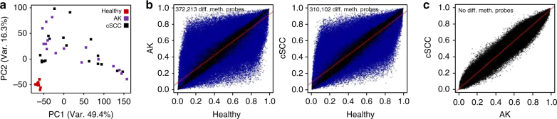
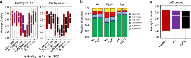
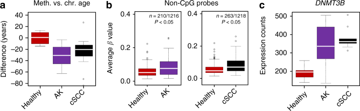
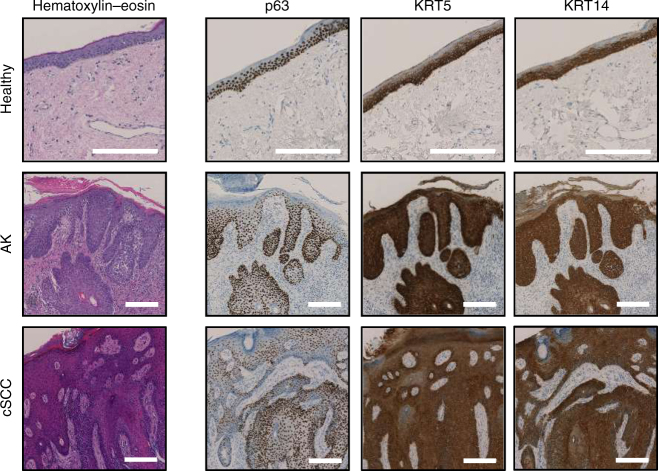
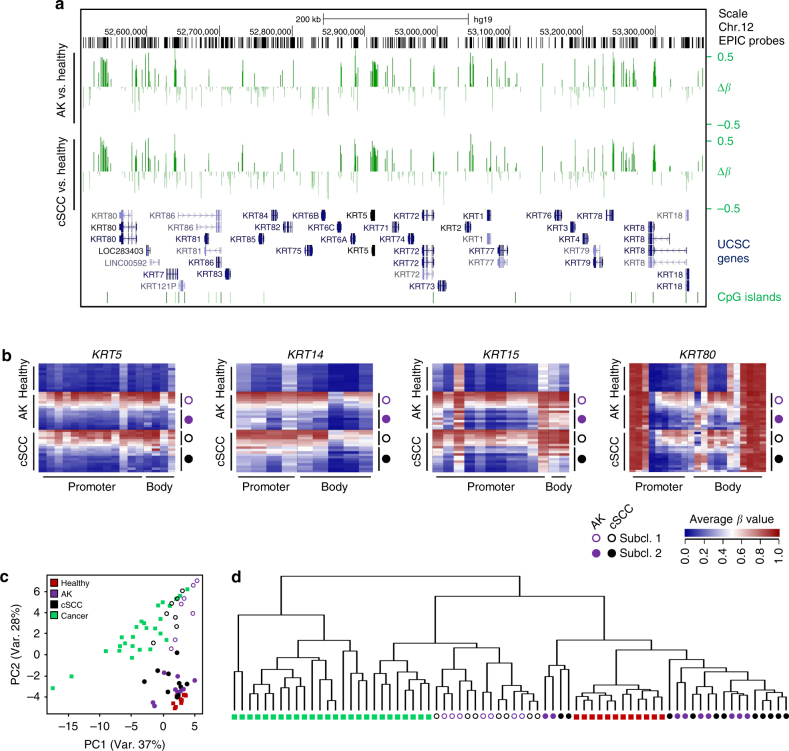
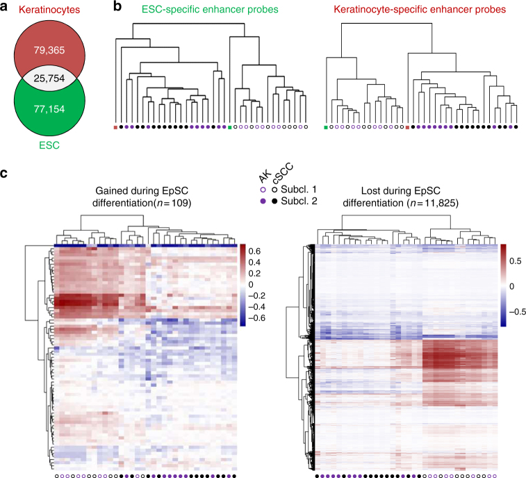
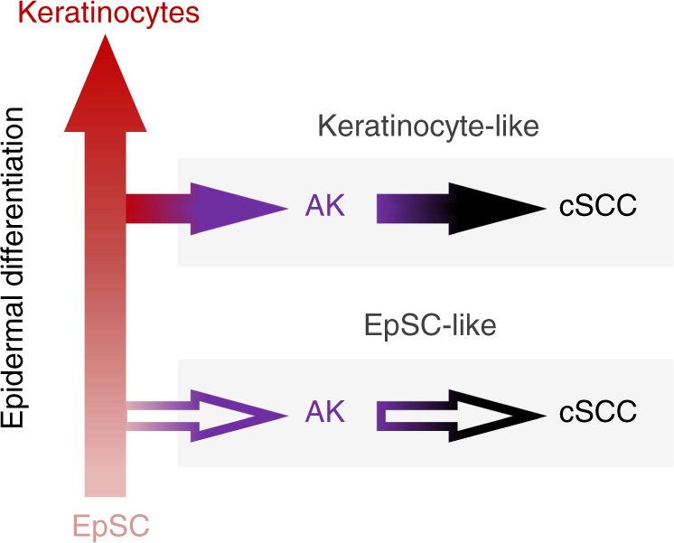
Cutaneous squamous cell carcinoma (cSCC) is the second most common skin cancer and usually progresses from a UV-induced precancerous lesion termed actinic keratosis (AK). Despite various efforts to characterize these lesions molecularly, the etiology of AK and its progression to cSCC remain partially understood. Here, we use Infinium MethylationEPIC BeadChips to interrogate the DNA methylation status in healthy, AK and cSCC epidermis samples. Importantly, we show that AK methylation patterns already display classical features of cancer methylomes and are highly similar to cSCC profiles. Further analysis identifies typical features of stem cell methylomes, such as reduced DNA methylation age, non-CpG methylation, and stem cell-related keratin and enhancer methylation patterns. Interestingly, this signature is detected only in half of the samples, while the other half shows patterns more closely related to healthy epidermis. These findings suggest the existence of two subclasses of AK and cSCC emerging from distinct keratinocyte differentiation stages.
Conflict of interest statement
E.W., K.S., S.G., H.W. and M.W. are employees of Beiersdorf AG. F.L. has received consultation fees from Beiersdorf AG.
Figures
Similar articles
-
MiR-204 silencing in intraepithelial to invasive cutaneous squamous cell carcinoma progression.Mol Cancer. 2016 Jul 25;15(1):53. doi: 10.1186/s12943-016-0537-z. Mol Cancer. 2016. PMID: 27457246 Free PMC article.
-
Gene expression landscape of cutaneous squamous cell carcinoma progression.Br J Dermatol. 2024 Oct 17;191(5):760-774. doi: 10.1093/bjd/ljae249. Br J Dermatol. 2024. PMID: 38867481
-
Targeted deep sequencing reveals genomic alterations of actinic keratosis/cutaneous squamous cell carcinoma in situ and cutaneous squamous cell carcinoma.Exp Dermatol. 2023 Apr;32(4):447-456. doi: 10.1111/exd.14730. Epub 2022 Dec 26. Exp Dermatol. 2023. PMID: 36533870
-
Telomeres and Telomerase in Cutaneous Squamous Cell Carcinoma.Int J Mol Sci. 2019 Mar 16;20(6):1333. doi: 10.3390/ijms20061333. Int J Mol Sci. 2019. PMID: 30884806 Free PMC article. Review.
-
Dermatopathologic features of cutaneous squamous cell carcinoma and actinic keratosis: Consensus criteria and proposed reporting guidelines.J Am Acad Dermatol. 2023 Jun;88(6):1317-1325. doi: 10.1016/j.jaad.2022.12.057. Epub 2023 Feb 24. J Am Acad Dermatol. 2023. PMID: 36841336 Review.
Cited by
-
Genetic Studies of Actinic Keratosis Development: Where Are We Now?Ann Dermatol. 2023 Dec;35(6):389-399. doi: 10.5021/ad.23.072. Ann Dermatol. 2023. PMID: 38086352 Free PMC article. Review.
-
Concomitant DNA methylation and transcriptome signatures define epidermal responses to acute solar UV radiation.Sci Rep. 2020 Jul 31;10(1):12918. doi: 10.1038/s41598-020-69683-8. Sci Rep. 2020. PMID: 32737342 Free PMC article. Clinical Trial.
-
Bioinformatic Methods and Bridging of Assay Results for Reliable Tumor Mutational Burden Assessment in Non-Small-Cell Lung Cancer.Mol Diagn Ther. 2019 Aug;23(4):507-520. doi: 10.1007/s40291-019-00408-y. Mol Diagn Ther. 2019. PMID: 31250328 Free PMC article.
-
DNA-methylation patterns imply a common cellular origin of virus- and UV-associated Merkel cell carcinoma.Oncogene. 2022 Jan;41(1):37-45. doi: 10.1038/s41388-021-02064-1. Epub 2021 Oct 19. Oncogene. 2022. PMID: 34667274 Free PMC article.
-
Viral Status Predicts the Patterns of Genome Methylation and Decitabine Response in Merkel Cell Carcinoma.J Invest Dermatol. 2022 Mar;142(3 Pt A):641-652. doi: 10.1016/j.jid.2021.07.173. Epub 2021 Aug 30. J Invest Dermatol. 2022. PMID: 34474081 Free PMC article.
References
Publication types
MeSH terms
LinkOut - more resources
Full Text Sources
Other Literature Sources
Medical
