. 2018 May 1;314(5):C603-C615.
doi: 10.1152/ajpcell.00177.2017. Epub 2018 Feb 7.

Platelet procoagulant phenotype is modulated by a p38-MK2 axis that regulates RTN4/Nogo proximal to the endoplasmic reticulum: utility of pathway analysis

Affiliations

Platelet procoagulant phenotype is modulated by a p38-MK2 axis that regulates RTN4/Nogo proximal to the endoplasmic reticulum: utility of pathway analysis

Özgün Babur et al. Am J Physiol Cell Physiol. .

Abstract

Upon encountering physiological cues associated with damaged or inflamed endothelium, blood platelets set forth intracellular responses to ultimately support hemostatic plug formation and vascular repair. To gain insights into the molecular events underlying platelet function, we used a combination of interactome, pathway analysis, and other systems biology tools to analyze associations among proteins functionally modified by reversible phosphorylation upon platelet activation. While an interaction analysis mapped out a relative organization of intracellular mediators in platelet signaling, pathway analysis revealed directional signaling relations around protein kinase C (PKC) isoforms and mitogen-activated protein kinases (MAPKs) associated with platelet cytoskeletal dynamics, inflammatory responses, and hemostatic function. Pathway and causality analysis further suggested that platelets activate a specific p38-MK2 axis to phosphorylate RTN4 (reticulon-4, also known as Nogo), a Bcl-xl sequestration protein and critical regulator of endoplasmic reticulum (ER) physiology. In vitro, we find that platelets drive a p38-MK2-RTN4-Bcl-xl pathway associated with the regulation of the ER and platelet phosphatidylserine exposure. Together, our results support the use of pathway tools in the analysis of omics data sets as a means to help generate novel, mechanistic, and testable hypotheses for platelet studies while uncovering RTN4 as a putative regulator of platelet cell physiological responses.

Keywords: Bcl-xl; CausalPath; MAPKAPK2; Pathway Commons; platelets.


Figures

Fig. 1.
Fig. 1.
Interactome analysis of the regulated platelet phosphoproteome. A protein-protein interaction (PPI) network of dynamically regulated platelet phosphoproteins identified by Beck et al. (16) was generated from an interaction query of Pathway Commons using ChiBE. Node colors indicate the relative intensity of the reported phosphorylation change (red = increase; blue = decrease). Selected proteins of interest to this study, including the MAP kinase p38 (MAPK14), the MAP kinase-activated protein kinase MK2 (MAPKAPK2), and RTN4 are indicated with stars.
Fig. 2.
Fig. 2.
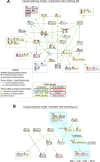
Pathway and causality analysis of the activated platelet phosphoproteome. Results from CausalPath identifying pathway fragments and associated changes in platelet protein phosphorylation to show potential cause-effect relations as visualized with ChiBE. The unrestricted model in A is based on generalized enzyme:substrate relations (i.e., phosphorylated/activated p38 is known to be involved in the phosphorylation and activation of MK2), independent of phosphorylation site localization details. The stringent model in B requires the matching of enzymatic events with phosphorylation site details (i.e., phosphorylation of p38 at Thr180,Tyr182 is known to be involved in MK2 Thr334 phosphorylation and activation). This model highlights a putative p38→MK2→RTN4 axis associated with platelet activation (light blue background). As indicated in the Legend, nodes represent proteins (conventionally labeled with gene names), and edges represent either causal phosphorylation (green) or dephosphorylation (red) events. Protein phosphorylation sites are shown with smaller “p” circles, where a green border indicates an activating site and red border indicates inactivating site. The background color of phosphorylation sites indicates their differential measurement from data, red indicating an increase and blue indicating a decrease. The grouped nodes in a compound node indicate that all members have the same graph topology and are grouped for complexity management. Relative targets of the p38 inhibitor (SB202190) and MK2 inhibitor (PF3644022) used in this study are indicated in the context of this model.
Fig. 3.
Fig. 3.
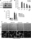
p38 and MK2 phosphorylation and function in platelets. A: replicate samples (n = 3) of washed human platelets (5 × 108/ml) were pretreated with the p38 inhibitor SB202190 (2 μM), the MK2 inhibitor PF3644022 (2 μM), a combination of the P2Y1 and P2Y12 antagonists MRS 2179 (10 μM) and AR-C 66096 (10 μM), or vehicle alone (0.1% DMSO) before stimulation with bovine thrombin (0.5 U/ml, 5 min), lysis into Laemmli sample buffer, SDS-PAGE, transfer to nitrocellulose, and Western blot analysis of phospho-p38 Thr180,Tyr182 (p-p38) and phospho-MK2 Thr334 (p-MK2) immunoreactivity. Total α-tubulin levels serve as a control for equal protein loading. Tick marks indicate the relative positions of 40-kDa molecular mass marker (for p-p38) 50-kDa molecular mass marker (for p-MK2 and α-tubulin). B: densitometry analysis of p38 (black bars) and MK2 (gray bars) phosphorylation (arbitrary units; AU). *P ≤ 0.05 relative to thrombin stimulated platelets. C: washed human platelets (2 × 108/ml) were incubated with vehicle (0.01% DMSO), SB 202190 (10 µM), PF 3644022 (10 µM), or Ro 31-8220 (5 µM) before activation with thrombin (1 U/ml) for 30 s. Platelet dense granule secretion was measured as function of ATP release generated by an ATP-luciferin-luciferase reaction. Values are mean ± SE of raw luminescence; (n = 4). *P ≤ 0.05, platelets compared with vehicle in the presence of thrombin (AU). D: washed human platelets (2 × 107/ml) were treated with SB202190 (2 µM), PF3644022 (2 µM), the lysine acetyltransferase inhibitor C646 [10 µM, a previously described inhibitor of platelet spreading on fibrinogen (8)], or vehicle alone (0.1% DMSO) before incubation on fibrinogen-coated coverglass (45 min, 37°C), fixation, staining and imaging by DIC, and fluorescence microscopy (n = 3). Scale bar = 10 µm.
Fig. 4.
Fig. 4.
RTN4 expression, localization and phosphorylation in platelets. A: MDA-MB-231 cell lysates, HUVEC lysates, human platelet lysates, and RTN4 and nonspecific IgM immunoprecipitates (IP) from platelets were separated by SDS-PAGE, transferred to nitrocellulose and examined for RTN4 immunoreactivity by Western blotting (WB). Positions of molecular weight (MW) markers are indicated. Representative results are shown (n = 3). B: replicate samples (n = 3) of washed human platelets were incubated on fibrinogen-coated coverglass in the absence or presence of 1 U/ml thrombin before fixation, staining for RTN4 (green) and PDI (red), and imaging by SR-SIM at wide field (scale bar = 10 µm) and ×100 (scale bar = 2 µm) magnification. C: replicate samples (n = 3) of washed human platelets were pretreated with SB202190 (SB; 2 µM), PF3644022 (PF; 2 µM), or vehicle alone (veh; 0.1% DMSO) before stimulation with thrombin (0.5 U/ml, 5 min) and lysis. RTN4 and nonspecific IgM immunoprecipitates (IP) were examined for total RTN4 and RTN4 phospho Ser107 (RXXpS) immunoreactivity by Western blotting (WB). Tick marks indicate position of 50-kDa molecular mass marker.
Fig. 5.
Fig. 5.
Proximity of Bcl-xl to p38, MK2, and RTN4 in platelet function. A: signaling and protein binding relations in the neighborhood vicinity of MK2 and RTN4 in Pathway Commons, as queried and rendered by ChiBE. Directed relations (arrows) indicate that the source protein controls a state change of the target protein. A state change can be a modification on the protein, on its location, or a change to the complex that the target protein is a member. Undirected edges (connecting lines) indicate that two proteins appear as members in the same complex. B: replicate samples (n = 3) of washed human platelets (1 × 109/ml, 1 ml) were incubated with DTBP cross-linker for 30 min at room temperature before lysis and processing for immunoprecipitation (IP). Following IP with RTN4 or control IgM antisera, samples were analyzed for RTN4 capture and Bcl-xl coimmunoprecipitation by Western blotting (WB). Tick marks indicate relative positions of 30- and 50-kDa molecular mass markers for Bcl-xl and RTN4, respectively. C: replicate samples (n = 3) of washed human platelets (5 × 108/ml) were pretreated with SB202190 (SB; 2 µM), PF3644022 (PF; 2 µM), or vehicle alone (veh.; 0.1% DMSO) before stimulation with thrombin (0.5 U/ml, 5 min). Following lysis into Laemmli sample buffer, samples were examined for Bcl-xl phospho-Ser62 (p-Bcl-xl) immunoreactivity by Western blotting. Total Bcl-xl levels serve as a control for equal protein loading. Tick marks indicate relative positions of 30-kDa molecular mass marker.
Fig. 6.
Fig. 6.
p38-MK2-RTN4-Bcl-xl axis in procoagulant platelet function. A: washed human platelets were pretreated with SB202190 (2 µM), PF3644022 (2 µM), or vehicle alone (0.1% DMSO) before incubation on fibrinogen-coated coverglass in the absence or presence of thrombin (0.5 U/ml). Following fixation in PFA, samples were processed for SR-SIM visualization of RTN4 (green) and Bcl-xl (red) at wide field (×63, scale bar = 10 µm) and ×100 magnification (scale bar = 2 µm). Results are representative of n = 3 experiments. B: washed human platelets (2 × 107/ml) were pretreated with SB202190 (2 µM), PF3644022 (2 µM), the intracellular calcium chelator BAPTA (40 µM), or vehicle alone (0.1% DMSO) before incubation on fibrinogen-coated cover glass in the absence or presence of 0.25 U/ml thrombin (37°C, 45 min). Following an additional 30 min in the presence of annexin V-Alexa Fluor 488 and 2.5 mM calcium, platelets were visualized for PS exposure and general morphology by DIC and fluorescence microscopy. Scale bar = 10 µm. Results are representative of n = 3 experiments.

Similar articles

Cited by

References

    1. Acevedo L, Yu J, Erdjument-Bromage H, Miao RQ, Kim JE, Fulton D, Tempst P, Strittmatter SM, Sessa WC. A new role for Nogo as a regulator of vascular remodeling. Nat Med 10: 382–388, 2004. doi:10.1038/nm1020. - DOI - PubMed
    1. Adam F, Kauskot A, Rosa JP, Bryckaert M. Mitogen-activated protein kinases in hemostasis and thrombosis. J Thromb Haemost 6: 2007–2016, 2008. doi:10.1111/j.1538-7836.2008.03169.x. - DOI - PubMed
    1. Agbani EO, Hers I, Poole AW. Temporal contribution of the platelet body and balloon to thrombin generation. Haematologica 102: e379–e381, 2017. doi:10.3324/haematol.2017.166819. - DOI - PMC - PubMed
    1. Andre P. Intracellular signaling as a potential target for antiplatelet therapy. Handb Exp Pharmacol 210: 339–367, 2012. doi:10.1007/978-3-642-29423-5_14. - DOI - PubMed
    1. Aslan JE. Platelet shape change. In: Platelets in Thrombotic and Non-Thrombotic Disorders, edited by Gresele P, López J, Kleiman N, Page C. New York: Springer, 2017. doi:10.1007/978-3-319-47462-5_24. - DOI

Publication types

MeSH terms

Substances

LinkOut - more resources