Characterization of interaction between Trim28 and YY1 in silencing proviral DNA of Moloney murine leukemia virus
- PMID: 29407374
- PMCID: PMC8456507
- DOI: 10.1016/j.virol.2018.01.012
Characterization of interaction between Trim28 and YY1 in silencing proviral DNA of Moloney murine leukemia virus
Abstract
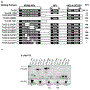
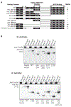
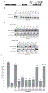
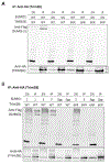
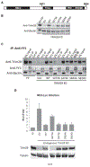
Moloney Murine Leukemia Virus (M-MLV) proviral DNA is transcriptionally silenced in embryonic cells by a large repressor complex tethered to the provirus by two sequence-specific DNA binding proteins, ZFP809 and YY1. A central component of the complex is Trim28, a scaffold protein that regulates many target genes involved in cell cycle progression, DNA damage responses, and viral gene expression. The silencing activity of Trim28, and its interactions with corepressors are often regulated by post-translational modifications such as sumoylation and phosphorylation. We defined the interaction domains of Trim28 and YY1, and investigated the role of sumoylation and phosphorylation of Trim28 in mediating M-MLV silencing. The RBCC domain of Trim28 was sufficient for interaction with YY1, and acidic region 1 and zinc fingers of YY1 were necessary and sufficient for its interaction with Trim28. Additionally, we found that residue K779 was critical for Trim28-mediated silencing of M-MLV in embryonic cells.
Keywords: Moloney murine leukemia virus; Phosphorylation; Protein-protein interaction; SUMO; Transcriptional silencing; Trim28; YY1.
Copyright © 2018 Elsevier Inc. All rights reserved.
Figures

Similar articles
-
Proviral silencing in embryonic cells is regulated by Yin Yang 1.Cell Rep. 2013 Jul 11;4(1):50-8. doi: 10.1016/j.celrep.2013.06.003. Epub 2013 Jun 27. Cell Rep. 2013. PMID: 23810560 Free PMC article.
-
EBP1, a novel host factor involved in primer binding site-dependent restriction of moloney murine leukemia virus in embryonic cells.J Virol. 2014 Feb;88(3):1825-9. doi: 10.1128/JVI.02578-13. Epub 2013 Nov 13. J Virol. 2014. PMID: 24227866 Free PMC article.
-
Differential control of retrovirus silencing in embryonic cells by proteasomal regulation of the ZFP809 retroviral repressor.Proc Natl Acad Sci U S A. 2017 Feb 7;114(6):E922-E930. doi: 10.1073/pnas.1620879114. Epub 2017 Jan 23. Proc Natl Acad Sci U S A. 2017. PMID: 28115710 Free PMC article.
-
TRIM28 and the control of transposable elements in the brain.Brain Res. 2019 Feb 15;1705:43-47. doi: 10.1016/j.brainres.2018.02.043. Epub 2018 Mar 6. Brain Res. 2019. PMID: 29522722 Review.
-
Post-entry restriction of retroviral infections.AIDS Rev. 2003 Jul-Sep;5(3):156-64. AIDS Rev. 2003. PMID: 14598564 Review.
Cited by
-
Comparing the expression levels of tripartite motif containing 28 in mild and severe COVID-19 infection.Virol J. 2022 Oct 3;19(1):156. doi: 10.1186/s12985-022-01885-0. Virol J. 2022. PMID: 36192760 Free PMC article.
-
Inhibition of HIV-1 gene transcription by KAP1 in myeloid lineage.Sci Rep. 2021 Jan 29;11(1):2692. doi: 10.1038/s41598-021-82164-w. Sci Rep. 2021. PMID: 33514850 Free PMC article.
-
An influenza virus-triggered SUMO switch orchestrates co-opted endogenous retroviruses to stimulate host antiviral immunity.Proc Natl Acad Sci U S A. 2019 Aug 27;116(35):17399-17408. doi: 10.1073/pnas.1907031116. Epub 2019 Aug 7. Proc Natl Acad Sci U S A. 2019. PMID: 31391303 Free PMC article.
-
TRIM5α self-assembly and compartmentalization of the HIV-1 viral capsid.Nat Commun. 2020 Mar 11;11(1):1307. doi: 10.1038/s41467-020-15106-1. Nat Commun. 2020. PMID: 32161265 Free PMC article.
-
Navigating the brain and aging: exploring the impact of transposable elements from health to disease.Front Cell Dev Biol. 2024 Feb 27;12:1357576. doi: 10.3389/fcell.2024.1357576. eCollection 2024. Front Cell Dev Biol. 2024. PMID: 38476259 Free PMC article. Review.
References
-
- Barklis E, Mulligan RC, Jaenisch R, 1986. Chromosomal position or virus mutation permits retrovirus expression in embryonal carcinoma cells. Cell 47, 391–399. - PubMed
-
- Bohren KM, Nadkarni V, Song JH, Gabbay KH, Owerbach D, 2004. A M55V polymorphism in a novel SUMO gene (SUMO-4) differentially activates heat shock transcription factors and is associated with susceptibility to type I diabetes mellitus. J. Biol. Chem 279, 27233–27238. - PubMed
-
- Bolderson E, Savage KI, Mahen R, Pisupati V, Graham ME, Richard DJ, Robinson PJ, Venkitaraman AR, Khanna KK, 2012. Kruppel-associated Box (KRAB)-associated co-repressor (KAP-1) Ser-473 phosphorylation regulates heterochromatin protein 1beta (HP1-beta) mobilization and DNA repair in heterochromatin. J. Biol. Chem 287, 28122–28131. - PMC - PubMed
-
- Bushmeyer S, Park K, Atchison ML, 1995. Characterization of functional domains within the multifunctional transcription factor, YY1. J. Biol. Chem 270, 30213–30220. - PubMed
-
- Cammas F, Mark M, Dolle P, Dierich A, Chambon P, Losson R, 2000. Mice lacking the transcriptional corepressor TIF1beta are defective in early postimplantation development. Development 127, 2955–2963. - PubMed
Publication types
MeSH terms
Substances
Grants and funding
LinkOut - more resources
Full Text Sources
Other Literature Sources
Miscellaneous

