. 2018 Feb 1;14(2):e1007203.
doi: 10.1371/journal.pgen.1007203. eCollection 2018 Feb.

Chinmo prevents transformer alternative splicing to maintain male sex identity

Affiliations

Chinmo prevents transformer alternative splicing to maintain male sex identity

Lydia Grmai et al. PLoS Genet. .

Abstract

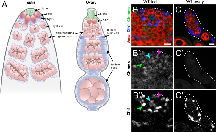
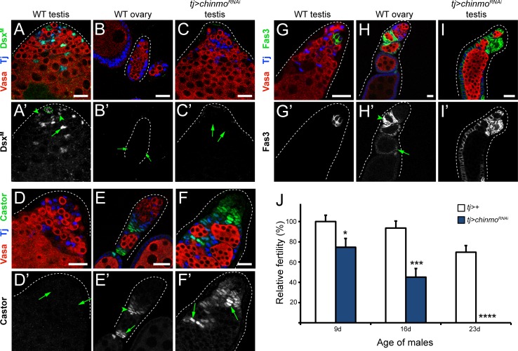
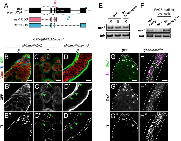
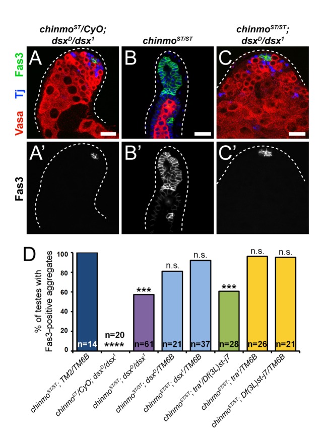
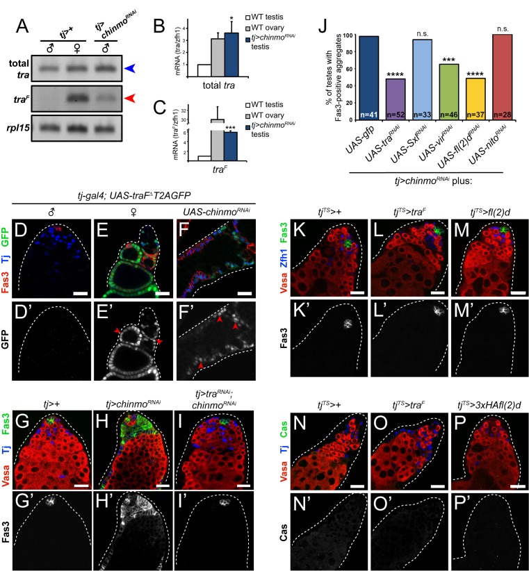
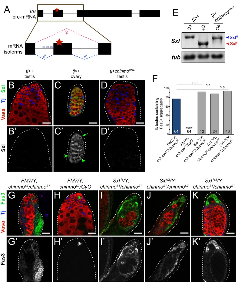
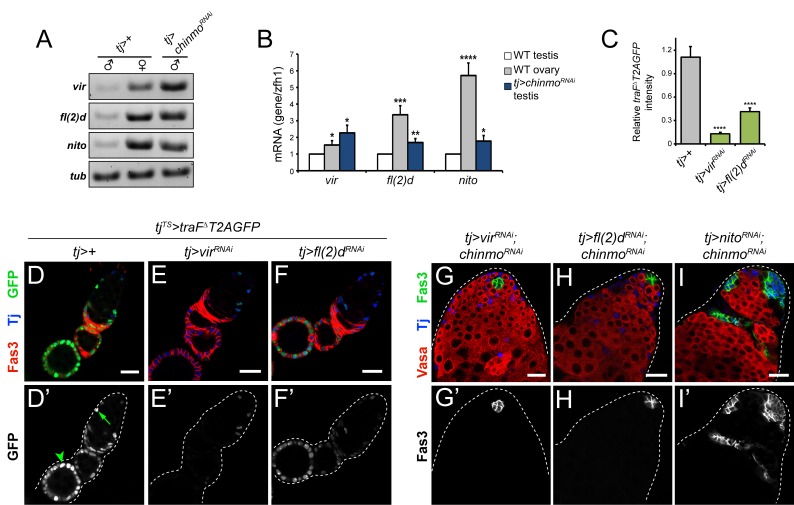
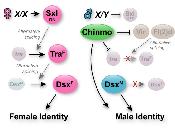
Reproduction in sexually dimorphic animals relies on successful gamete production, executed by the germline and aided by somatic support cells. Somatic sex identity in Drosophila is instructed by sex-specific isoforms of the DMRT1 ortholog Doublesex (Dsx). Female-specific expression of Sex-lethal (Sxl) causes alternative splicing of transformer (tra) to the female isoform traF. In turn, TraF alternatively splices dsx to the female isoform dsxF. Loss of the transcriptional repressor Chinmo in male somatic stem cells (CySCs) of the testis causes them to "feminize", resembling female somatic stem cells in the ovary. This somatic sex transformation causes a collapse of germline differentiation and male infertility. We demonstrate this feminization occurs by transcriptional and post-transcriptional regulation of traF. We find that chinmo-deficient CySCs upregulate tra mRNA as well as transcripts encoding tra-splice factors Virilizer (Vir) and Female lethal (2)d (Fl(2)d). traF splicing in chinmo-deficient CySCs leads to the production of DsxF at the expense of the male isoform DsxM, and both TraF and DsxF are required for CySC sex transformation. Surprisingly, CySC feminization upon loss of chinmo does not require Sxl but does require Vir and Fl(2)d. Consistent with this, we show that both Vir and Fl(2)d are required for tra alternative splicing in the female somatic gonad. Our work reveals the need for transcriptional regulation of tra in adult male stem cells and highlights a previously unobserved Sxl-independent mechanism of traF production in vivo. In sum, transcriptional control of the sex determination hierarchy by Chinmo is critical for sex maintenance in sexually dimorphic tissues and is vital in the preservation of fertility.


Conflict of interest statement

The authors have declared that no competing interests exist.

Figures

Fig 1
Fig 1. Chinmo is expressed dimorphically in Drosophila gonads.
(A) Schematic of the adult Drosophila testis (left) and ovary (right). In the testis, the niche (green) supports two populations of stem cells, germline stem cells (GSCs, dark pink) and somatic cyst stem cells (CySCs, dark blue). The GSC divides to produce differentiating daughter cells (light pink) that undergo four transit-amplifying divisions. The CySC divides to produce cyst daughter cells (light blue) that exit the cell cycle and ensheath the differentiating GSC daughter. Cyst cells continue to ensheath the associated spermatogonial cyst during transit-amplifying divisions. In the ovary, the niche (green) supports GSCs (dark pink), which divide to give rise to differentiating daughters (light pink) that undergo 4 mitotic divisions. The developing germline cyst is ensheathed by an epithelial layer of follicle cells (light blue). Follicle cells are proliferative descendants of follicle stem cells (FSCs, dark blue) located in the anterior part of the ovary. (B) Chinmo (green) is present in CySCs (B’, green arrowhead), GSCs (B’, magenta arrowhead), and niche cells (B’, cyan arrowhead) in a wild type testis. (C) Chinmo is not expressed in follicle cells of a wild type ovary. Vasa (red) marks the germline and Zfh1 (blue) marks somatic cells in the testis and ovary. Scale bars = 10 μm.
Fig 2
Fig 2. Chinmo is required in CySCs for male somatic sex identity and non-autonomously for germline maintenance.
(A) In a wild type testis, DsxM (green) is present in niche cells (A’, outlined with dotted green line), CySCs (A’, arrowheads) and cyst cells (A’, arrow). (B) DsxM is not expressed in a wild type ovary (B’, arrows indicate early follicle cells). (C) DsxM is lost in the CySC lineage in a tj>chinmoRNAi testis (C’, arrows indicate early somatic cells). (D) Castor (Cas, green) is absent from a wild type testis (D’, arrows indicate early somatic cells). (E) In a wild type ovary, Cas (green) is expressed in early follicle cells (E’, arrowhead) and stalk cells (E’, arrow). (F) Cas (green) is ectopically expressed in feminizing somatic cells in the testis (F’, arrows) upon loss of Chinmo. (G) Fasciclin-3 (Fas3, green) is normally restricted to niche cells in a wild type testis. (H) In a wild type ovary, Fas3 (green) is high in early follicle cells (H’, arrowhead) and is lower in mature follicle cells (H’, arrow). (I) Fas3 (green) is ectopically expressed upon loss of Chinmo in CySC lineage. (J) Relative fertility of tj>chinmoRNAi males (blue bars) is decreased at 9, 16 or 23 days (d) post eclosion compared with control tj>+ males (white bars). tj>chinmoRNAi males are completely sterile by 23 days post eclosion (* denotes p<0.05; *** denotes p<0.001; **** denotes p<0.0001 as determined by single-factor ANOVA). Error bars represent SEM. In A-I, Vasa (red) marks the germline and Tj (blue) marks cyst cells. Scale bars = 20 μm. Time point in A-I is 7 days post-eclosion.
Fig 3
Fig 3. DsxF protein is synthesized in chinmo-mutant CySCs.
(A) Schematic of dsx pre-mRNA splicing. dsxF and dsxM share the first three coding exons and differ at their C-termini. Exon 4 contains a non-canonical splice acceptor, and thus under default splicing conditions, exon 3 is adjoined with exon 5 to yield the male-specific dsxM isoform (blue). In XX somatic cells, TraF is expressed and binds tandem TraF-binding sites (white dashed line) in exon 4. The TraF complex then recruits the spliceosome, leading to synthesis of the female-specific dsxF isoform (pink). Pink and blue stars indicate female and male stop codons, respectively. (B) A knock-in transcriptional reporter for dsx (dsx-gal4) activates UAS-GFP expression (green) in the CySC lineage of a wild type testis. (C) dsx-gal4 activates GFP expression in both escort cells (C’, arrow) and follicle cells (C’, arrowheads) of wild type ovary. (D) dsx-gal4 activates GFP expression (D’, arrowheads) in the somatic lineage of a chinmoST/chinmoST testis. Note that the feminized soma in this testis is organized at the periphery, adjacent to the muscle sheath. (E) Semi-quantitative RT-PCR on homogenized wild type testes (left lane), wild type ovaries (middle lane), and tj>chinmoRNAi testes (right lane). As determined by primers that recognize both dsx mRNA isoforms (dsxCOMMON, or dsxC), dsx transcripts are present in both wild type testes and wild type ovaries (left and middle lanes). dsx mRNA is also expressed in tj>chinmoRNAi testes (right lane). α-tub (tub) was used as a loading control. Flies were aged 9–20 days prior to dissection. (F) Semi-quantitative RT-PCR on RNA extracts from FACS-purified cyst cells from control tj>+ or tj>chinmoRNAi testes. Whole adult ovaries from yw females (labeled “WT ovary”) were homogenized as a positive control for dsxF mRNA detection. dsxF is detected in wild type ovaries (left lane) and in tj>chinmoRNAi cyst cells (right lane) but not in tj>+ cyst cells (middle lane). β-tubulin (tub) was used as a loading control. (G,H) Dsx (green), as detected by the DsxC antibody that recognizes both DsxF and DsxM, is present in somatic cells of a wild type testis (G) and a tj>chinmoRNAi testis (H). Tj (magenta) marks cyst cells. In B-D, Vasa (red) marks germ cells and Tj (blue) marks cyst cells. Scale bars = 20 μm.
Fig 4
Fig 4. DsxF is required for feminization in chinmo-mutant CySCs.
(A-C) Representative confocal images of testes from chinmoST/chinmoST; dsxD/dsx1 flies. In a testis from a chinmoST/CyO; dsxD/dsx1 sibling male, only niche cells express Fas3 (A’). By contrast, in a chinmoST/chinmoST testis most of the somatic lineage is positive for Fas3 (B’). In a testis from a “rescued” chinmoST/chinmoST; dsxD/dsx1 male, Fas3 is again restricted to niche cells (C’). Results are quantified in Fig 4D and S1 Table. Vasa (red) marks germ cells and Tj (blue) marks cyst cells. Scale bars = 20 μm.(D) Quantification of Fas3-positive aggregates as a readout of feminization. 100% of chinmoST/chinmoST; TM2/TM6B testes contain Fas3-positive aggregates (dark blue bar) at 7 days post-eclosion. By contrast, none of the testes from chinmoST/CyO; dsxD/dsx1 sibling males contain aggregates (second bar). There is a significant reduction in the percentage of testes from chinmoST/chinmoST; dsxD/dsx1 (purple bar) or chinmoST/chinmoST; tra1/Df(3L)st-j7 (green bar) containing Fas3-positive aggregates, indicating a partial rescue of the feminization. However, testes from the various sibling controls are still feminized (light blue and yellow bars). Sample sizes are indicated within bars. *** denotes p<0.001 and **** denotes p<0.0001 as determined by two-tailed Student’s t-test (for qRT-PCR, compared with tj>+ controls) or Fisher’s Exact Test (for quantifications, compared with chinmoST/chinmoST). n.s. means not significant. See S1 Table for percentage values.
Fig 5
Fig 5. The female sex determinant TraF is required for feminization of chinmo-mutant CySCs.
(A) Semi-quantitative RT-PCR of total tra and traF mRNA in control tj>+ testes (first lane), control tj>+ ovaries (second lane) and tj>chinmoRNAi testes (third lane). All three samples contain a band for total tra (first row, blue arrowhead). However, a traF band is detected in ovaries, as expected, and in tj>chinmoRNAi testes (second row, red arrowhead) but not in control tj>+ testes. rpl15 was used as a loading control (third row). Flies were aged 9–20 days prior to dissection. (B-C) qRT-PCR of total tra (B) or traF (C) in control tj>+ testes (white bars), control tj>+ ovaries (gray bars) and tj>chinmoRNAi testes (blue bars). There is significantly more total tra mRNA (B) and traF (C) in tj>chinmoRNAi testes compared to control tj>+ testes. Values represent the average of three biological replicates. * denotes p<0.05; *** denotes p<0.001 as determined by two-tailed Student’s t-test (compared with tj>+ testes). Error bars represent SEM. Flies were aged 9–20 days prior to dissection. (D-F) GFP caused by alternative splicing of traFΔT2AGFP pre-mRNA is not observed in control tj>traFΔT2AGFP testes (D’). By contrast, GFP indicative of traF splicing is robustly observed in follicle cells of a control tj>traFΔT2AGFP ovary (E’, arrowheads) and in somatic cells of a tj>traFΔT2AGFP; chinmoRNAi testis (F’, arrowheads). Tj (blue) marks somatic cells. Fas3 (red) marks hub cells in wild type testes, follicle cells in wild type ovaries and feminized cyst cells in tj>chinmoRNAi testes. (G-I) In a control tj>+ testis, only niche cells express Fas3 (G’). By contrast, in a tj>chinmoRNAi testis, most of the somatic lineage is positive for Fas3 (H’). In a “rescued” tj>traRNAi; chinmoRNAi testis, Fas3 is again restricted to niche cells (I’). Vasa (red) marks germ cells, Tj (blue) marks cyst cells, and Fas3 (green) marks niche cells and feminizing somatic cells. Results are quantified in Fig 4J and S1 Table. (J) Quantification of CySC feminization in tj>GFP; chinmoRNAi testes and various genotypes. 100% of tj>GFP; chinmoRNAi testes contain Fas3-positive aggregates (dark blue bar). There is a significant reduction in the percentage of feminized testes when tra is concomitantly depleted from tj>chinmoRNAi testes (purple bar). There is also a significant reduction when vir or fl(2)d is depleted from tj>chinmoRNAi testes (green and yellow bars, respectively). However, there is no rescue of male sex identity in tj>chinmoRNAi testes when Sxl, or nito is concomitantly depleted (light blue and red bars, respectively). Sample sizes are indicated within bars. *** denotes p<0.001; **** denotes p<0.0001 as determined by Fisher’s Exact Test (for rescue quantifications, compared with tj>GFP; chinmoRNAi). n.s. means not significant. See S1 Table for percentage values. (K-M) Fas3 is not ectopically expressed in CySCs from control (tjTS>+) testes (K’), testes with somatic mis-expression of traF (tjTS>traF) (L’), or testes with somatic mis-expression of fl(2)d (tjTS>fl(2)d) (M’). (N-P) Cas is not expressed in control (tjTS>+) (N’), tjTS>traF testes (O’), or tjTS>fl(2)d testes (P’). In K-P, Vasa (red) marks germ cells, and Zfh1 or Tj (blue) mark cyst cells. Scale bars = 20 μm.
Fig 6
Fig 6. Sxl is not required for feminization of chinmo-mutant CySCs.
(A) Schematic of tra pre-mRNA splicing. The poly(U) tract upstream of exon 2 is bound by the RRM domain of Sxl in females, causing skipping of exon 2. In wild type males, exons 1–4 comprise tra mRNA and translation terminates at the early stop codon in exon 2 (red star). Pink dashed lines indicate female-specific alternative splicing and blue dashed lines indicate non-sex-specific default splicing. (B-D) Sxl is not expressed in a control tj>+ testis (B’) but is expressed in follicle cells (C’, arrowheads) and in an early germ cell (C’, arrow) of a control tj>+ ovary. Sxl protein is not detected in a tj>chinmoRNAi testis (D’). (E) Semi-quantitative RT-PCR on Sxl in homogenized control tj>+ testes (left lane), control tj>+ ovaries (middle lane), and tj>chinmoRNAi testes (right lane). Control tj>+ testes express SxlM transcripts (blue arrowhead), while control tj>+ ovaries express SxlF transcripts (red arrowhead). SxlM is still present and SxlF is undetectable in tj>chinmoRNAi testes (right lane). SxlJYR primers were used to differentiate between SxlM and SxlF mRNA isoforms in this experiment. α-tubulin (tub) was used as a loading control. (F) Quantification of CySC feminization in Sxl; chinmoST backgrounds. Sample sizes are indicated within bars. **** denotes p<0.0001 as determined by Fisher’s Exact Test (compared to FM7/Y; chinmoST/chinmoST). See S1 Table for percentage values. (G-K) Representative images for Sxl; chinmoST epistasis experiments. Genetic loss of Sxl by 3 different alleles–Sxl f1 (I), Sxl f2 (J), or Sxl f18 (K)–does not prevent feminization of chinmoST/chinmoST cyst cells (G’), defined by the accumulation of Fas3-positive aggregates. Control FM7/Y; chinmoST/CyO cyst cells do not feminize (H’). Scale bars = 20 μm.
Fig 7
Fig 7. Vir and Fl(2)d, but not Nito, are required for feminization of chinmo-deficient CySCs.
(A) Semi-quantitative RT-PCR of vir, fl(2)d, and nito in homogenized control tj>+ testes (left lane), control tj>+ ovaries (middle lane) and tj>chinmoRNAi testes (right lane). vir, fl(2)d, and nito are expressed at higher levels in ovaries (middle lane) than in tj>+ testes (left lane). vir, fl(2)d, and nito are expressed at higher levels in tj>chinmoRNAi testes (right lane) than in control tj>+ testes (left lane). α-tubulin (tub) was used as a loading control. Flies were aged 9–20 days prior to dissection. (B) qRT-PCR analysis of vir, fl(2)d, and nito in homogenized control tj>+ testes (white bars), control tj>+ ovaries (gray bars) and tj>chinmoRNAi testes (blue bars). vir, fl(2)d, and nito are expressed at significantly higher levels in ovaries and tj>chinmoRNAi testes compared to control testes. * denotes p<0.05; ** denotes p<0.01; *** denotes p<0.001; **** denotes p<0.0001 as determined by two-tailed Student’s t-test. Error bars represent SEM. Flies were aged 9–20 days prior to dissection. (C) Quantification of GFP levels (synthesized from UAS-traFΔT2AGFP transgene) in tj>+, tj>virRNAi, and tj>fl(2)dRNAi ovaries represented in D-F. Errors represent SEM. **** denotes p<0.0001 as determined by Student’s t-test. (D-F) Representative images of control tj>+ (D’), tj>virRNAi (E’), and tj>fl(2)dRNAi (F’) ovaries in a UAS-traFΔT2AGFP background. GFP expressed from UAS-traFΔT2AGFP is detectable in escort cells (D’, arrow) and follicle cells (D’, arrowhead). GFP levels are dramatically reduced upon vir or fl(2)d depletion (E’ and F’, respectively) compared with wild type follicle cells (D’). Tj (blue) marks escort/follicle cells and Fas3 (red) marks follicle cells. (G-I) Depletion of nito (I’) does not reduce Fas3-positive (green) aggregates (a readout for feminization) in tj>chinmoRNAi testes. By contrast, depletion of vir (G’) or fl(2)d (H’) in tj>chinmoRNAi reduces the percentage of testes with these aggregates. Results are quantified in Fig 5J and S1 Table. Vasa (red) marks germ cells and Tj (blue) marks somatic cells. Scale bars = 20 μm.
Fig 8
Fig 8. Model for adult somatic sex maintenance in the Drosophila somatic gonad.
Left: In XX animals, the production of Sxl leads to alternative splicing of tra pre-mRNA into traF. TraF protein then alternatively splices dsx pre-mRNA into dsxF. The DsxF protein promotes female-specific transcriptional changes. Right: In XY animals, Sxl is not produced. Neither tra nor dsx pre-mRNA are alternatively spliced, resulting in the production of DsxM protein, which ensures male-specific transcription of target genes. In addition to the absence of Sxl in XY cells, adult somatic stem cells of the Drosophila testis have an extra level of insurance of male sex identity. Chinmo, which is expressed only in male but not female somatic gonadal cells, represses expression of tra, vir and fl(2)d in CySCs. This safeguards male identity by reducing the availability of tra pre-mRNA and of factors (i.e., Vir and Fl(2)d)) that can splice it into traF. Thus, in addition to the canonical sex determination pathway that establishes male and female programs from early development, adult male, sexually dimorphic cells protect their sexual identity by transcriptional repression of tra and its splice factors.

Similar articles

Cited by

References

    1. Lee G, Hall JC, Park JH. Doublesex gene expression in the central nervous system of Drosophila melanogaster. Journal of neurogenetics. 2002;16(4):229–48. . - PubMed
    1. Rideout EJ, Dornan AJ, Neville MC, Eadie S, Goodwin SF. Control of sexual differentiation and behavior by the doublesex gene in Drosophila melanogaster. Nature neuroscience. 2010;13(4):458–66. doi: 10.1038/nn.2515 ; PubMed Central PMCID: PMC3092424. - DOI - PMC - PubMed
    1. Rideout EJ, Narsaiya MS, Grewal SS. The Sex Determination Gene transformer Regulates Male-Female Differences in Drosophila Body Size. PLoS genetics. 2015;11(12):e1005683 doi: 10.1371/journal.pgen.1005683 ; PubMed Central PMCID: PMC4692505. - DOI - PMC - PubMed
    1. Hudry B, Khadayate S, Miguel-Aliaga I. The sexual identity of adult intestinal stem cells controls organ size and plasticity. Nature. 2016;530(7590):344–8. doi: 10.1038/nature16953 ; PubMed Central PMCID: PMC4800002. - DOI - PMC - PubMed
    1. Mauvais-Jarvis F. Sex differences in metabolic homeostasis, diabetes, and obesity. Biology of sex differences. 2015;6:14 doi: 10.1186/s13293-015-0033-y ; PubMed Central PMCID: PMC4559072. - DOI - PMC - PubMed

Publication types

MeSH terms