Cytomegalovirus induces HLA-class-II-restricted alloreactivity in an acute myeloid leukemia cell line
- PMID: 29377903
- PMCID: PMC5788343
- DOI: 10.1371/journal.pone.0191482
Cytomegalovirus induces HLA-class-II-restricted alloreactivity in an acute myeloid leukemia cell line
Abstract
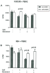
Cytomegalovirus (HCMV) reactivation is found frequently after allogeneic hematopoietic stem cell transplantation (alloSCT) and is associated with an increased treatment-related mortality. Recent reports suggest a link between HCMV and a reduced risk of cancer progression in patients with acute leukemia or lymphoma after alloSCT. Here we show that HCMV can inhibit the proliferation of the acute myeloid leukemia cell line Kasumi-1 and the promyeloid leukemia cell line NB4. HCMV induced a significant up-regulation of HLA-class-II-molecules, especially HLA-DR expression and an increase of apoptosis, granzyme B, perforin and IFN-γ secretion in Kasumi-1 cells cocultured with peripheral blood mononuclear cells (PBMCs). Indolamin-2,3-dioxygenase on the other hand led only to a significant dose-dependent effect on IFN-γ secretion without effects on proliferation. The addition of CpG-rich oligonucleotides and ganciclovir reversed those antiproliferative effects. We conclude that HCMV can enhance alloreactivity of PBMCs against Kasumi-1 and NB4 cells in vitro. To determine if this phenomenon may be clinically relevant further investigations will be required.
Conflict of interest statement
Figures


Similar articles
-
Cytomegalovirus Reactivation after Allogeneic Hematopoietic Stem Cell Transplantation is Associated with a Reduced Risk of Relapse in Patients with Acute Myeloid Leukemia Who Survived to Day 100 after Transplantation: The Japan Society for Hematopoietic Cell Transplantation Transplantation-related Complication Working Group.Biol Blood Marrow Transplant. 2015 Nov;21(11):2008-16. doi: 10.1016/j.bbmt.2015.07.019. Epub 2015 Jul 26. Biol Blood Marrow Transplant. 2015. PMID: 26211985
-
Cytomegalovirus induces apoptosis in acute leukemia cells as a virus-versus-leukemia function.Leuk Lymphoma. 2015;56(11):3189-97. doi: 10.3109/10428194.2015.1032968. Epub 2015 May 15. Leuk Lymphoma. 2015. PMID: 25818505
-
Evaluation of human cytomegalovirus-specific CD8+ T-cells in allogeneic haematopoietic stem cell transplant recipients using pentamer and interferon-γ-enzyme-linked immunospot assays.J Clin Virol. 2013 Oct;58(2):427-31. doi: 10.1016/j.jcv.2013.07.006. Epub 2013 Jul 30. J Clin Virol. 2013. PMID: 23910932
-
Allogeneic stem cell transplantation in acute myeloid leukemia: a risk-adapted approach.Blood Rev. 2008 Nov;22(6):293-302. doi: 10.1016/j.blre.2008.03.008. Epub 2008 May 1. Blood Rev. 2008. PMID: 18455284 Review.
-
Alternative donors hematopoietic stem cells transplantation for adults with acute myeloid leukemia: Umbilical cord blood or haploidentical donors?Best Pract Res Clin Haematol. 2010 Jun;23(2):207-16. doi: 10.1016/j.beha.2010.06.002. Best Pract Res Clin Haematol. 2010. PMID: 20837332 Review.
Cited by
-
The Immune Response Against Human Cytomegalovirus Links Cellular to Systemic Senescence.Cells. 2020 Mar 20;9(3):766. doi: 10.3390/cells9030766. Cells. 2020. PMID: 32245117 Free PMC article. Review.
-
Different recovery patterns of CMV-specific and WT1-specific T cells in patients with acute myeloid leukemia undergoing allogeneic hematopoietic cell transplantation: Impact of CMV infection and leukemia relapse.Front Immunol. 2023 Feb 7;13:1027593. doi: 10.3389/fimmu.2022.1027593. eCollection 2022. Front Immunol. 2023. PMID: 36824620 Free PMC article.
-
Human Cytomegalovirus Glycoprotein-Initiated Signaling Mediates the Aberrant Activation of Akt.J Virol. 2020 Jul 30;94(16):e00167-20. doi: 10.1128/JVI.00167-20. Print 2020 Jul 30. J Virol. 2020. PMID: 32493823 Free PMC article.
-
Human Cytomegalovirus Decreases Major Histocompatibility Complex Class II by Regulating Class II Transactivator Transcript Levels in a Myeloid Cell Line.J Virol. 2020 Mar 17;94(7):e01901-19. doi: 10.1128/JVI.01901-19. Print 2020 Mar 17. J Virol. 2020. PMID: 31915281 Free PMC article.
-
Apoptosis Disorder, a Key Pathogenesis of HCMV-Related Diseases.Int J Mol Sci. 2021 Apr 15;22(8):4106. doi: 10.3390/ijms22084106. Int J Mol Sci. 2021. PMID: 33921122 Free PMC article. Review.
References
-
- Boeckh M, Geballe AP. Cytomegalovirus: pathogen, paradigm, and puzzle. J Clin Invest. 2011; 121: 1673–1680. doi: 10.1172/JCI45449 - DOI - PMC - PubMed
-
- Drylewicz J, Schellens IM, Gaiser R, Nanlohy NM, Quakkelaar ED, Otten H, et al. Rapid reconstitution of CD4 T cells and NK cells protects against CMV-reactivation after allogeneic stem cell transplantation. J Transl Med. 2016; 14: 230–239. doi: 10.1186/s12967-016-0988-4 - DOI - PMC - PubMed
-
- Lisboa LF, Tong Y, Kumar D, Pang XL, Asberg A, Hartmann A, et al. Analysis and clinical correlation of genetic variation in cytomegalovirus. Transpl Infect Dis. 2012; 14:132–140. doi: 10.1111/j.1399-3062.2011.00685.x - DOI - PubMed
-
- Schmidt-Hieber M, Labopin M, Beelen D, Volin L, Ehninger G, Finke J, et al. CMV serostatus still has an important prognostic impact in de novo acute leukemia patients after allogeneic stem cell transplantation: a report from the Acute Leukemia Working Party of EBMT. Blood. 2013; 122: 3359–3364. doi: 10.1182/blood-2013-05-499830 - DOI - PubMed
-
- Einsele H, Ehninger G, Steidle M, Fischer I, Bihler S, Gerneth F, et al. Lymphocytopenia as an unfavorable prognostic factor in patients with cytomegalovirus infection after bone marrow transplantation. Blood. 1993; 82: 1672–1678. - PubMed
Publication types
MeSH terms
Substances
Grants and funding
LinkOut - more resources
Full Text Sources
Other Literature Sources
Medical
Research Materials

