

Regulation of inflammatory responses by dynamic subcellular localization of RNA-binding protein Arid5a
- PMID: 29358370
- PMCID: PMC5819453
- DOI: 10.1073/pnas.1719921115
Regulation of inflammatory responses by dynamic subcellular localization of RNA-binding protein Arid5a
Abstract
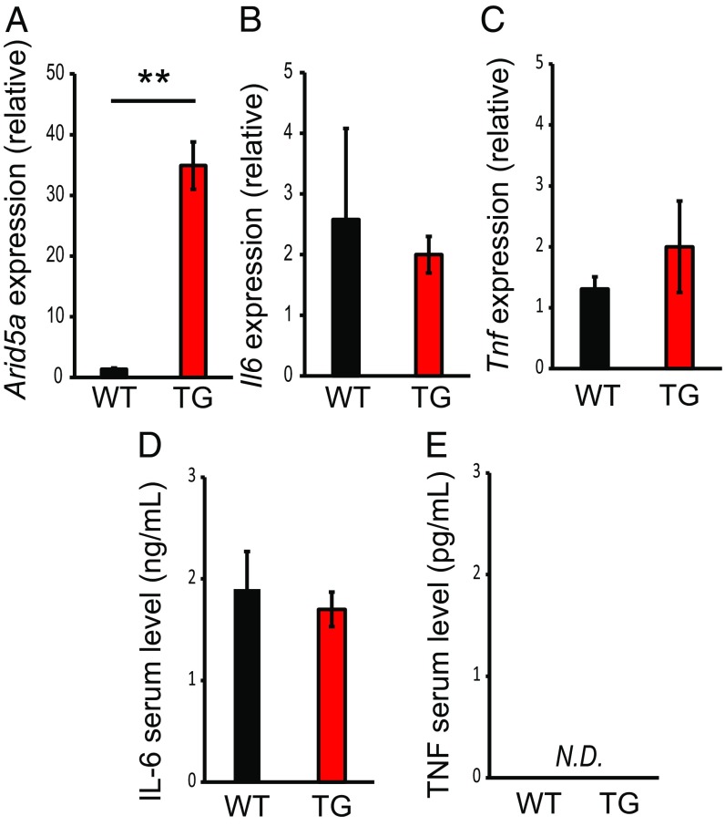
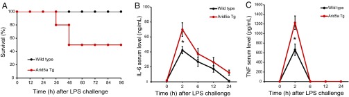
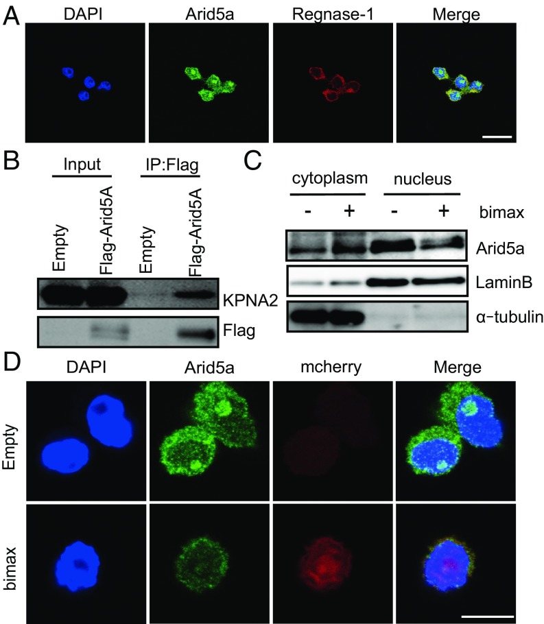
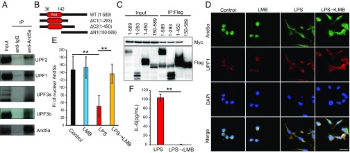
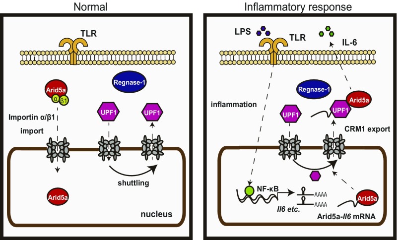
Adenine-thymine (AT)-rich interactive domain 5a (Arid5a) is an RNA-binding protein found in the cytoplasm and nucleus of normally growing cells. Although Arid5a is known to play an important role in immune regulation, whether and how Arid5a subcellular localization impacts immune regulation has remained unclear. In this study, we generated Arid5a transgenic (TG) mice to address this question. While ectopic Arid5a overexpression did not affect expression of inflammatory cytokines under unstimulated conditions, significantly higher levels of inflammatory cytokines, such as IL-6, were produced in response to lipopolysaccharide (LPS) stimulation. Consistent with this, TG mice were more sensitive to LPS treatment than wild-type mice. We also found that Arid5a is imported into the nucleus via a classical importin-α/β1-mediated pathway. On stimulation, nuclear Arid5a levels were decreased, while there was a concomitant increase in cytoplasmic Arid5a. Arid5a is associated with up-frameshift protein 1, and its nuclear export is regulated by a nuclear export receptor, chromosomal region maintenance 1. Taken together, these data indicate that Arid5a is a dynamic protein that translocates to the cytoplasm from the nucleus so as to properly exert its dual function in mRNA stabilization and transcriptional regulation during inflammatory conditions.
Keywords: Arid5a; IL-6; LPS; cytoplasm; inflammation.
Copyright © 2018 the Author(s). Published by PNAS.
Conflict of interest statement
The authors declare no conflict of interest.
Figures

Similar articles
-
Arid5a exacerbates IFN-γ-mediated septic shock by stabilizing T-bet mRNA.Proc Natl Acad Sci U S A. 2016 Oct 11;113(41):11543-11548. doi: 10.1073/pnas.1613307113. Epub 2016 Sep 26. Proc Natl Acad Sci U S A. 2016. PMID: 27671645 Free PMC article.
-
TLR4-induced NF-κB and MAPK signaling regulate the IL-6 mRNA stabilizing protein Arid5a.Nucleic Acids Res. 2017 Mar 17;45(5):2687-2703. doi: 10.1093/nar/gkx064. Nucleic Acids Res. 2017. PMID: 28168301 Free PMC article.
-
Arid5a/IL-6/PAI-1 Signaling Is Involved in the Pathogenesis of Lipopolysaccharide-Induced Kidney Injury.Biol Pharm Bull. 2023;46(12):1753-1760. doi: 10.1248/bpb.b23-00482. Biol Pharm Bull. 2023. PMID: 38044094
-
A Potential Therapeutic Target RNA-binding Protein, Arid5a for the Treatment of Inflammatory Disease Associated with Aberrant Cytokine Expression.Curr Pharm Des. 2018;24(16):1766-1771. doi: 10.2174/1381612824666180426103753. Curr Pharm Des. 2018. PMID: 29701145 Review.
-
Arid5a Regulation and the Roles of Arid5a in the Inflammatory Response and Disease.Front Immunol. 2019 Dec 5;10:2790. doi: 10.3389/fimmu.2019.02790. eCollection 2019. Front Immunol. 2019. PMID: 31867000 Free PMC article. Review.
Cited by
-
Feedback regulation of Arid5a and Ppar-γ2 maintains adipose tissue homeostasis.Proc Natl Acad Sci U S A. 2019 Jul 23;116(30):15128-15133. doi: 10.1073/pnas.1906712116. Epub 2019 Jul 9. Proc Natl Acad Sci U S A. 2019. PMID: 31289228 Free PMC article.
-
RNA Metabolism Governs Immune Function and Response.Adv Exp Med Biol. 2024;1444:145-161. doi: 10.1007/978-981-99-9781-7_10. Adv Exp Med Biol. 2024. PMID: 38467978 Review.
-
The RNA binding protein Arid5a drives IL-17-dependent autoantibody-induced glomerulonephritis.J Exp Med. 2024 Sep 2;221(9):e20240656. doi: 10.1084/jem.20240656. Epub 2024 Jul 26. J Exp Med. 2024. PMID: 39058386 Free PMC article.
-
The Splicing Factor hnRNP M Is a Critical Regulator of Innate Immune Gene Expression in Macrophages.Cell Rep. 2019 Nov 5;29(6):1594-1609.e5. doi: 10.1016/j.celrep.2019.09.078. Cell Rep. 2019. PMID: 31693898 Free PMC article.
-
Novel ratio-expressions of genes enables estimation of wound age in contused skeletal muscle.Int J Legal Med. 2024 Jan;138(1):197-206. doi: 10.1007/s00414-023-03095-x. Epub 2023 Oct 7. Int J Legal Med. 2024. PMID: 37804331
References
-
- Dubey PK, et al. Arid5a-deficient mice are highly resistant to bleomycin-induced lung injury. Int Immunol. 2017;29:79–85. - PubMed
Publication types
MeSH terms
Substances
LinkOut - more resources
Full Text Sources
Other Literature Sources
Molecular Biology Databases
Research Materials
Miscellaneous
