. 2018 Jan 22;14(1):e1006868.
doi: 10.1371/journal.ppat.1006868. eCollection 2018 Jan.

Interferon regulatory factor 8 regulates caspase-1 expression to facilitate Epstein-Barr virus reactivation in response to B cell receptor stimulation and chemical induction

Affiliations

Interferon regulatory factor 8 regulates caspase-1 expression to facilitate Epstein-Barr virus reactivation in response to B cell receptor stimulation and chemical induction

Dong-Wen Lv et al. PLoS Pathog. .

Abstract

Interferon regulatory factor 8 (IRF8), also known as interferon consensus sequence-binding protein (ICSBP), is a transcription factor of the IRF family. IRF8 plays a key role in normal B cell differentiation, a cellular process that is intrinsically associated with Epstein-Barr virus (EBV) reactivation. However, whether IRF8 regulates EBV lytic replication remains unknown. In this study, we utilized a CRISPR/Cas9 genomic editing approach to deplete IRF8 and found that IRF8 depletion dramatically inhibits the reactivation of EBV upon lytic induction. We demonstrated that IRF8 depletion suppresses the expression of a group of genes involved in apoptosis and thus inhibits apoptosis induction upon lytic induction by B cell receptor (BCR) stimulation or chemical induction. The protein levels of caspase-1, caspase-3 and caspase-8 all dramatically decreased in IRF8-depleted cells, which led to reduced caspase activation and the stabilization of KAP1, PAX5 and DNMT3A upon BCR stimulation. Interestingly, caspase inhibition blocked the degradation of KAP1, PAX5 and DNMT3A, suppressed EBV lytic gene expression and viral DNA replication upon lytic induction, suggesting that the reduced caspase expression in IRF8-depleted cells contributes to the suppression of EBV lytic replication. We further demonstrated that IRF8 directly regulates CASP1 (caspase-1) gene expression through targeting its gene promoter and knockdown of caspase-1 abrogates EBV reactivation upon lytic induction, partially through the stabilization of KAP1. Together our study suggested that, by modulating the activation of caspases and the subsequent cleavage of KAP1 upon lytic induction, IRF8 plays a critical role in EBV lytic reactivation.


Conflict of interest statement

The authors have declared that no competing interests exist.

Figures

Fig 1
Fig 1. IRF8 depletion inhibits the reactivation of EBV in Akata (EBV+) cells.
A. The locations of two sgRNAs (sg1 and sg2) used for IRF8 depletion. B. Western blot was performed to check the knockdown efficiency of IRF8 by sg1 and sg2 compared with the non-targeting control sgRNA (NC). C-G.IRF8-depleted (sg1 and sg2) and control (NC) Akata (EBV+) cells were either untreated (0 hr) or treated with anti-IgG for 24 and 48 hrs to induce lytic replication. The cell pellets and supernatant was harvested 24 and 48 hrs after anti-IgG stimulation. Protein extracts were analyzed by western blot using antibodies against IRF8 and EBV immediate-early (ZTA) and early (BGLF4) proteins (C and D). RT-qPCR showing the suppression of EBV immediate-early (ZTA and RTA) and late (BGLF2) genes expression upon IRF8 depletion (sg1 and sg2) (E). qPCR showing the reduction of intracellular viral DNA (F) and extracellular virion-associated DNA (G) copy numbers upon IRF8 depletion. The EBV genome copy number was measured by qPCR using primers specific to EBV BALF5. The intracellular EBV copy number was normalized by qPCR using specific primers to β-actin. Data are presented as means ± standard deviations (n = 3). * p<0.05 and ** p<0.01.
Fig 2
Fig 2. IRF8 depletion suppresses the expression of genes involved in apoptosis.
A. Schematic representation of RNA-seq analyses of Akata (EBV+) cells carrying control (NC) or IRF8-sg2 sgRNAs, RNAs were extracted from cells derived from three distinct lentiviral transductions. Using 2-fold change as a cutoff, 196 and 57 genes were down- or up-regulated upon IRF8 depletion, respectively. Gene Ontology analysis showing that 19 genes involved in “positive regulation of apoptosis” (red dots) were down-regulated by IRF8 depletion. B. Fold changes of the 19 apoptosis-related genes and the validation of 8 of them by RT-qPCR analysis of RNAs from cells derived from three distinct lentiviral transductions. C. IRF8 depletion (sg2) suppresses caspase-1 expression and the generation of cleaved caspase substrates upon lytic induction by anti-IgG cross-linking. Western blot analysis of protein extracts from Fig 1D using antibodies against caspase-1, PARP, and cleaved caspase substrates (Peptides containing [DE(T/S/A)D] motif) as indicated.
Fig 3
Fig 3. IRF8 depletion suppresses caspase activation and caspase activation is required for EBV lytic replication.
A. IRF8 depletion suppresses caspase activation. Western blot analysis of protein extracts from Fig 1D using antibodies against caspase-3, cleaved caspase-3, caspase-8, cleaved caspase-8, caspase-7, cleaved caspase-7, caspase-9, cleaved caspass-9, caspase-2 and Bcl-2 as indicated. B. Caspase inhibition suppresses EBV lytic gene expression. Akata (EBV+) cells were untreated or pre-treated with pan-caspase inhibitor (Z-VAD-FMK) for 1 hr and then anti-IgG was added for 48 hrs. RNA was extracted and EBV lytic gene expression was analyzed by RT-qPCR. Data are presented as means ± standard deviations of triplicate assays. ** p<0.01 (compared with the second bar). C. Caspase inhibition suppresses EBV DNA replication. Protein extracts from cells treated as Panel B were analyzed by western blot using antibodies against cleaved-PARP, EBV ZTA and BGLF4 as indicated. β-actin was used as loading controls. Genomic DNA was extracted and relative EBV DNA copy numbers was measured by qPCR using primers specific to EBV BALF5. The EBV copy number was normalized by qPCR using specific primers to β-actin. Data are presented as means ± standard deviations of triplicate assays. ** p<0.01 (compared with the second bar). D. IRF8 depletion suppresses the degradation of KAP1, PAX5 and DNMT3A upon lytic induction. Western blot analysis of protein extracts from Fig 1D using antibodies against KAP1, PAX5, DNMT3A and STAT3 as indicated. E. Caspase inhibition restores the expression of KAP1, PAX5 and DNMT3A. Protein extracts from Panel C were analyzed by western blot using antibodies against KAP1, PAX5, DNMT3A and STAT3 as indicated. The longer exposure of KAP1 blot revealed two cleaved KAP1 products upon lytic induction (lane 2, arrow heads).
Fig 4
Fig 4. IRF8 depletion suppresses EBV reactivation in LCL and P3HR-1 cells upon lytic induction.
A and B. Control (NC) and IRF8-depleted (sg1 and sg2) LCL cells were either untreated (0 hr) or treated with 1 μg/mL gemcitabine (A) or 20 μg/mL α-IgM (B) for 48 hrs to induce lytic replication. Western blot analyses showing IRF8, ZTA, caspase-1 and cleaved-PARP level as indicated. C. Control (NC) and IRF8-depleted (sg1 and sg2) P3HR-1 cells were either untreated (0 hr) or treated with TPA (20 ng/ml)/sodium butyrate (NaBu, 3 mM) for 48 hrs to induce lytic replication. Western blot analyses showing IRF8, ZTA and caspase-1 level as indicated.
Fig 5
Fig 5. IRF8 regulates CASP1 promoter activities.
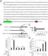
A. Schematic representation of the promoter of human CASP1. IRF8 consensus binding site is highlighted in green. The ATG of CASP1 is highlighted in red. B. The pGL2-CASP1p constructs (with or without IRF8 consensus site) and the IRF8 consensus site mutated construct were co-transfected into 293T cells with either vector control or IRF8 expression vectors. Luciferase assays were performed 36 hrs post-transfection. The value of cells transfected with empty vectors was set as 1. The results were presented as mean ± standard deviation of triplicate assays. C. The pGL2-CASP1p1 construct was co-transfected into 293T cells with either vector control, wild-type IRF8 (WT) or IRF8 DNA binding mutant (K108E) expression vectors and luciferase assays were performed 36 hrs post-transfection. The value of cells transfected with empty vectors was set as 1. The results were presented as mean ± standard deviation of triplicate assays. D. The pGL2-CASP1p1 construct was co-transfected into 293T cells with either vector control or IRF8 and IRF1 expression vectors and luciferase assays were performed 36 hrs post-transfection. The value of cells transfected with empty vectors was set as 1. The results were presented as mean ± standard deviation of triplicate assays. *** p<0.001.
Fig 6
Fig 6. CASP1 depletion inhibits the reactivation of EBV in Akata (EBV+) cells.
A. The locations of two sgRNAs (sg1 and sg2) used for CASP1 depletion. B-D. CASP1-depleted (sg1 and sg2) and control (NC) Akata (EBV+) cells were either untreated (0 hr) or treated with anti-IgG for 24 and 48 hrs to induce lytic replication. The cell pellets were harvested 24 and 48 hrs after anti-IgG stimulation. Protein extracts were analyzed by western blot using antibodies against CASP1 and EBV immediate-early (ZTA and RTA) proteins and β-actin (B). RT-qPCR showing the suppression of EBV immediate-early (ZTA and RTA) and late (BGLF2) genes expression upon CASP1 depletion (sg1 and sg2) (C). qPCR showing the reduction of intracellular viral DNA copy numbers upon CASP1 depletion (D). The EBV genome copy number was measured by qPCR using primers specific to EBV BALF5. The intracellular EBV copy number was normalized by qPCR using specific primers to β-actin. Data are presented as means ± standard deviations (n = 3). ** p<0.01.
Fig 7
Fig 7. CASP1 depletion suppresses EBV reactivation in LCL and P3HR-1 cells upon lytic induction.
A and B. Control (NC) and CASP1-depleted (sg1 and sg2) LCL cells were either untreated (0 hr) or treated with 1 μg/mL gemcitabine (A) or 20 μg/mL α-IgM (B) for 48 hrs to induce lytic replication. Western blot analyses showing caspase-1, ZTA and RTA level as indicated. C. Control (NC) and CASP1-depleted (sg1 and sg2) P3HR-1 cells were either untreated (0 hr) or treated with TPA (20 ng/ml)/sodium butyrate (NaBu, 3 mM) for 48 hrs to induce lytic replication. Western blot analyses showing caspase-1, ZTA and RTA level as indicated.
Fig 8
Fig 8. Caspase-1 promotes EBV reactivation partially through KAP1 cleavage.
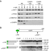
A. Caspase-1 depletion suppresses KAP1 degradation. Protein extracts form Fig 5B were analyzed by western blot using antibodies against KAP1, PAX5, DNMT3A and Caspase-8 (CASP8). B. Caspase-1 and -8 cleave KAP1 in vitro. HA-KAP1 and the antibody recognition sites are labeled as indicated. HA-tagged KAP1 was immuoprecipitated from transfected 293T cells using HA magnetic beads. The beads-bound HA-KAP1 was incubated with individual recombinant caspase for 2 hrs at 37°C. WB was performed using either anti-HA or anti-KAP1 antibodies. The relative positions of cleaved fragments were labeled as indicated.
Fig 9
Fig 9. KAP1 depletion facilitates EBV reactivation upon lytic induction.
A. Control (NC) and KAP1-depleted (sg1 and sg2) Akata (EBV+)-CASP1-sg1 cells were untreated (0 hr) or treated with α-IgG (1:200) for 24 and 48 hrs to induce lytic replication. Western blot analyses showing KAP1, ZTA and RTA level as indicated. β-actin blot was included as loading controls. B. Intracellular viral DNA from cells treated as in (A) was measured by qPCR using primers to EBV BALF5. The value of NC control at 0 hr (lane 1) was set as 1. Data are presented as means ± standard deviations of triplicate assays. ** p<0.01.
Fig 10
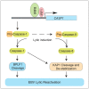
Fig 10. Hypothesized model by which IRF8 contributes to EBV lytic replication.
IRF8 regulates the protein levels of caspase-1 and caspase-8. BCR stimulation triggers the activation of caspases and subsequent BPLF1 cleavage and the destabilization of KAP1, which leads to enhanced viral gene expression and DNA replication.

Similar articles

Cited by

References

    1. Young LS, Yap LF, Murray PG (2016) Epstein-Barr virus: more than 50 years old and still providing surprises. Nature Reviews Cancer 16: 789–802. doi: 10.1038/nrc.2016.92 - DOI - PubMed
    1. Hammerschmidt W, Sugden B (2013) Replication of Epstein–Barr Viral DNA. Cold Spring Harbor perspectives in biology 5: a013029 doi: 10.1101/cshperspect.a013029 - DOI - PMC - PubMed
    1. Zalani S, Holley-Guthrie E, Kenney S (1995) The Zif268 cellular transcription factor activates expression of the Epstein-Barr virus immediate-early BRLF1 promoter. J Virol 69: 3816–3823. - PMC - PubMed
    1. Wu FY, Wang SE, Chen H, Wang L, Hayward SD, et al. (2004) CCAAT/enhancer binding protein alpha binds to the Epstein-Barr virus (EBV) ZTA protein through oligomeric interactions and contributes to cooperative transcriptional activation of the ZTA promoter through direct binding to the ZII and ZIIIB motifs during induction of the EBV lytic cycle. J Virol 78: 4847–4865. doi: 10.1128/JVI.78.9.4847-4865.2004 - DOI - PMC - PubMed
    1. Robinson AR, Kwek SS, Kenney SC (2012) The B-cell specific transcription factor, Oct-2, promotes Epstein-Barr virus latency by inhibiting the viral immediate-early protein, BZLF1. PLoS Pathog 8: e1002516 doi: 10.1371/journal.ppat.1002516 - DOI - PMC - PubMed

Publication types

MeSH terms

Substances