. 2018 Jan 19;14(1):e1006794.
doi: 10.1371/journal.ppat.1006794. eCollection 2018 Jan.

Leishmania proteophosphoglycans regurgitated from infected sand flies accelerate dermal wound repair and exacerbate leishmaniasis via insulin-like growth factor 1-dependent signalling

Affiliations

Leishmania proteophosphoglycans regurgitated from infected sand flies accelerate dermal wound repair and exacerbate leishmaniasis via insulin-like growth factor 1-dependent signalling

Emilie Giraud et al. PLoS Pathog. .

Abstract

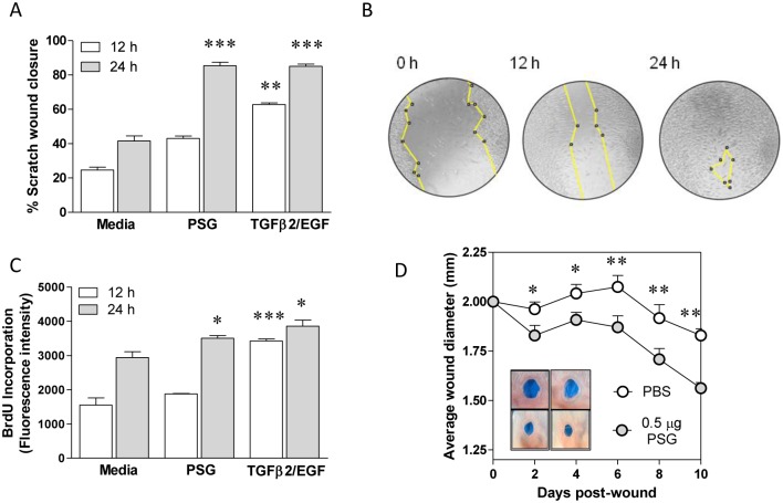
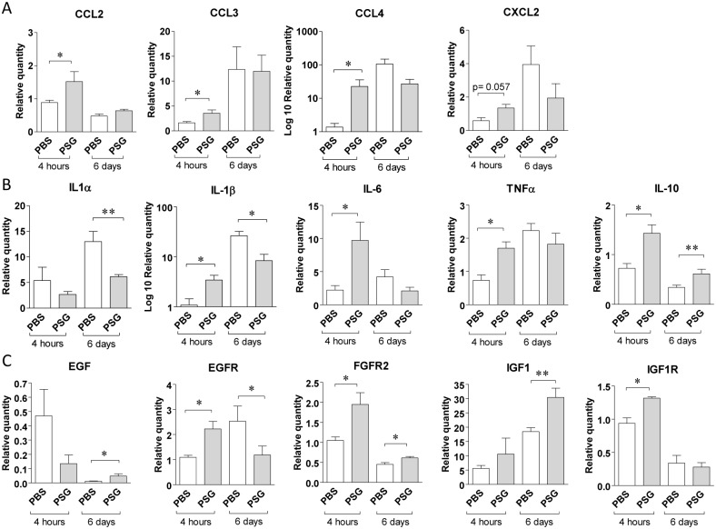
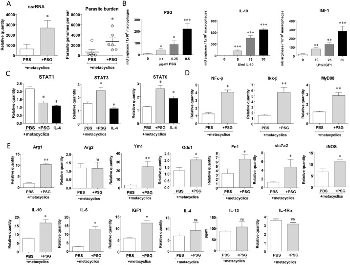
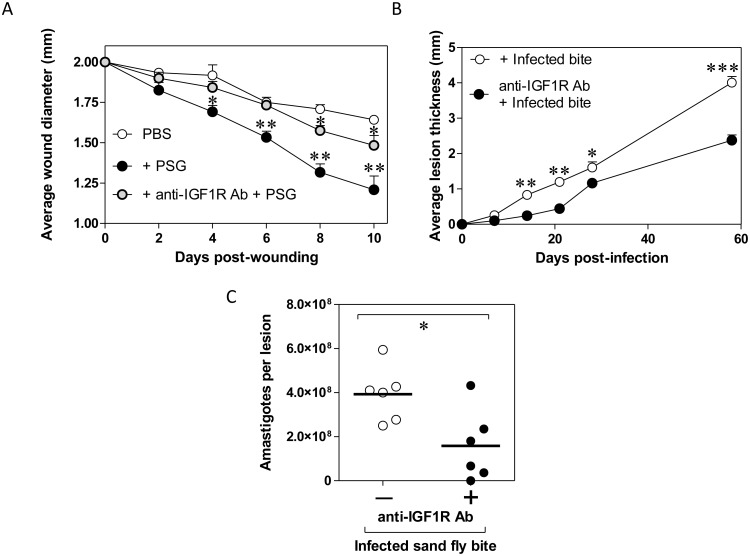
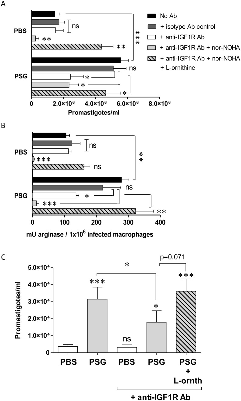
Leishmania parasites are transmitted to vertebrate hosts by female phlebotomine sand flies as they bloodfeed by lacerating the upper capillaries of the dermis with their barbed mouthparts. In the sand fly midgut secreted proteophosphoglycans from Leishmania form a biological plug known as the promastigote secretory gel (PSG), which blocks the gut and facilitates the regurgitation of infective parasites. The interaction between the wound created by the sand fly bite and PSG is not known. Here we nanoinjected a sand fly egested dose of PSG into BALB/c mouse skin that lead to the differential expression of 7,907 transcripts. These transcripts were transiently up-regulated during the first 6 hours post-wound and enriched for pathways involved in inflammation, cell proliferation, fibrosis, epithelial cell differentiation and wound remodelling. We found that PSG significantly accelerated wound healing in vitro and in mice; which was associated with an early up-regulation of transcripts involved in inflammation (IL-1β, IL-6, IL-10, TNFα) and inflammatory cell recruitment (CCL2, CCL3, CCL4, CXCL2), followed 6 days later by enhanced expression of transcripts associated with epithelial cell proliferation, fibroplasia and fibrosis (FGFR2, EGF, EGFR, IGF1). Dermal expression of IGF1 was enhanced following an infected sand fly bite and was acutely responsive to the deposition of PSG but not the inoculation of parasites or sand fly saliva. Antibody blockade of IGF1 ablated the gel's ability to promote wound closure in mouse ears and significantly reduced the virulence of Leishmania mexicana infection delivered by an individual sand fly bite. Dermal macrophages recruited to air-pouches on the backs of mice revealed that IGF1 was pivotal to the PSG's ability to promote macrophage alternative activation and Leishmania infection. Our data demonstrate that through the regurgitation of PSG Leishmania exploit the wound healing response of the host to the vector bite by promoting the action of IGF1 to drive the alternative activation of macrophages.


Conflict of interest statement

The authors have declared that no competing interests exist.

Figures

Fig 1
Fig 1. PSG exacerbates Leishmania in skin over a wide physiological range and induces different gene modulation at different time points post-infection in skin.
(A-C) BALB/c mice were infected with 1 x 10³ L. mexicana metacyclic promastigotes with or without 1.0 μg (A), 0.5 μg (B) or 0.03 (C) μg L. mexicana PSG, s.c in to the left footpad and the evolution of the lesions, compared to the contralateral foot, recorded over 80 days. Final average parasite burdens ± S.D. for these lesions: PBS: 5.167 x 10⁷ ± 9.79 x 10⁶; 1.0 μg PSG: 4.74 x 10⁸ ± 3.21 x 10⁸; 0.5 μg PSG: 3.27 x 10⁸ ± 3.59 x 10⁸; 0.03 μg PSG: 1.96 x 10⁸ ± 3.05 x 10⁸ amastigotes per footpad. BALB/c mice received co-inoculation of either PBS or 0.03 μg PSG with 1 x 10³ L. mexicana metacyclic promastigotes between the dermal sheets of the ear pinna using a nanoinjector. D) Heat map representation of normalised expression values. Genes shown are the top 50 differentially expressed at 6 hours (FC >1.5 at 5% FDR). E) Heat map representation of the Pearson correlation coefficients between replicates for all modulated genes. The colour gradient extends from red, representing perfect correlation (correlation distance, r, of 1), to cyan for low correlation. Chip coding: Experiment/Time/Replicate number, e.g. MT6-2 represents the 2nd chip from a control ear injected with PBS and parasites, sampled at 6 hours post-infection; chips coded PSG are from PSG-inoculated ears. (F and G) Venn diagrams of the number of overlapping genes differentially expressed (FC > 1.5 at 5% FDR) at 6, 24 and 48 hours between ears injected with PBS and metacyclic promastigotes versus PSG and metacyclic promastigotes. F) Total number of modulated genes. G) Genes involved in different phases of wound healing. H) Validation of microarray gene expression following i.d. injection of 0.5 μg PSG per ear. Real-time quantitative PCR of 34 randomly selected up- and down-regulated genes, among the differentially expressed gene list of 6 hours p.i. (FC>1.5, FDR 5%).
Fig 2
Fig 2. PSG improves wound healing in vitro and in skin.
Monolayers of L929 fibroblasts were scratched in the presence of culture media supplemented with or without 0.5 μg/ml L. mexicana PSG. Positive controls were treated with 10 μg/ml TGFβ2 and 10 μg/ml EGF. A) At 0, 12 and 24 hours post-wound, photomicrographs were taken and scratch closure was determined from using ImageJ. B) Representative images for the closure of a PSG-treated scratch. C) In replicate experiments, cell proliferation of fibroblasts was determined by measuring the incorporation of the fluorescent thymidine analogue, BrdU. Statistical analyses were performed between Media vs. PSG at each time point. Each in condition was performed in quadruplicate, data is pooled from 3 experiments. D) Daily wound closure following intradermal injection of PBS (open symbols) or 0.5 μg L. mexicana PSG (closed symbols) in the ears of BALB/c mice. Ears were wounded with a 2 mm diameter full-thickness ear punch 6 hours after the intra-dermal injection of PBS or PSG. Inset photographs are ears representative of each group at day 10 post-wound. Average wound closure ±SEM is shown from 12 mice per group (*: p<0.05; **: p<0.005; ***: p<0.0005 by Mann Whitney t-test); data is pooled from duplicate experiments.
Fig 3
Fig 3. The presence of PSG in a dermal wound differentially modulates the expression of genes involved in various stages of healing.
Ears of BALB/c mice were intra-dermally inoculated with 0.5 μg L. mexicana PSG (grey bars) or PBS (white bars) 4 hours prior to a full-thickness 2 mm diameter punch biopsy. A-C) 4 hours and 6 days post-wound ears were measured for transcripts involved in the inflammation and cell proliferation phases of wound healing by real-time quantitative PCR. A) Chemokines: CCL2, CCL3, CCL4 and CXCL2, B) pro-inflammatory-modulating cytokines: IL-1α, IL-1β, IL-6, IL-10 and TNFα, and C) epidermal growth factors and receptors: EGF, IGF1, EGFR, IGF1R and FGFR2. Relative expression was normalised to the housekeeping genes nono and l19 and is presented as the mean ±SD with 9–12 mice per group (*: p<0.05, **: p<0.005 by Mann Whitney t-test).
Fig 4
Fig 4. The presence of PSG during an early Leishmania infection promotes the expression of genes and signalling pathways involved in macrophage alternative activation.
Ears of BALB/c mice were intra-dermally infected with 1 x 10³ L. mexicana metacyclic promastigotes with 0.5 μg L. mexicana PSG (grey bars) or PBS (white bars), or 5 x 10⁵ BMMΦ from BALB/c mice were exposed to L. mexicana PSG or PBS with or without L. mexicana metacyclic promastigotes (MOI 5:1). A) 48h post-infection expression of Leishmania ssrRNA in ears and the extrapolated parasite burdens, n = 6/group. B) Arginase activity of uninfected BMMΦ exposed to increasing concentrations of PSG, IL-10 or IGF1 for 48 hours. C) 48 h expression of signal transducer and activator of transcription (STAT) factors 1, 3 and 6 in 5 x 10⁵ infected BMMΦ in response to PBS, 0.5 μg/ml PSG or uninfected BMMΦ exposed to 20 U/ml recombinant murine IL-4. D) 48 h expression of STAT-signalling transcription factors: NF-κB, Ikk-β and MyD88 in infected BALB/c macrophages co-incubated with PBS or 0.5μg PSG.E) 48 h expression of markers of macrophage alternative activation or arginine metabolism measured in infected BALB/c ears in response to intradermal injection of PBS or 0.5 μg/ml PSG by real-time quantitative PCR. IL-13 production was determined from whole cell lysates using Luminex. Data is representative of triplicate experiments. Relative expression was normalised to the housekeeping genes ywhaz and nono and is presented as the mean ±SD (*: p<0.05, **: p<0.005 by Mann Whitney t-test).
Fig 5
Fig 5. PSG does not require IL-4Rα signalling to exacerbate cutaneous leishmaniasis in mice.
A and B) Footpad infections of WT (A) and IL-4Rα-/- (B) BALB/c mice with 1 x 10³ L. mexicana metacyclic promastigotes intradermally injected with PBS or 0.5 μg L. mexicana PSG, 6 mice per group. C) Final parasite burdens of footpads from B) at the end of the experiment (WT: 80 d post-infection, IL-4Rα-/-: 151 d post-infection). Data pooled from two replicate experiments. A&B) Averages ±SEM are shown, C) Each point represent individual mice, bars indicate the mean (*: p<0.05; **: p<0.005; ***: p<0.0005 by Mann Whitney t-test).
Fig 6
Fig 6. Insulin-like growth factor 1-signalling controls the wound healing and Leishmania-exacerbating properties of PSG in mice.
A) Rate of 2 mm diameter wound closure in BALB/c ears preconditioned with PBS, 0.5 μg L. mexicana PSG or 0.5 μg L. mexicana PSG + 1:50 anti-IGF1R antibody, 12 mice per group. B) Single L. mexicana-infected sand fly bite infections in footpads of BALB/c mice treated with or without 1:50 anti-IGF1R antibody, 6 mice per group. C) Final parasite burdens of footpads from B) at the end of the experiment. Data pooled from two replicate experiments. A&B) Averages ±SEM are shown, C) Each point represent individual mice, bars indicate the mean (*: p<0.05; **: p<0.005; ***: p<0.0005 by Mann Whitney t-test).
Fig 7
Fig 7. PSG enhances IGF1 expression in skin following an infected fly bite and enhances macrophage alternative activation.
A) IGF1, IGF1R and IL-10 expression in BALB/c mouse ears 6 hours post-bite from single uninfected or L. mexicana-infected Lu. longipalpis sand flies (Day 8 post-infection), one bite per ear (12 uninfected bites, 35 infected bites). B) IGF1, IGF1R and IL-10 expression in bitten ears in relation to the dose of transmitted parasites, determined from Leishmania ssrRNA expression in ears. C) IGF1, IGF1R and IL-10 expression in BALB/c ears 6 hours following intradermal injection of with PBS, 0.5 μg L. mexicana PSG, 0.5 μg Lu. longipalpis saliva or PSG and saliva combined. Data is pooled from duplicate experiments with 12 mice per group. Relative expression was normalised to the housekeeping genes nono and l19 and is presented as the mean ±SD (*: p<0.05, **: p<0.005 by Mann Whitney t-test).
Fig 8
Fig 8. PSG manipulates macrophage arginases to promote intracellular Leishmania growth in vitro and in vivo via IGF1 signalling.
A) Viable amastigote burdens of macrophages infected in vitro was determined by transformation assay of amastigotes into promastigotes liberated from 5 x 10⁶ BALB/c BMMΦ. Macrophages were infected with L. mexicana metacyclic promastigotes (MOI 1:1) in the presence of ±1 μg L. mexicana PSG; ±1:100 anti-IGF1R Ab or isotype control; ±100 μM nor-NOHA; ±100 μM L-ornithine for 48 hours. B) Arginase activity of duplicate infected macrophages from (A). C) Parasite burden of BALB/c dermal air-pouches infected with 1x10⁶ L. mexicana metacyclic promastigotes ±1 μg PSG, ±1:50 anti-IGF1R Ab; ±100 μg L-ornithine for 48 hours. Macrophages were recovered from infected air-pouches and separated by adherence to plastic. Data is representative of triplicate experiments (A&B) or pooled from duplicate experiments (C, 8 mice per group). Unless they are linked with a bar all test groups are compared to their relevant saline control; (ns, not significant p>0.05; *: p<0.05; **: p<0.005; ***: p<0.0005 by Mann Whitney t-test).

Similar articles

Cited by

References

    1. Sacks D, Kamhawi S (2001) Molecular aspects of parasite-vector and vector-host interactions in leishmaniasis. Annu Rev Microbiol 55: 453–483. doi: 10.1146/annurev.micro.55.1.453 - DOI - PubMed
    1. Lestinova T, Rohousova I, Sima M, de Oliveira CI, Volf P (2017) Insights into the sand fly saliva: Blood-feeding and immune interactions between sand flies, hosts, and Leishmania. PLoS Negl Trop Dis. 11: e0005600 doi: 10.1371/journal.pntd.0005600 - DOI - PMC - PubMed
    1. Titus RG, Ribeiro JM (1988) Salivary gland lysates from the sand fly Lutzomyia longipalpis enhanced Leishmania infectivity. Science 239: 1306–1308. - PubMed
    1. Kamhawi S, Belkaid Y, Modi G, Rowton E, Sacks DL (2000) Protection against cutaneous leishmaniasis resulting from bites of uninfected sand flies. Science 290: 1351–1354. - PubMed
    1. Oliveira F, Lawyer PG, Kamhawi S, Valenzuela JG (2008) Immunity to distinct sand fly salivary proteins primes the anti-Leishmania immune response toward protection or exacerbation of disease. PLoS Negl Trop Dis 2: e226 doi: 10.1371/journal.pntd.0000226 - DOI - PMC - PubMed

Publication types

MeSH terms