. 2018 Jan 19;9(2):47.
doi: 10.1038/s41419-017-0067-7.

Nucleolin and ErbB2 inhibition reduces tumorigenicity of ErbB2-positive breast cancer

Affiliations

Nucleolin and ErbB2 inhibition reduces tumorigenicity of ErbB2-positive breast cancer

Eya Wolfson et al. Cell Death Dis. .

Abstract

ErbB2, a member of the ErbB family of receptor tyrosine kinases, is an essential player in the cell's growth and proliferation signaling pathways. Amplification or overexpression of ErbB2 is observed in ∼30% of breast cancer patients, and often drives cellular transformation and cancer development. Recently, we have shown that ErbB2 interacts with the nuclear-cytoplasmic shuttling protein nucleolin, an interaction which enhances cell transformation in vitro, and increases mortality risk and disease progression rate in human breast cancer patients. Given these results, and since acquired resistance to anti-ErbB2-targeted therapy is a major obstacle in treatment of breast cancer, we have examined the therapeutic potential of targeting the ErbB2-nucleolin complex. The effect of the nucleolin-specific inhibitor GroA (AS1411) on ErbB2-positive breast cancer was tested in vivo, in a mouse xenograft model for breast cancer; as well as in vitro, alone and in combination with the ErbB2 kinase-inhibitor tyrphostin AG-825. Here, we show that in vivo treatment of ErbB2-positive breast tumor xenografts with GroA reduces tumor size and leads to decreased ErbB2-mediated signaling. Moreover, we found that co-treatment of breast cancer cell lines with GroA and the ErbB2 kinase-inhibitor tyrphostin AG-825 enhances the anti-cancer effects exerted by GroA alone in terms of cell viability, mortality, migration, and invasiveness. We, therefore, suggest a novel therapeutic approach, consisting of combined inhibition of ErbB2 and nucleolin, which has the potential to improve breast cancer treatment efficacy.


Conflict of interest statement

The authors declare that they have no competing financial interests.

Figures

Fig. 1
Fig. 1. Nucleolin affects growth and development of ErbB2-positive breast tumors in mice
a Left, comparison of the growth rates and volumes of SKBR3 tumor xenografts, expressing either GFP or GFP-nucleolin (GFP and NCL, respectively; means ± SE; ***p < 0.005; n = 5). Right, changes in volume of either GFP or NCL tumors following treatment with the nucleolin-specific inhibitor GroA (AS1411) (means ± SE; ***p < 0.005—compared to the respective untreated tumors; n = 5). b Upon experiment termination (~20 days), GFP and NCL tumors, either untreated or treated with GroA, were dissected and compared in size. Lower panel, final tumor volumes are presented as means ± SE (*p < 0.05, ***p < 0.005—treated tumors compared to their respective, untreated, controls; ^p < 0.05—comparison between GFP and NCL tumors; n = 5). c Phosphorylation levels of ErbB2 (upper and lower panels) and Erk (upper panel) in dissected tumors, either untreated (upper) or treated with GroA (lower; NCL), were determined using anti-phospho-ErbB2 and anti-phopsho-Erk antibodies, as indicated (means ± SE; ***p < 0.005; n = 5)
Fig. 2
Fig. 2. GroA reduces ErbB2-mediated signaling in vivo through disruption of ErbB2–nucleolin complexes in ErbB2-positive breast tumors
a Phosphorylation levels of Erk in dissected GFP or NCL tumors were determined by immunostaining using anti-phopsho-Erk antibodies (means ± SE). b Co-immunoprecipitation (co-IP) analysis of ErbB2 and nucleolin in dissected GFP tumors following GroA treatment. Left, representative blots are presented. Right, quantification of the results (means ± SE); ErbB2 background levels, obtained from control pulldowns (right lane), were subtracted from ErbB2 levels, obtained through nucleolin pulldown (left and middle lanes); the resulting ErbB2 protein levels were normalized to nucleolin pulldown levels in each sample. *p < 0.05, ***p < 0.005, n = 5
Fig. 3
Fig. 3. GroA and AG-825 disrupt ErbB2–nucleolin complexes, and GroA specifically inhibits viability of breast cancer cells
a Left panel, visualization of the interaction between ErbB2 and nucleolin (red dots) in SKBR3 breast cancer cells either untreated or treated with GroA or tyrphostin AG-825, as indicated, was performed using a proximity ligation assay (PLA). Right panel, differences between signal intensity in cells represented as the number of dots per cell (means ± SE). b Viability of MCF10A breast cells, and of MCF7 and SKBR3 breast cancer cells following the indicated GroA treatment, as detected by methylene blue analysis (means ± SD; ***p < 0.005—untreated cells compared to treated cells of the same cell line; ^^^p < 0.005—comparison between MCF10A cells and MCF7/SKBR3 cells; n > 3)
Fig. 4
Fig. 4. Co-inhibition of nucleolin and ErbB2 reduces breast cancer cell viability
a SKBR3 and MCF7 cells were treated with GroA (10 and 5 μM, respectively) and AG-825 (40 or 65 μM, respectively), and cell viability was measured by the methylene blue assay at the indicated time points (means ± SD). b SKBR3 cells were treated with GroA (10 μM) and anti-ErbB2 siRNA, and cell viability was measured by the methylene blue assay (means ± SD). c SKBR3 and MCF7 cells were pre-treated with GroA and AG-825 as indicated, and total area of colonies formed was determined (means ± SD). *p < 0.05, **p < 0.01, ***p < 0.005—co-treated cells compared to untreated or single-agent treated cells; ^p < 0.05, ^^^p < 0.005—GroA/AG-825/anti-ErbB2 siRNA treated cells compared to untreated cells; n > 3
Fig. 5
Fig. 5. Co-treatment with GroA and AG-825 reduces ErbB2 activation
SKBR3 and MCF7–ErbB2 (ErbB2-overexpressing clones) were treated with GroA, AG-825 or both, as indicated, and ErbB2 phosphorylation levels were determined using anti-phospho-ErbB2 antibody (means ± SD; *p < 0.05, **p < 0.01, ***p < 0.005—co-treated cells compared to untreated or single-agent treated cells; ^p < 0.05, ^^^p < 0.005—GroA/AG-825 treated cells compared to untreated cells; n > 3). Inset, immunoblot analysis of ErbB2 expression in MCF7–ErbB2 cells compared with naïve MCF7 cells; numbers below bands indicate average fold induction of naïve MCF7 cells
Fig. 6
Fig. 6. GroA and AG-825 lead to cell proliferation impairment and increased cell death
a SKBR3 cells were treated with GroA with or without AG-825, as indicated, incubated with 5′-bromo-2′-deoxyuridine (BrdU) and subjected to immunostaining with anti-BrdU antibodies. Left panel, representative images; right panel, percentage of mitotic cells was estimated by counting the number of BrdU-positive cells compared to the number of total cells (mean ± SE). b SKBR3 and MCF7 cells were treated with GroA, with or without AG-825, at the indicated concentrations. The cells were stained with bisbenzimide (Hoechst) and propidium iodide (PI) to assess the number of dying cells. Left panel, representative images; right panel, percentage of dying cells was estimated by counting the number of PI-positive cells compared to the number of total cells (Hoechst-positive; mean ± S.D). *p < 0.05, **p < 0.01, ***p < 0.005—co-treated cells compared to untreated or single-agent treated cells; ^p < 0.05, ^^p < 0.01, ^^^p < 0.005—GroA/AG-825 treated cells compared to untreated cells; n > 3
Fig. 7
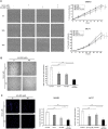
Fig. 7. Combined treatment with GroA and AG-825 impairs breast cancer cell tumorigenicity
a Migration rate of SKBR3 and MCF7 cells in the presence of GroA, AG-825 or both was determined using the scratch assay. Left panel, representative images of SKBR3 cells 0, 20 and 40 h post-wound infliction; right panel, cell migration rate during 40 h post-wound infliction, represented as percent of wound confluence (results from representative experiments are shown; means ± SD; n > 3). b SKBR3 cells ability to grow in an anchorage-independent manner in the presence of GroA, AG-825 or both was examined using the 3D basement membrane culture assay. c SKBR3 and MCF7 cells were co-treated with GroA and AG-825, and subjected to cell invasion analysis. Left panel, representative images of SKBR3 cells; middle and right panels, number of cells that successfully penetrated the Cultrex basement membrane layer (means ± SD). *p < 0.05, ***p < 0.005—co-treated cells compared to untreated or single-agent treated cells; ^^^p < 0.005—GroA/AG-825 treated cells compared to untreated cells; n > 3

Similar articles

Cited by

References

    1. Roskoski R., Jr. ErbB/HER protein-tyrosine kinases: structures and small molecule inhibitors. Pharmacol. Res. 2014;87:42–59. doi: 10.1016/j.phrs.2014.06.001. - DOI - PubMed
    1. Bertelsen V, Stang E. The mysterious ways of ErbB2/HER2 trafficking. Membranes. 2014;4:424–446. doi: 10.3390/membranes4030424. - DOI - PMC - PubMed
    1. Olayioye MA, Neve RM, Lane HA, Hynes NE. The ErbB signaling network: receptor heterodimerization in development and cancer. EMBO J. 2000;19:3159–3167. doi: 10.1093/emboj/19.13.3159. - DOI - PMC - PubMed
    1. Spencer KS, Graus-Porta D, Leng J, Hynes NE, Klemke RL. ErbB2 is necessary for induction of carcinoma cell invasion by ErbB family receptor tyrosine kinases. J. Cell Biol. 2000;148:385–397. doi: 10.1083/jcb.148.2.385. - DOI - PMC - PubMed
    1. Revillion F, Bonneterre J, Peyrat JP. ERBB2 oncogene in human breast cancer and its clinical significance. Eur. J. Cancer. 1998;34:791–808. doi: 10.1016/S0959-8049(97)10157-5. - DOI - PubMed

Publication types

MeSH terms