

doi: 10.1038/nmeth.4509.
Epub 2017 Nov 20.
Systematic characterization of maturation time of fluorescent proteins in living cells
Affiliations
- PMID: 29320486
- PMCID: PMC5765880
- DOI: 10.1038/nmeth.4509
Item in Clipboard
Systematic characterization of maturation time of fluorescent proteins in living cells
Nat Methods.
2018 Jan.
Abstract
The slow maturation time of fluorescent proteins (FPs) limits the temporal accuracy of measurements of rapid processes such as gene expression dynamics and effectively reduces fluorescence signal in growing cells. We used high-precision time-lapse microscopy to characterize the maturation kinetics of 50 FPs that span the visible spectrum at two different temperatures in Escherichia coli cells. We identified fast-maturing FPs from this set that yielded the highest signal-to-noise ratio and temporal resolution in individual growing cells.
Conflict of interest statement
The authors declare no competing financial interests.
Figures
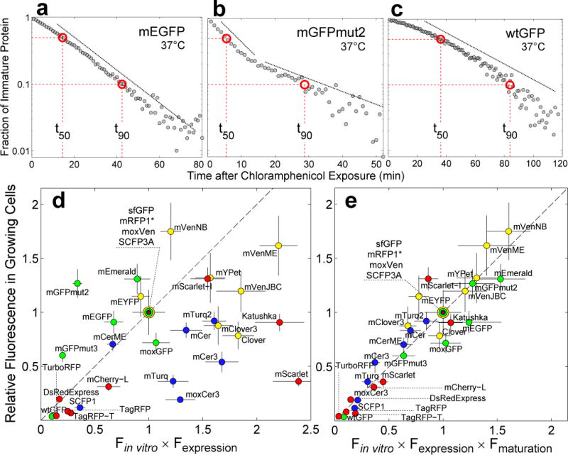
(a), (b) and (c). Fraction of immature protein as a function of time after translational arrest with chloramphenicol in single cells. (a) mEGFP maturation kinetics shows a single exponential decay (dashed line). (b) mGFPmut2 exhibits a more complex maturation process with two kinetic steps (dashed lines). (c) wtGFP matures with a slow non-exponential rate that increases with time. Dashed red lines indicate the time it takes for 50%, t50, and 90%, t90, of FP to mature, respectively. (d) Fluorescence signal in growing cells vs Fin vitro=QY·ε multiplied by Fexpression=amount of protein. Fluorescence signal is the mean fluorescence of single-cells (exponential growth in single-cell chemostat, M9 rich media, 37°C, mean from 70±20 cells, ±SEM). QY and ε are the values reported when the FPs were first published except for mVenusME and mCeruelanME; for these two FPs, we used our own in vitro data, see Online Methods. Fexpression was estimated using SDS-PAGE gel densitometry (±SD, see Online Methods). For green FPs, data normalized by sfGFP data; for yellow FPs, by moxVenus data; for blue FPs, by SCFP3A data; and for red FPs by mRFP1*. Dotted line is the identity. Dilution is given by the doubling time of E. coli, tgr=28.5±2min. (e) Fluorescence signal in growing cells vs Fin vitro × Fexpression multiplied by Fmat=1/(1 + t50/tgr). Fluorescence signal data is the same as in (d). QY and ε were quantified in our laboratory independently, except for in vitro data of red FPs, see Online Methods. We have assumed that the in vitro brightness of mRFP1* and mCherry-L is the same as the in vitro brightness of mRFP1 and mCherry, respectively, because amino acid differences are not part of the β-barrel. Error bars calculated by propagation of QY, ε and t50 errors.
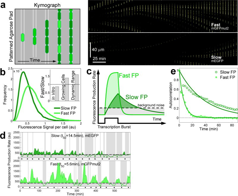
In growing cells, fluorescence signal associated with fast-maturing FPs can exhibit greater intensity and dynamic range than equally bright slow-maturing FPs. (a) Left: cartoon of a kymograph of a linear colony. Kymograph composed from the fluorescence channel of the time-lapse movie. Cells express stochastic bursts of an FP; intensity decreases because of dilution due to growth, thus creating the dark and bright bands in the kymograph. Right: kymograph made from the fluorescence channel showing that a fast FP reports brighter transcriptional events than a slower FP even when both fluorophores have the same in vitro brightness. Upper half, fast FP (mGFPmut2, ε·QY=39.3, t50=5.6min); lower half, slow FP (mEGFP, ε·QY=45.5, t50=14.5min); expression driven by the repressed PlacZ promoter. (b) Distribution of fluorescence signal per cell for the fast (light green, ncell=2489) and the slow FP (dark green, ncell= 2310). Signal from the fast FP is always higher than that from the slow FP (Supplementary Fig. 14). Inset. In vitro brightness, fluorescence signal in growing cells, and dynamic range of the fast relative to the slow FP (Supplementary Fig. 15). (c) Black solid line: transcriptional burst that yields the same amount of either fast- or slow-maturing FP. (d) Time traces of the fluorescence production rate of the repressed PlacZ using the fast or the slow FP. The detection limit (dashed line) is 3σ units above autofluorescence production rate; black dots indicate cell division, tdiv=33min at 37 °C. Shaded bands indicate periods of promoter activity below the detection limit. (e) Autocorrelation of fluorescence production rate for the slow and the fast FP variants (characteristic decay times tslowFP=24.5min and tfastFP=6.3min). Both FPs are driven by the same promoter thus using the fast FP increases temporal resolution.
Similar articles
-
Incomplete proteasomal degradation of green fluorescent proteins in the context of tandem fluorescent protein timers.Mol Biol Cell. 2016 Jan 15;27(2):360-70. doi: 10.1091/mbc.E15-07-0525. Epub 2015 Nov 25. Mol Biol Cell. 2016. PMID: 26609072 Free PMC article.
-
SPLIFF: A Single-Cell Method to Map Protein-Protein Interactions in Time and Space.Methods Mol Biol. 2015;1346:151-68. doi: 10.1007/978-1-4939-2987-0_11. Methods Mol Biol. 2015. PMID: 26542721
-
High variation of fluorescence protein maturation times in closely related Escherichia coli strains.PLoS One. 2013 Oct 14;8(10):e75991. doi: 10.1371/journal.pone.0075991. eCollection 2013. PLoS One. 2013. PMID: 24155882 Free PMC article.
-
The bacterial divisome: more than a ring?Curr Genet. 2017 May;63(2):161-164. doi: 10.1007/s00294-016-0630-2. Epub 2016 Jul 8. Curr Genet. 2017. PMID: 27387519 Review.
-
Development and use of fluorescent protein markers in living cells.Science. 2003 Apr 4;300(5616):87-91. doi: 10.1126/science.1082520. Science. 2003. PMID: 12677058 Review.
Cited by
-
An intermediate Rb-E2F activity state safeguards proliferation commitment.Nature. 2024 Jul;631(8020):424-431. doi: 10.1038/s41586-024-07554-2. Epub 2024 Jun 26. Nature. 2024. PMID: 38926571 Free PMC article.
-
The best of both worlds: Chemigenetic fluorescent sensors for biological imaging.Cell Chem Biol. 2024 Sep 19;31(9):1652-1664. doi: 10.1016/j.chembiol.2024.08.002. Epub 2024 Sep 4. Cell Chem Biol. 2024. PMID: 39236713 Review.
-
The dynamic-process characterization and prediction of synthetic gene circuits by dynamic delay model.iScience. 2024 Feb 5;27(3):109142. doi: 10.1016/j.isci.2024.109142. eCollection 2024 Mar 15. iScience. 2024. PMID: 38384832 Free PMC article.
-
Empirical Bayes method using surrounding pixel information for number and brightness analysis.Biophys J. 2021 Jun 1;120(11):2156-2171. doi: 10.1016/j.bpj.2021.03.033. Epub 2021 Apr 1. Biophys J. 2021. PMID: 33812845 Free PMC article.
-
The GATA factor ELT-3 specifies endoderm in Caenorhabditis angaria in an ancestral gene network.Development. 2022 Nov 1;149(21):dev200984. doi: 10.1242/dev.200984. Epub 2022 Oct 24. Development. 2022. PMID: 36196618 Free PMC article.
References
Publication types
MeSH terms
Substances
Grants and funding
LinkOut - more resources
Full Text Sources
Other Literature Sources
Research Materials
Miscellaneous
