. 2018 Jan 8;12(1):e0006191.
doi: 10.1371/journal.pntd.0006191. eCollection 2018 Jan.

A tetravalent virus-like particle vaccine designed to display domain III of dengue envelope proteins induces multi-serotype neutralizing antibodies in mice and macaques which confer protection against antibody dependent enhancement in AG129 mice

Affiliations

A tetravalent virus-like particle vaccine designed to display domain III of dengue envelope proteins induces multi-serotype neutralizing antibodies in mice and macaques which confer protection against antibody dependent enhancement in AG129 mice

Viswanathan Ramasamy et al. PLoS Negl Trop Dis. .

Abstract

Background: Dengue is one of the fastest spreading vector-borne diseases, caused by four antigenically distinct dengue viruses (DENVs). Antibodies against DENVs are responsible for both protection as well as pathogenesis. A vaccine that is safe for and efficacious in all people irrespective of their age and domicile is still an unmet need. It is becoming increasingly apparent that vaccine design must eliminate epitopes implicated in the induction of infection-enhancing antibodies.

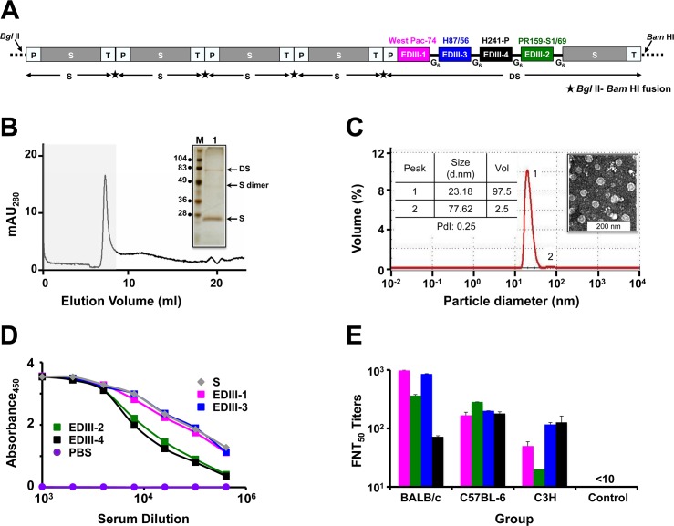
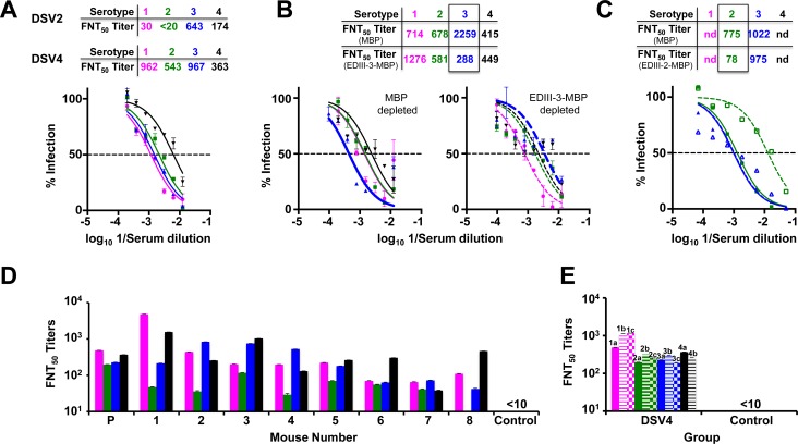
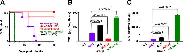
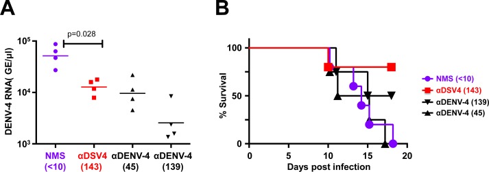
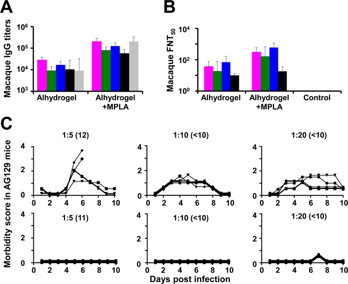
Methodology/principal findings: We report a Pichia pastoris-expressed dengue immunogen, DSV4, based on DENV envelope protein domain III (EDIII), which contains well-characterized serotype-specific and cross-reactive epitopes. In natural infection, <10% of the total neutralizing antibody response is EDIII-directed. Yet, this is a functionally relevant domain which interacts with the host cell surface receptor. DSV4 was designed by in-frame fusion of EDIII of all four DENV serotypes and hepatitis B surface (S) antigen and co-expressed with unfused S antigen to form mosaic virus-like particles (VLPs). These VLPs displayed EDIIIs of all four DENV serotypes based on probing with a battery of serotype-specific anti-EDIII monoclonal antibodies. The DSV4 VLPs were highly immunogenic, inducing potent and durable neutralizing antibodies against all four DENV serotypes encompassing multiple genotypes, in mice and macaques. DSV4-induced murine antibodies suppressed viremia in AG129 mice and conferred protection against lethal DENV-4 virus challenge. Further, neither murine nor macaque anti-DSV4 antibodies promoted mortality or inflammatory cytokine production when passively transferred and tested in an in vivo dengue disease enhancement model of AG129 mice.

Conclusions/significance: Directing the immune response to a non-immunodominant but functionally relevant serotype-specific dengue epitope of the four DENV serotypes, displayed on a VLP platform, can help minimize the risk of inducing disease-enhancing antibodies while eliciting effective tetravalent seroconversion. DSV4 has a significant potential to emerge as a safe, efficacious and inexpensive subunit dengue vaccine candidate.


Conflict of interest statement

All authors have declared that no competing interests exist. Further, LJW, MMM and REJ, who were employees of Global Vaccines, Inc. (a not-for-profit vaccine company), at the time of this work, declare that neither any of them nor the company has any proprietary or monetary interest in the DSV4 vaccine.

Figures

Fig 1
Fig 1. DSV4 assembles into immunogenic VLPs.
(A) Schematic representation of DSV4 antigen construct. Magenta, green, blue, and black blocks represent EDIII-encoding domains of DENV-1, -2, -3 and -4, respectively (the specific strains are indicated on the top of respective blocks). EDIIIs were linked to each other and to fusion partner Hepatitis B surface antigen (S; adw serotype; grey colored block), in frame, through hexa-glycyl (G6) linkers to encode Dengue-HBsAg (DS) protein. Four expression cassettes of S and one expression cassette of DS were assembled in pAO815 vector between Bgl II and Bam HI sites. Each expression cassette consists of 5’ AOX1 promoter (P), the recombinant gene (S or DS) and transcription terminator sequences (T). All the expression cassettes are in tandem, with the black star between adjacent expression cassettes representing Bgl II-Bam HI fusion site. (B) Elution of purified DSV4 in void volume (grey shaded region) of a Superose 6 column. The inset shows a silver stained gel picture of the material eluted in the void volume (lane ‘1’) with positions of DS, S and dimer of S indicated by arrows on the right. Protein size markers were run in lane ‘M’; their sizes (in kDa) are shown on the left. (C) Volume distribution profile of DSV4 using DLS. The left inset shows DLS parameter values (size, volume and PdI) for peaks 1 and 2. The right inset shows transmission electron microscopic image of DSV4 VLPs. (D) Indirect ELISA reactivity of pooled serum (n = 6) collected 15 days after completion of immunization of BALB/c mice (immunized on days 0, 30 and 90) with DSV4 adsorbed on alhydrogel. Serum was analyzed using purified recombinant EDIII-1 (magenta curve), EDIII-2 (green curve), EDIII-3 (blue curve), EDIII-4 (black curve) and HBsAg (grey curve) as capture antigen. PBS-immunized sera (purple curve) served as negative control. Each data point represents the average of duplicates. (E) Groups (n = 6) of three different mouse strains (BALB/c, C57BL-6 and C3H) were immunized with DSV4 VLPs as described in methods. A fourth group (n = 6, BALB/c) was immunized with S VLPs to serve as negative control. DSV4 and S VLPs were formulated in a mixture of alhydrogel plus MPLA for this experiment alone. Immune sera from all groups were analyzed for nAb titers (FNT50 titers) using the FACS assay against WHO reference strains of DENV-1 (magenta bars), DENV-2 (green bars), DENV-3 (blue bars) and DENV-4 (black bars). Mann-Whitney test-derived p values for DENV-1 (0.130), DENV-2 (0.959), DENV-3 (0.160) and DENV-4 (0.573) indicated no statistically significant difference in nAb titers between BALB/c and C57BL-6 mice. C3H mice elicited significantly lower nAb titers against DENV-1 (p = 0.007) and DENV-2 (p = 0.004), compared to BALB/c mice, and against DENV-2 (p = 0.006), compared to C57BL-6 mice.
Fig 2
Fig 2. DSV4 elicits tetravalent seroconversion in BALB/c mice.
(A) Virus neutralizing activity of anti-DSV4 antiserum (pooled from 6 mice) against WHO reference strains of DENV-1 (magenta), DENV-2 (green), DENV-3 (blue) and DENV-4 (black) as a function of serum dilution, determined using the FACS-based assay. The dashed horizontal line indicates 50% neutralization of virus infectivity. Data points represent mean (n = 6) values; the error bars represent SD. Tables above the graph indicate neutralizing antibody titers (FNT50) elicited by DSV4 (tetravalent immunogen) and DSV2 (bivalent immunogen). Mann-Whitney test-derived p values comparing nAb titers elicited by DSV4 in panel A above (alhydrogel alone as adjuvant) and DSV4 in Fig 1E (alhydrogel + MPLA mixture as adjuvant) against DENV-1 (0.901), -2 (0.710), -3 (0.804) and -4 (0.382) showed no significant differences between the two adjuvant groups. (B) Virus nAb titers against the same four DENV strains indicated in panel A in anti-DSV4 antiserum post-depletion using either MBP (solid curves, left panel) or EDIII-3-MBP (dashed curves, right panel). The four DENV serotypes are indicated using the same colors as in panel A. The table on the top indicates calculated FNT50 titers post MBP (middle row) and EDIII-3-MBP (bottom row) depletion. (C). Similar experiment as in panel B except that depletion was with either MBP (solid curves) or EDIII-2-MBP (dashed curves), followed by FNT50 titer determination against DENV-2 (green) and DENV-3 (blue). In panels ‘B’ and ‘C’, nAb titers post-depletion with the MBP carrier or specific EDIII-MBP fusion protein are boxed to highlight specific depletion; nd: not done. (D) Determination of FNT50 titers against DENV-1, DENV-2, DENV-3 and DENV-4 in individual (1–8) and pooled (P, n = 8) antisera from BALB/c mice immunized with DSV4 VLPs. Pooled antisera from a group of BALB/c mice (n = 6) immunized with S VLPs served as the control. DENVs used and the colors to indicate the different serotypes are the same as in panel A. (E) Pooled sera from BALB/c mice (n = 6 per group) immunized with DSV4 VLPs and control mice, immunized with 1x PBS, were analyzed for FNT50 titers against various genotypes corresponding to each of the four DENV serotypes. DENV-1 (magenta) genotypes analyzed were West-Pac 74 (1a), UNC1036 (1b) and UNC1017 (1c); DENV-2 (green) genotypes were S-16803 (2a), IQT2133 (2b) and UNC2037 (2c); DENV-3 (blue) genotypes were CH53489 (3a), UNC3043 (3b) and UNC3001 (3c); and DENV-4 (black) genotypes were TVP360 (4a) and UNC4019 (4b). Data are average of two replicate determinations with error bars shown. Control mice sera did not manifest discernible FNT50 titers (<10) even at the lowest serum dilution tested against any of the DENVs.
Fig 3
Fig 3. Evaluation of enhancement by antibodies elicited by DSV4 VLPs in vitro and in vivo.
(A) DENV-1 (magenta), DENV-2 (green), DENV-3 (blue) and DENV-4 (black) were pre-incubated with serial three-fold dilutions of anti-DSV4 antiserum followed by FACS determination of infection of K562 cells in culture. (B) AG129 mice received neat NMS (n = 6), anti-DSV4 antiserum, αDSV4 (n = 9) or anti-DENV-2 antiserum, αDENV-2 (n = 4) on day -1. An aliquot of serum was collected from the AG129 mice just before sub-lethal challenge with DENV-2 S221 on day 0 for determination of pre-challenge FNT50 titers indicated in parenthesis along the horizontal axis. On day 3, serum viremia was determined by real time analysis. (C) Mice in panel ‘B’ were monitored daily for survival. Pre-challenge FNT50 titers are indicated in parenthesis adjacent to the groups. The difference in survival of the NMS and αDSV4 was not significant (p>0.999), while those between NMS and αDENV-2 (p = 0.016) and αDSV4 and αDENV-2 (p = 0.004) groups were significant, based on the Mantel-Cox test. (D) Experimental groups (n = 5) were similar to those in panel C, with multiple dilutions of αDSV4 and αDENV-2 antisera (pre-challenge FNT50 titers indicated in parenthesis) being used for passive transfer on day -1. A control group received mAb 4G2. All were challenged as before on day 0, and monitored for survival. Survival curves for different groups (panels C and D) at the 100% mark have been slightly displaced to make them visible. The anti-DSV4 antisera used in the in vitro and in vivo experiments were from different immunizations. The difference in survival of the NMS and αDSV4 was not significant (p>0.999), while the survival rates of NMS group (p = 0.0002), αDSV4, all dilution groups (p = 0.002), αDENV-2, 45–84 group (p = 0.014), and αDENV-2, 150–160 group (p = 0.004), compared to that of the 4G2 group, were significantly higher, based on the Mantel-Cox test.
Fig 4
Fig 4. Lack of ADE by anti-DSV4 antibodies correlates with protective efficacy in AG129 mice.
(A) Groups (n = 6) of AG129 mice were challenged with sub-lethal dose of DENV-2 S221 pre-incubated with normal mouse serum (NMS) or in vitro-generated ICs of the same sub-lethal dose of DENV-2 using either sub-neutralizing amounts of anti-DSV4 antiserum [αDSV4 (30%)], or fully neutralizing amounts of anti-DSV4 antiserum [αDSV4 (100%)], anti-DENV-2 antiserum [αDENV-2 (100%)] or mAb 4G2 [4G2 (100%)], and monitored for survival. Survival curves for different groups at the 100% mark have been slightly displaced to make them visible. Survival rates of αDSV4 (30%) IC group (p = 0.0009) and αDSV4 (100%) IC group (p = 0.0009), compared to that of the 4G2 (100%) group was significant, based on the Log-Rank (Mantel-Cox) test; with respect to the NMS group (challenged with free DENV-2 S221) also, survival was significantly higher for αDSV4 (30%) group (p = 0.006) and the αDSV4 (100%) group (p = 0.0006). (B) Groups of AG129 mice (n = 3) were challenged as described in panel A with the exception that the αDSV4 (30%) IC group was replaced with an untreated group to serve as the negative control (NC). Three days post-inoculation mice were euthanized, small intestines dissected out after perfusion and homogenized. Clarified homogenates were used for the determination of TNF-α using a commercial ELISA kit with purified recombinant murine TNF-α as the reference. (C) The same experiment as in panel B, except that the clarified homogenates were used for IL-6 determination using a commercial ELISA kit with purified recombinant murine IL-6 as the reference. Data shown in panels ‘B’ and ‘C’ are mean values (n = 3) with the bars denoting standard deviation. Unpaired t test was used to derive the p values shown in panels B and C.
Fig 5
Fig 5. Passively transferred anti-DSV4 BALB/c antiserum suppresses viremia and confers protection to AG129 mice against lethal DENV-4 challenge.
(A) Groups (n = 4) of AG129 mice received NMS, anti-DSV4 antiserum (α-DSV4), or anti-DENV-4 antiserum (αDENV-4) at 2 dilutions (to result in two different pre-challenge nAb titers) on day -1. Pre-challenge FNT50 titers are shown in parenthesis below. On day 0 the mice were challenged with a lethal dose of DENV-4 703–4 virus. Serum viremia was determined on day 3. The difference in viremia between the NMS and αDSV4 groups (p = 0.028), NMS and αDENV-4, 1:3 dilution group (p = 0.028), and αDENV-4, 1:10 dilution group (p = 0.028) were significant (Mann-Whitney test). (B) Mice in ‘A’ were monitored for survival. The difference in survival between NMS and αDSV4 groups (p = 0.027), αDSV4 and αDENV-4, 1:10 dilution group (p = 0.036) were significant (Mantel-Cox test).
Fig 6
Fig 6. Evaluation of DSV4 in macaques.
(A) Serum IgG titers were determined in macaques (n = 6) which had been immunized with DSV4 formulated either in alhydrogel alone (Alhydrogel) or in alhydrogel with MPLA (Alhydrogel+MPLA) by indirect ELISA, using purified recombinant EDIII-1 (magenta), EDIII-2 (green), EDIII-3 (blue), EDIII-4 (black) or S (grey) protein as the coating antigen. ELISA titers (GMTs) obtained using the double adjuvant formulation, were significantly higher than those obtained using the single adjuvant formulation, for each of the five coating antigens. Differences in ELISA GMTs between the two adjuvant groups for EDIII-1, EDIII-2, EDIII-3, EDIII-4 and S coating antigens were significant, with p values of 0.002, 0.002, 0.002, 0.008 and 0.002, respectively, using the Mann-Whitney test. (B) Neutralizing antibody titers (FNT50) against WHO reference strains of DENV-1 (magenta), DENV-2 (green), DENV-3 (blue) and DENV-4 (black) in macaque immune sera from the two adjuvant groups in panel A were determined using the FACS-based assay. Differences in neutralizing GMTs between the two adjuvant groups were significant for DENV-1 (p = 0.004), DENV-2 (p = 0.015) and DENV-3 (p = 0.002), but not DENV-4 (p = 0.225), by the Mann-Whitney test. In both panels ‘A’ and ‘B’, data shown are geometric mean values (n = 6); error bars denote SD. (C) ADE potential of macaque immune sera was analyzed in AG129 mice. Data show the morbidity scores of AG129 mice (n = 5; symbols denote individual mice, monitored over 10 days) passively transferred with pooled macaque anti-TviDV antiserum (top panels) or macaque anti-DSV4 antiserum (bottom panels) at three dilutions, as indicated and challenged a day later with a sub-lethal dose of DENV-2 S221. Pre-challenge FNT50 titres are indicated in parenthesis adjacent to the corresponding dilutions used. Morbidity scores were as follows: 0.5, mild ruffled fur; 1, ruffled fur; 1.5, loose stools, eyes compromised; 2, lethargy; 2.5, limited mobility from stimulation, hunching; 3, Not moving, or >20% initial weight loss; 4, moribund. Mice were euthanized when score was 3 or higher.

Similar articles

Cited by

References

    1. Bhatt S, Gething PW, Brady OJ, Messina JP, Farlow AW, Moyes CL, et al. The global distribution and burden of dengue. Nature 2013; 496: 504–507. doi: 10.1038/nature12060 - DOI - PMC - PubMed
    1. World Health Organization. Sustaining the drive to overcome the global impact of neglected tropical diseases: Second WHO report on Neglected tropical diseases WHO, 2013; WHO/HTM/NTD/2013.1.
    1. Lindenbach BD, Murray CL, Thiel HJ, Rice CM. Flaviviridae In: Knipe DM, Howley PM, editors-in-chief. Fields Virology, 6e. Philadelphia: Wolters Kluwer and Lippincott Williams & Wilkins; 2013. Pp. 712–746.
    1. Holmes EC, Twiddy SS. The origin, emergence and evolutionary genetics of dengue virus. Infection Genetic & Evolution 2003; 3: 19–28. - PubMed
    1. Rico-Hesse R. Molecular evolution and distribution of dengue viruses type 1 and 2 in nature. Virology 1990; 174: 479–493. - PubMed

Publication types

MeSH terms