. 2017 Jan;13(1):84-98.
doi: 10.1166/jbn.2017.2341.

Cellular Immune Correlates Preventing Disease Against Respiratory Syncytial Virus by Vaccination with Virus-Like Nanoparticles Carrying Fusion Proteins

Affiliations

Cellular Immune Correlates Preventing Disease Against Respiratory Syncytial Virus by Vaccination with Virus-Like Nanoparticles Carrying Fusion Proteins

Young-Tae Lee et al. J Biomed Nanotechnol. 2017 Jan.

Abstract

Cellular immune correlates conferring protection against respiratory syncytial virus (RSV) but preventing vaccine-enhanced respiratory disease largely remain unclear. We investigated cellular immune correlates that contribute to preventing disease against human respiratory syncytial virus (RSV) by nanoparticle vaccine delivery. Formalin-inactivated RSV (FI-RSV) vaccines and virus-like nanoparticles carrying RSV fusion proteins (F VLP) were investigated in mice. The FI-RSV vaccination caused severe weight loss and histopathology by inducing interleukin (IL)-4+, interferon (IFN)-γ+, IL-4+IFN-γ+ CD4+ T cells, eosinophils, and lung plasmacytoid dendritic cells (DCs), CD103+ DCs, and CD11b+ DCs. In contrast, the F VLP-immune mice induced protection against RSV without disease by inducing natural killer cells, activated IFN-γ+, and IFN-γ+ tumor necrosis factor (TNF)-α+ CD8+ T cells in the lung and bronchiolar airways during RSV infection but not disease-inducing DCs and effector T cells. Clodronate-mediated depletion studies provided evidence that alveolar macrophages that were present at high levels in the F VLP-immune mice play a role in modulating protective cellular immune phenotypes. There was an intrinsic difference between the F VLP and FI-RSV treatments in stimulating proinflammatory cytokines. The F VLP nanoparticle vaccination induced distinct innate and adaptive cellular subsets that potentially prevented lung disease after RSV infection.

Keywords: Alveolar Macrophages; Clodronate Liposome; Formalin-Inactivated RSV; Fusion Protein Nanoparticles; Respiratory Syncytial Virus (RSV).


Conflict of interest statement

Conflicts of Interest The authors declare no conflicts of interest.

Figures

Figure 1
Figure 1
Immunization with F VLPs induces Th1 type dominant antibody production. Mice (n = 10 in each set) were immunized with F VLP (10 μg/mouse) or FI-RSV (2 μg/mouse) at week 0 and boosted at week 4. RSV specific antibody levels were determined in the sera 3 weeks after the prime and boost immunization against inactivated RSV with ELISA and measured at OD 450 nm. Total IgG (a and e), IgG2a isotype (b and f), IgG1 isotype (c and g), and the ratio of IgG2a to IgG1 (d and h) were determined from mice immunized with FI-RSV (top) or RSV F VLP (bottom). An unpaired two-tailed Student’s t test was used to determine the statistical significance. Error bars indicate means± SEM. *P < 0.05; ***P < 0.001.
Figure 2
Figure 2
F VLP-immune mice do not show pulmonary histopathology. The lung tissues were harvested from animals 5 days after RSV challenge (n = 5). The tissues were fixed with 10% neutral buffered formalin solution. (a) The tissues were embedded in paraffin and 5 μm sections were stained with hematoxylin and eosin (H&E), PAS, and congo red. (b) The inflammation scores on a scale of 0 to 3 were determined around the airways, interstitial areas, and blood vessels in the H&E stained tissue sections. (c) The bronchiolar mucus production scores were determined around the airways in the PAS stained tissues. (d) Eosinophilia was determined by congo red staining. The scale bars indicate 100 μm. H&E:hematoxylin and eosin; PAS: Periodic acid–Schiff. The data were reproducible with two independent experiments (n = 3). The one-way ANOVA test was used to determine statistical significance. Error bars indicate means± SEM. *P < 0.05; **P < 0.01; *** < 0.001.
Figure 3
Figure 3
F VLP-immune mice recruit low levels of respiratory dendritic cell subsets into the lungs. The BALF and lung tissues from each group of mice (n = 5) were collected 5 days after RSV challenge. Distinct DC subsets were separated by flow cytometry analysis using the surface markers CD45, CD11c, CD11b, B220, and F4/80. B220+, CD103+, and CD11b+ DCs. Plasmacytoid DCs (pDCs, B220+CD11c+F4/80CD45+), CD103+ DCs (CD103+CD11c+F4/80CD45+), and CD11b+ DCs (CD11b+CD11c+F4/80CD45+) were analyzed in the BALF (a), lungs (b), and MLN (c). BAL:bronchoalveolar lavage; MLN: mediastinal lymph nodes. The data are representative of two independent experiments. A One-way ANOVA was used to determine the statistical significance. Error bars indicate means±SEM. *P < 0.05; **P < 0.01; ***P < 0.001.
Figure 4
Figure 4
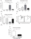
RSV infection significantly recruits eosinophils but down-regulates alveolar macrophages in FI-RSV immune mice. Immune cells were harvested from the BALF and lungs of mice 5 days post RSV challenge (n = 5). Flow cytometry was used to analyze the immune cell infiltration. (a) The eosinophil (CD11cCD11b+SiglecF+) and (b) total AM numbers (CD11c+CD11bF4/80+) were determined in each mouse group. (c) The AM depletion in the F VLP-immune mice was confirmed by flow cytometry. (d) Clodronate treatment depletes AMs from F VLP-immune mice. At 6 weeks after boost immunization, mice immunized with F VLP (n = 5/group) were treated with clodronate liposome (CL) 4 hours prior to RSV infection. Phosphate-buffered saline (PBS) treated mice were included as a control in each group. One-way ANOVA or an unpaired two-tailed Student’s t tests were used to determine the statistical significance. Data are representative of two independent experiments. Error bars indicate means±SEM. #P < 0.05 (between FI-RSV and F VLP-immune groups treated with PBS). *P < 0.05.
Figure 5
Figure 5
Alveolar macrophages contribute to protection in F VLP-immune mice against weight loss. Unimmunized naïve, FI-RSV-immune, PBS- and CL-treated F VLP-immune mice were challenged with RSV (1 × 10⁶ PFU). Lung samples from each mouse group (n = 5) were harvested 5 days post RSV challenges. (a) Body weights were monitored daily. (b) Viral titers in the lungs (individual, n = 5 were determined with an immunoplaque assay. (c) Body weights from the F VLP-immune mice were compared between mice that were or were not CL treated. (d) Lung viral loads were compared between the PBS- and CL-treated F VLP mice. Representative data of two independent experiments (n = 5 in each set) are presented. One-way ANOVA or an unpaired two-tailed Student’s t tests were used to determine the statistical significance. Error bars indicate means ± SEM. *P < 0.05; ** P < 0.01; ***P < 0.001.
Figure 6
Figure 6
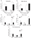
F VLP-immune mice recruit natural killer cells, but not eosinophils, into the airways. Immune cells were harvested from the BALF and lungs of CL-treated and PBS (mock control)-treated F VLP mice 5 days post RSV challenge (n = 5). Flow cytometry was used to analyze the immune cell infiltration. (a) Eosinophils (CD11b+CD11cSiglecF+). The eosinophil frequencies and numbers were analyzed by using CD11c and Siglec F antibodies. (b) CD69+ NK cells. The CD49b marker was used to gate NK cells. Activated NK cells were further analyzed by evaluating CD69 marker expression. Plasmacytoid DCs (pDCs, B220+CD11c+F4/80CD45+), CD103+ DCs (CD103+CD11c+F4/80CD45+), and CD11b+ DCs (CD11b+CD11c+F4/80CD45+) were analyzed in the BALF (c) and lungs (d) of F VLP- immune mice after CL treatment. (e) The inflammation scores on a scale of 0 to 3 were determined around the airways, interstitial areas, and blood vessels in the H&E stained tissue sections. Representative data are shown from two independent experiments. The unpaired two-tailed Student’s t test was used to determine the statistical significance. Error bars indicate means± SEM. *P < 0.05; ** < P < 0.01; *** < P < 0.001.
Figure 7
Figure 7
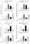
F VLP vaccination induces high CD8+ T cells in the airways and low CD4+ T cells in the lungs upon RSV infection. Immune cells were isolated from the BALF and lungs 5 days post RSV infection and used for flow cytometry analysis (n = 5/group). (a) The total number of CD3+CD4+ T cells was quantified in the BALF and lungs. Activated CD3+CD4+ T cells were further determined by using CD69 as an activation marker. (b) The total number of CD3+CD8+ T cells was analyzed in the BALF and lungs and further separated into activated cells by evaluating CD69 expression. Representative data from two independent experiments (n = 5 in each set) are presented. CL: CL-treated F VLP-immune mice. One-way ANOVA or an unpaired two-tailed Student’s t tests were used to determine the statistical significance. Error bars indicate means± SEM. *P < 0.05; **P < 0.01; ***P < 0.001.
Figure 8
Figure 8
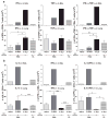
FI-RSV and F VLP vaccines differentially modulate antigen-specific effector CD4+ and CD8+ T cell responses in the airways and lungs. Immune cells were collected from the BALF and lungs of vaccinated mice 5 days after RSV challenge (n = 5/group). The cells were in vitro stimulated in the presence of RSV F85–93 (KYKNAVTEL) or RSV G183–195 (WAICKRIPNKKPG) peptides. Cytokine-producing cells were determined by an intracellular cytokine staining flow cytometry assay using anti-IL-4, -IFN-γ, and -TNF-α antibodies. After F or G peptide stimulation, the total cytokine-producing CD8+ T (a) or CD4+ T (b) cell numbers were determined in the BALF and lung samples of each group. The cells from the BALF of each group were pooled (n = 53. CL: CL-treated F VLP-immune mice. Representative data from two independent experiments (n = 5 in each set) are presented. One-way ANOVA or an unpaired two-tailed Student’s t tests were used to determine the statistical significance. Error bars indicate means± SEM. *P < 0.05; **P < 0.01; ***P < 0.001.
Figure 9
Figure 9
FI-RSV and F VLP vaccines show intrinsic differences in stimulating dendritic cells and macrophages to produce inflammatory cytokines. Bone marrow cells from BALB/c and C57BL/6 mice were harvested and cultured in the presence of GM-CSF or M-CSF to differentiate the cells into BMDCs or BMDMs, respectively. (a) BMDMs and (b) BMDCs from BALB/c mice were further stimulated with purified FI-RSV or F VLP vaccines and cytokine levels (TNF-α and IL-6) were determined with ELISAs from the culture supernatants. (c) TNF-α and (d) IL-6 levels were analyzed from BMDCs that were obtained from C57BL/6 and MyD88 deficient mice. The data were pooled from 2 independent experiments (n = 3 each set). An unpaired two-tailed Student’s t test was used to determine the statistical significance. Error bars indicate means± SEM. ***P < 0.001.

Similar articles

Cited by

References

    1. Smyth RL, Openshaw PJ. Lancet. 2006;368:312. - PubMed
    1. Collins PL, Graham BS. J Virol. 2008;82:2040. - PMC - PubMed
    1. Kapikian AZ, Mitchell RH, Chanock RM, Shvedoff RA, Stewart CE. Am J Epidemiol. 1969;89:405. - PubMed
    1. Kim HW, Canchola JG, Brandt CD, Pyles G, Chanock RM, Jensen K, Parrott RH. Am J Epidemiol. 1969;89:422. - PubMed
    1. Johnson TR, Graham BS. J Virol. 1999;73:8485. - PMC - PubMed