Comparative Study
. 2018 Feb 23;293(8):2687-2700.
doi: 10.1074/jbc.M117.803411. Epub 2018 Jan 3.

HspB1 and Hsc70 chaperones engage distinct tau species and have different inhibitory effects on amyloid formation

Affiliations
Comparative Study

HspB1 and Hsc70 chaperones engage distinct tau species and have different inhibitory effects on amyloid formation

Hannah E R Baughman et al. J Biol Chem. .

Abstract

The microtubule-associated protein tau forms insoluble, amyloid-type aggregates in various dementias, most notably Alzheimer's disease. Cellular chaperone proteins play important roles in maintaining protein solubility and preventing aggregation in the crowded cellular environment. Although tau is known to interact with numerous chaperones, it remains unclear how these chaperones function mechanistically to prevent tau aggregation and how chaperones from different classes compare in terms of mechanism. Here, we focused on the small heat shock protein HspB1 (also known as Hsp27) and the constitutive chaperone Hsc70 (also known as HspA8) and report how each chaperone interacts with tau to prevent its fibril formation. Using fluorescence and NMR spectroscopy, we show that the two chaperones inhibit tau fibril formation by distinct mechanisms. HspB1 delayed tau fibril formation by weakly interacting with early species in the aggregation process, whereas Hsc70 was highly efficient at preventing tau fibril elongation, possibly by capping the ends of tau fibrils. Both chaperones recognized aggregation-prone motifs within the microtubule-binding repeat region of tau. However, HspB1 binding remained transient in both aggregation-promoting and non-aggregating conditions, whereas Hsc70 binding was significantly tighter under aggregation-promoting conditions. These differences highlight the fact that chaperones from different families play distinct but complementary roles in the prevention of pathological protein aggregation.

Keywords: 70 kilodalton heat shock protein (Hsp70); aggregation; amyloid; chaperone; small heat shock protein (sHsp); tau.


Conflict of interest statement

The authors declare that they have no conflicts of interest with the contents of this article

Figures

Figure 1.
Figure 1.
Domain organization of tau, HspB1, and Hsc70. Top, full-length tau2N4R is natively unstructured and consists of four domains. The tau4RD construct used in this work contains residues 244–372, which include the four microtubule-binding repeats. The sequences 275VQIINK280 and 306VQIVYK311 at the beginning of the second and third repeats drive tau fibril formation. Shaded areas, regions that can be alternatively spliced. Middle, HspB1 contains three domains. The core ACD has a β-sandwich fold and forms a stable dimer. The CTR is intrinsically disordered and contains an IXI motif that interacts with the ACD in the context of large oligomers. The NTR is partially disordered and involved in oligomerization, and it contains three serine residues that are phosphorylated in response to cellular stress. The “D3” and “dimer” constructs, described under “Results,” affect the oligomerization state of HspB1. Bottom, Hsc70 contains two domains: an N-terminal nucleotide binding domain with ATPase activity and a C-terminal substrate binding domain that interacts with client proteins.
Figure 2.
Figure 2.
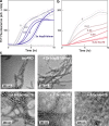
Effects of Hsc70 and HspB1 on tau4RD fibril formation. a, ThT fluorescence traces over the course of a representative fibril formation reaction. Tau4RD (3.5 μm) fibrillizes over the course of 10 h following the addition of the aggregation inducer heparin (6 μm, average molecular mass 3 kDa) and follows expected unseeded amyloid formation kinetics. HspB1dimer delays the onset of tau4RD fibril formation in a dose-dependent manner but does not alter the final ThT fluorescence signal. Each trace is the average of 6 wells in a 96-well plate. For clarity, error bands (S.E.) are shown only for the highest and lowest chaperone concentrations. b, Hsc70 is a highly potent substoichiometric inhibitor and decreases the extent of fibril formation in a dose-dependent manner. c, representative negative stain electron micrographs of end products of ThT fibril formation reactions. Tau4RD forms long fibrils in the absence of chaperone and is still able to form long, fully developed fibrils in the presence of HspB1dimer. It forms smaller oligomeric species in the presence of Hsc70, indicating that Hsc70 holds much of the protein in a non-fibrillar state.
Figure 3.
Figure 3.
Mechanisms of chaperone inhibition. a, intensity from 1D NMR spectra monitoring the disappearance of small, soluble tau4RD species during aggregation. In the absence of chaperone (gray circles), 50 μm tau4RD loses some intensity following the addition of 120 μm heparin (drop between first and second data points) and then loses intensity in a sigmoidal manner as it becomes incorporated in NMR-invisible fibrils. HspB1dimer slows this intensity loss in a dose-dependent manner (blue squares and triangles). b, Hsc70 is inhibitory even at a 1:50 molar ratio, making it a highly potent substoichiometric inhibitor. At a 1:1 ratio, it causes a rapid decrease in intensity, probably due to enhanced binding in the presence of heparin, followed by a plateau. c, SDS-PAGE analysis of end products of 1D NMR experiments. End products were centrifuged, and total end products (T), supernatant (S), and pellet (P) were run on a gel. In the absence of chaperone and in the presence of 1 molar eq of HspB1dimer, most of the tau4RD ends up in the insoluble fraction. In the presence of 1 molar eq of Hsc70, almost all of the tau4RD remains soluble. This indicates that the rapid intensity loss in the presence of Hsc70 is due to binding rather than aggregation. d, ThT fluorescence traces over the course of a tau4RD fibril formation reaction. HspB1dimer (1 molar eq) was added to 3.5 μm tau4RD either at the beginning of the reaction (dark blue) or 0.5, 1, or 2.5 h after the reaction had been initiated by the addition of 6 μm heparin (lighter blues). HspB1dimer was still effective at delaying aggregation when added during the lag phase of the reaction but was no longer effective when added during the elongation phase of the reaction. Adding buffer instead of chaperone at any time point did not alter aggregation kinetics (gray curves). Curves are averages of six replicates. For clarity, error bands (S.E.) are shown only for the 0 h experiments. e, Hsc70 (0.05 molar eq) was added to 3.5 μm tau4RD either at the beginning of the reaction (dark red) or 0.5, 1, or 2.5 h after the reaction had been started by the addition of 6 μm heparin (lighter reds). The addition of Hsc70 during the lag phase did not alter its effect on the aggregation kinetics, and addition during the elongation phase prevented any further increase in ThT fluorescence. Adding buffer instead of chaperone at any time point did not alter aggregation kinetics (gray curves). Curves are averages of four or six replicates.
Figure 4.
Figure 4.
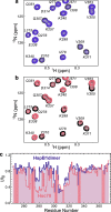
Chaperone binding sites. a, 15N HSQC TROSY of 15N-labeled tau4RD alone (black) and in the presence of 2 molar eq of HspB1dimer (blue). b, 15N HSQC TROSY of 15N-labeled tau4RD alone (black) and in the presence of 2 molar eq of Hsc70 (red). c, peak broadening from a and b, quantified by dividing the intensity of a peak in the bound spectrum (I) by the intensity of the same peak in the unbound spectrum (I0). Broadening due to HspB1dimer maps primarily to the aggregation-prone motif in the third repeat of tau4RD (306VQIVYK311), whereas broadening due to Hsc70 maps to the aggregation-prone motifs at the start of both the second and third repeats of tau4RD (275VQIINK280 and 306VQIVYK311).
Figure 5.
Figure 5.
Chaperone binding affinities. FCS-derived changes in hydrodynamic radius of tau4RD∼A488, measured in the presence of increasing concentrations of Hsc70 (solid circles) or HspB1dimer (empty circles). The Hsc70 data are well fit by a one-site binding model with KD = 99 ± 28 μm, whereas HspB1dimer does not appear to saturate up to a chaperone concentration of 750 μm. Error bars, S.E.
Figure 6.
Figure 6.
Effects of heparin on chaperone binding. a, 15N HSQC TROSY of 15N-labeled tau4RD alone (black) and in the presence of 2.4 molar eq of heparin (gray). b, chemical-shift perturbations (CSP) of each peak in A plotted as a function of residue number. The highest chemical-shift perturbations map to the aggregation-prone motif at the start of the second repeat of tau4RD, 275VQIINK280. c, 15N HSQC TROSY of 15N-labeled tau4RD alone (black), in the presence of 2.4 molar eq of heparin (gray), in the presence of 2 molar eq of HspB1dimer (blue), and in the presence of both 2.4 eq of heparin and 2 eq of HspB1dimer (yellow). HspB1dimer binding is largely unchanged by the presence of heparin. d, 15N HSQC TROSY of 15N-labeled tau4RD alone (black), in the presence of 2.4 molar eq of heparin (gray), in the presence of 2 molar eq of Hsc70 (red), and in the presence of both 2.4 eq of heparin and 2 eq of Hsc70 (yellow). Hsc70 binding is enhanced by the presence of heparin, as indicated by the enhanced broadening in the yellow spectrum relative to the red. e, comparison of residues Val-309, Tyr-310, and Lys-311 in the HspB1dimer-bound and Hsc70-bound spectra in the presence of heparin. Although HspB1dimer and Hsc70 cause a similar amount of broadening in these peaks in the absence of heparin, in the presence of heparin, Hsc70 causes significantly more broadening in these peaks than HspB1dimer, indicating that heparin enhances Hsc70 binding in a way that it does not for HspB1dimer.
Figure 7.
Figure 7.
Effects of HspB1WT and HspB1D3 on tau4RD. a, intensity from 1D NMR spectra monitoring the disappearance of small, soluble tau4RD species during aggregation in the presence of HspB1WT. HspB1WT (green squares and triangles) slows this intensity loss in a manner similar to HspB1dimer (Fig. 3a). b, intensity from 1D NMR spectra monitoring tau4RD aggregation in the presence of HspB1D3 (teal squares and triangles). HspB1D3 slows intensity loss in a manner similar to HspB1dimer and WT. c, 15N HSQC TROSY of 15N-labeled tau4RD alone (black) and in the presence of 4 molar eq of HspB1WT (green). There is very little significant broadening in the spectrum with HspB1WT, suggesting that binding is very weak. d, 15N HSQC TROSY of 15N-labeled tau4RD alone (black) and in the presence of 4 molar eq of HspB1D3 (teal). There is slight broadening in peaks corresponding to 306VQIVYK311, indicating some binding. e, 15N HSQC TROSY of 15N-labeled tau4RD alone (black) and in the presence of 4 molar eq of HspB1dimer (blue). Peak broadening is much more substantial, indicating that HspB1WT and D3 both bind more weakly than HspB1dimer. f, quantification of tau 4RD peak intensity loss due to HspB1WT, HspB1D3, and HspB1dimer binding.
Figure 8.
Figure 8.
Model of chaperone effects on tau aggregation. HspB1 and Hsc70 interact with different species along the tau amyloid formation pathway. HspB1 interacts with early species and delays the formation of tau oligomers and fibrils (1D NMR and ThT results). Hsc70 has enhanced affinity for aggregation-prone, heparin-bound tau (1D and 2D NMR results); can hold tau in an oligomeric state (EM results); and can prevent fibril elongation (ThT results).

Similar articles

Cited by

References

    1. Drubin D. G., and Kirschner M. W. (1986) Tau protein function in living cells. J. Cell Biol. 103, 2739–2746 10.1083/jcb.103.6.2739 - DOI - PMC - PubMed
    1. Stamer K., Vogel R., Thies E., Mandelkow E., and Mandelkow E. M. (2002) Tau blocks traffic of organelles, neurofilaments, and APP vesicles in neurons and enhances oxidative stress. J. Cell Biol. 156, 1051–1063 10.1083/jcb.200108057 - DOI - PMC - PubMed
    1. Dawson H. N., Ferreira A., Eyster M. V., Ghoshal N., Binder L. I., and Vitek M. P. (2001) Inhibition of neuronal maturation in primary hippocampal neurons from tau deficient mice. J. Cell Sci. 114, 1179–1187 - PubMed
    1. Goedert M., Spillantini M. G., Jakes R., Rutherford D., and Crowther R. A. (1989) Multiple isoforms of human microtubule-associated protein tau: sequences and localization in neurofibrillary tangles of Alzheimer's disease. Neuron 3, 519–526 10.1016/0896-6273(89)90210-9 - DOI - PubMed
    1. Wolfe M. S. (2009) Tau mutations in neurodegenerative diseases. J. Biol. Chem. 284, 6021–6025 10.1074/jbc.R800013200 - DOI - PubMed

Publication types

MeSH terms