

Genetic contribution of retinoid-related genes to neural tube defects
- PMID: 29297599
- PMCID: PMC5839987
- DOI: 10.1002/humu.23397
Genetic contribution of retinoid-related genes to neural tube defects
Abstract
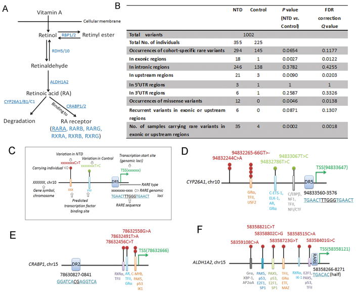
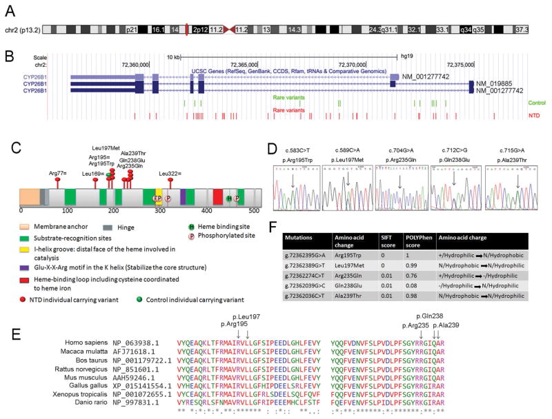
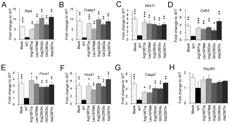
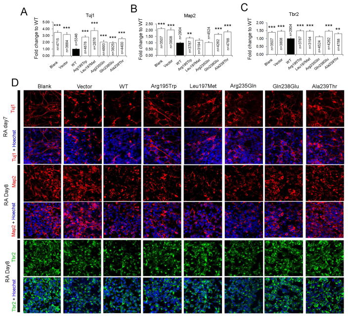
Rare variants are considered underlying causes of complex diseases. The complex and severe group of disorders called neural tube defects (NTDs) results from failure of the neural tube to close during early embryogenesis. Neural tube closure requires the coordination of numerous signaling pathways, including the precise regulation of retinoic acid (RA) concentration, which is controlled by enzymes involved in RA synthesis and degradation. Here, we used a case-control mutation screen study to reveal rare variants in retinoid-related genes in a Han Chinese NTD population by sequencing six genes in 355 NTD cases and 225 controls. More specific rare variants were found in exonic and upstream regions in NTD cases. The RA-responsive genes CYP26A1, CRABP1, and ALDH1A2 harbored NTD-specific rare variants in their upstream regions. Unexpectedly, the majority of missense variants in NTD cases were found in CYP26B1, which encodes a RA degradation enzyme, whereas no missense variants in this gene were found in controls. Functional analysis indicated that the CYP26B1 NTD variants were inefficient in the degradation of RA using assays of RA-induced transcription and RA-initiated neuronal differentiation. Our study supports the contribution of rare variants in RA-related genes to the etiology of human NTDs.
Keywords: CYP26B1; DNA sequencing; NTD; neural tube defects; retinoid-related genes.
© 2018 Wiley Periodicals, Inc.
Conflict of interest statement
DISCLOSURE STATEMENT
The authors declare no conflict of interest.
Figures
Similar articles
-
Retinoid machinery in distinct neural stem cell populations with different retinoid responsiveness.Stem Cells Dev. 2013 Oct 15;22(20):2777-93. doi: 10.1089/scd.2012.0422. Epub 2013 Jul 24. Stem Cells Dev. 2013. PMID: 23734950 Free PMC article.
-
Expression of the retinoic acid catabolic enzyme CYP26B1 in the human brain to maintain signaling homeostasis.Brain Struct Funct. 2016 Jul;221(6):3315-26. doi: 10.1007/s00429-015-1102-z. Epub 2015 Sep 15. Brain Struct Funct. 2016. PMID: 26374207 Free PMC article.
-
Replication and exploratory analysis of 24 candidate risk polymorphisms for neural tube defects.BMC Med Genet. 2014 Oct 8;15:102. doi: 10.1186/s12881-014-0102-9. BMC Med Genet. 2014. PMID: 25293959 Free PMC article.
-
Etiology, pathogenesis and prevention of neural tube defects.Congenit Anom (Kyoto). 2006 Jun;46(2):55-67. doi: 10.1111/j.1741-4520.2006.00104.x. Congenit Anom (Kyoto). 2006. PMID: 16732763 Review.
-
Roles of retinoic acid signaling in normal and abnormal development of the palate and tongue.Congenit Anom (Kyoto). 2014 May;54(2):69-76. doi: 10.1111/cga.12049. Congenit Anom (Kyoto). 2014. PMID: 24666225 Review.
Cited by
-
ALDH1A2-related disorder: A new genetic syndrome due to alteration of the retinoic acid pathway.Am J Med Genet A. 2023 Jan;191(1):90-99. doi: 10.1002/ajmg.a.62991. Epub 2022 Oct 19. Am J Med Genet A. 2023. PMID: 36263470 Free PMC article.
-
Up-regulation of miR-10a-5p expression inhibits the proliferation and differentiation of neural stem cells by targeting Chl1.Acta Biochim Biophys Sin (Shanghai). 2024 Jun 5;56(10):1483-1497. doi: 10.3724/abbs.2024078. Acta Biochim Biophys Sin (Shanghai). 2024. PMID: 38841745 Free PMC article.
-
Zebrafish as a Model to Study Retinoic Acid Signaling in Development and Disease.Biomedicines. 2023 Apr 15;11(4):1180. doi: 10.3390/biomedicines11041180. Biomedicines. 2023. PMID: 37189798 Free PMC article. Review.
-
Association between rare variants in specific functional pathways and human neural tube defects multiple subphenotypes.Neural Dev. 2020 Jul 10;15(1):8. doi: 10.1186/s13064-020-00145-7. Neural Dev. 2020. PMID: 32650820 Free PMC article.
-
Time Course Transcriptome Analysis of Spina Bifida Progression in Fetal Rats.Brain Sci. 2021 Nov 30;11(12):1593. doi: 10.3390/brainsci11121593. Brain Sci. 2021. PMID: 34942894 Free PMC article.
References
Publication types
MeSH terms
Substances
Grants and funding
LinkOut - more resources
Full Text Sources
Other Literature Sources
Medical
