. 2017 Dec 22;8(1):2282.
doi: 10.1038/s41467-017-02338-x.

Generic wound signals initiate regeneration in missing-tissue contexts

Affiliations

Generic wound signals initiate regeneration in missing-tissue contexts

Suthira Owlarn et al. Nat Commun. .

Abstract

Despite the identification of numerous regulators of regeneration in different animal models, a fundamental question remains: why do some wounds trigger the full regeneration of lost body parts, whereas others resolve by mere healing? By selectively inhibiting regeneration initiation, but not the formation of a wound epidermis, here we create headless planarians and finless zebrafish. Strikingly, in both missing-tissue contexts, injuries that normally do not trigger regeneration activate complete restoration of heads and fin rays. Our results demonstrate that generic wound signals have regeneration-inducing power. However, they are interpreted as regeneration triggers only in a permissive tissue context: when body parts are missing, or when tissue-resident polarity signals, such as Wnt activity in planarians, are modified. Hence, the ability to decode generic wound-induced signals as regeneration-initiating cues may be the crucial difference that distinguishes animals that regenerate from those that cannot.


Conflict of interest statement

The authors declare no competing financial interests.

Figures
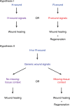
Fig. 1
Fig. 1
Two hypotheses for generating regeneration initiation signals. Hypothesis I: regeneration-initiation signals are induced exclusively after R-wounds, such as amputations; H-wounds, such as incisions or other epidermal wounds, activate different signals leading to wound healing but not to a subsequent regenerative response. Hypothesis II: the same signals are induced generically after all types of injuries; the tissue contexts in which the signals are induced determine whether regeneration occurs
Fig. 2
Fig. 2
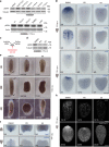
Transient inhibition of ERK activation blocks regeneration initiation in planarians. a Phosphorylated ERK levels dramatically increased within 15 min post-amputation. b Amputation-induced ERK activation occurred even after depletion of stem cells by γ-irradiation (1 or 2 days post-irradiation (d.p.irr)) and after inhibition of protein synthesis with cycloheximide (CHX). c MEK activates ERK by phosphorylation (pERK); MEK inhibitors PD0325901 (PD) and U0126 (U0) prevent ERK activation by inhibiting MEK. d Treatment with PD and U0 effectively reduced pERK levels. e, f Inhibition of ERK activity severely (PD heads 44/100, trunks 33/100, tails 3/120; U0 heads 43/70, trunks 47/70, tails 11/70) or completely (PD heads 56/100, trunks 67/100, tails 117/120; U0 heads 27/70, trunks 23/70, tails 59/70) inhibited formation of blastemas (e) and anterior (notum expression in lower panel) and posterior (wnt1 expression in upper panel) regeneration poles (f). Black arrows: regeneration of new tissues in the blastema; red arrows: remodeling of existing tissues. gh Other key regenerative responses, such as expression of early wound-induced genes (g), as well as stem cell proliferation (pH3, phospho-Histone H3 as a marker for mitotic cells) and accumulation of stem cells and progeny (SMEDWI-1) (h) were also severely affected by ERK inhibition. Sample numbers indicated in each image. h.p.a., hours post-amputation; d.p.a., days post-amputation; DMSO was used as the solvent control; scale bars: 200 μm
Fig. 3
Fig. 3
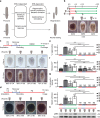
H-wounds inflicted on dormant tails induce regeneration in an ERK-dependent manner. a Distinct cellular responses occur in planarians undergoing wound healing (after H-wounds) or regeneration (after R-wounds). b “Dormant” tails that were re-amputated, whether immediately or 5 days after drug removal, fully regenerated. c Tail fragments did not regenerate after a 5-day PD treatment and recovery period unless re-injured; as in the case of re-amputations, incisions that cut through the animal along the DV axis but do not involve tissue loss led to the formation of the anterior regeneration pole (marked by notum; sample numbers indicated in each image) and full regeneration of heads and medial structures (see Supplementary Fig. 6 for representative images of immediate wounds). During the recovery period (blue in scheme), animals were kept in DMSO-containing water. d Induction of regeneration through re-injury was inhibited by PD treatment. e Animals that had formed blastemas were considered to be “regenerating” if they later also regenerated eyes. fg Corresponding proliferative responses at 4 d.p.a. (f) and functional recovery (g) were observed. Bars in graphs represent mean ± s.d.; two-sided t test (compared with uninjured PD-removed animals (first bar, dark gray), unless indicated otherwise), *p < 0.05, **p < 0.01, ***p < 0.001, NS, not significant. h Increased pERK levels were observed at 3 h post-wounding in conditions that induced regeneration. d.p.w. days post-wounding, scale bars: 200 μm
Fig. 4
Fig. 4
H-wounds inflicted on planarians with altered levels of Wnt signaling induce patterned outgrowths. a, b Simple incisions applied to β-catenin-1 RNAi planarians induced accessory heads, which were not observed when ERK activation was inhibited by PD, or in control RNAi animals. Sample numbers indicated in each image. In contrast, ectopic heads formed in existing posterior tissues were unaffected by PD treatment. Arrow, ectopic eyes; sFRP-1, anterior marker; ndk, marker for the brain region. c Similarly, ectopic lateral outgrowths but not posterior heads in homeostatic β-catenin-1 RNAi animals were inhibited by treatment with PD. de Outgrowths were also induced by lateral incisions inflicted on APC (d) or patched (e) RNAi planarians, which undergo direct or indirect ectopic activation of Wnt signaling. To increase the penetrance of outgrowth formation in (a), (d), and (e), two incisions were made instead of one. Numbers refer to injured areas (two per animal); scale bars: 100 μm
Fig. 5
Fig. 5
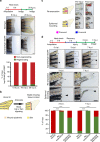
Epidermal wounds inflicted on dormant zebrafish fins induce regeneration. a Transient overexpression of a dominant-negative Fgf receptor-1 (dnFgfr1-GFP) until 8 days post-amputation (d.p.a.) blocked fin regeneration, which spontaneously resumed only in a minority of fin rays after relief from transgene expression. White arrowheads mark the amputation plane, n = 9 fish, 163 rays. b A stable missing-tissue context (dormant fin) can be produced by transient blockage of Fgf or Wnt/β−catenin signaling, which allows for wound epidermis formation but stably blocks fin ray regeneration. c Injury regimes applied to dormant fins. Re-amputation represents an R-wound, epidermal wounding an H-wound. Arrowhead points at epidermal wound. d Re-amputation (asterisk), as well as epidermal wounding (black arrowhead) re-initiated regenerative growth. Lower panels show magnification of boxed areas. The graph depicts the fraction of non-injured, re-amputated and epidermally wounded rays displaying regenerative growth at 3 and 7 days post-injury (d.p.i.). ***p < 0.001 χ 2 test. n (non-injured) = 449 rays, 61 fish, 3 experiments; n (re-amputated) = 30 rays, 28 fish, 1 experiment, n (epidermally wounded) = 84 rays, 61 fish, 3 experiments. Note that uninjured rays located next to regenerating rays frequently exhibited bystander growth (white arrowhead in box) in both wounding scenarios (see also Supplementary Fig. 9). Scale bars: 500 μm
Fig. 6
Fig. 6
The wound epidermis of dormant fins is not a barrier to regeneration. a Transient inhibition of Wnt/β-catenin signaling using overexpression of Axin1 in hsp70l:Axin1-YFP transgenic fish resulted in a stable blockage of fin ray regeneration. n = 9 fish. b Amputation and heat-shock regime used to produce recessed rays in dormant fins. Note that interray skin healed distally to non-regenerating recessed rays (asterisk) despite Axin1-YFP expression. c Skin wounding applied to the skin distal to recessed dormant rays (black arrowheads) induced regenerative growth beyond the primary amputation plane. Asterisks indicate non-injured control rays. Graph depicts the fraction of regenerating rays. ***p < 0.001 χ 2 test. n (non-injured) = 48 rays, 48 fish, two experiments, n (wounded) = 69 rays, 50 fish, two experiments. Scale bars: 500 µm
Fig. 7
Fig. 7
Generic wound signals initiate regeneration in missing-tissue contexts. a Injuries that normally do not trigger regeneration (H-wounds) do initiate regeneration in planarian dormant tails and zebrafish dormant fins (missing-tissue contexts). b Signals that are generically induced after all injuries have the capacity to initiate regeneration, but only when induced in missing-tissue contexts

Similar articles

Cited by

References

    1. Petrie TA, Strand NS, Yang CT, Rabinowitz JS, Moon RT. Macrophages modulate adult zebrafish tail fin regeneration. Development. 2014;141:2581–2591. doi: 10.1242/dev.098459. - DOI - PMC - PubMed
    1. Yokoyama H. Initiation of limb regeneration: the critical steps for regenerative capacity. Dev. Growth Differ. 2008;50:13–22. doi: 10.1111/j.1440-169X.2007.00973.x. - DOI - PubMed
    1. Wenemoser D, Reddien PW. Planarian regeneration involves distinct stem cell responses to wounds and tissue absence. Dev. Biol. 2010;344:979–991. doi: 10.1016/j.ydbio.2010.06.017. - DOI - PMC - PubMed
    1. Wurtzel O, et al. A generic and cell-type-specific wound response precedes regeneration in planarians. Dev. Cell. 2015;35:632–645. doi: 10.1016/j.devcel.2015.11.004. - DOI - PMC - PubMed
    1. Pellettieri J, et al. Cell death and tissue remodeling in planarian regeneration. Dev. Biol. 2010;338:76–85. doi: 10.1016/j.ydbio.2009.09.015. - DOI - PMC - PubMed

Publication types

LinkOut - more resources