The mito::mKate2 mouse: A far-red fluorescent reporter mouse line for tracking mitochondrial dynamics in vivo
- PMID: 29243279
- PMCID: PMC5818295
- DOI: 10.1002/dvg.23087
The mito::mKate2 mouse: A far-red fluorescent reporter mouse line for tracking mitochondrial dynamics in vivo
Abstract
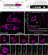
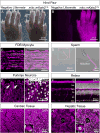
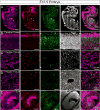
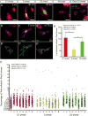
Mitochondria are incredibly dynamic organelles that undergo continuous fission and fusion events to control morphology, which profoundly impacts cell physiology including cell cycle progression. This is highlighted by the fact that most major human neurodegenerative diseases are due to specific disruptions in mitochondrial fission or fusion machinery and null alleles of these genes result in embryonic lethality. To gain a better understanding of the pathophysiology of such disorders, tools for the in vivo assessment of mitochondrial dynamics are required. It would be particularly advantageous to simultaneously image mitochondrial fission-fusion coincident with cell cycle progression. To that end, we have generated a new transgenic reporter mouse, called mito::mKate2 that ubiquitously expresses a mitochondria localized far-red mKate2 fluorescent protein. Here we show that mito::mKate2 mice are viable and fertile and that mKate2 fluorescence can be spectrally separated from the previously developed Fucci cell cycle reporters. By crossing mito::mKate2 mice to the ROSA26R-mTmG dual fluorescent Cre reporter line, we also demonstrate the potential utility of mito::mKate2 for genetic mosaic analysis of mitochondrial phenotypes.
Keywords: far-red fluorescence; live imaging; mitochondria.
© 2017 Wiley Periodicals, Inc.
Figures


Similar articles
-
[Labeling of liver cancer cell for fluorescence imaging study by far-red fluorescence protein reporter gene mKate2].Zhonghua Yi Xue Za Zhi. 2011 May 24;91(19):1344-7. Zhonghua Yi Xue Za Zhi. 2011. PMID: 21756763 Chinese.
-
Mouse lines with photo-activatable mitochondria to study mitochondrial dynamics.Genesis. 2012 Nov;50(11):833-43. doi: 10.1002/dvg.22050. Epub 2012 Aug 11. Genesis. 2012. PMID: 22821887 Free PMC article.
-
α-MHC MitoTimer mouse: In vivo mitochondrial turnover model reveals remarkable mitochondrial heterogeneity in the heart.J Mol Cell Cardiol. 2016 Jan;90:53-8. doi: 10.1016/j.yjmcc.2015.11.032. Epub 2015 Dec 2. J Mol Cell Cardiol. 2016. PMID: 26654779 Free PMC article.
-
Mitochondrial fusion/fission dynamics in neurodegeneration and neuronal plasticity.Neurobiol Dis. 2016 Jun;90:3-19. doi: 10.1016/j.nbd.2015.10.011. Epub 2015 Oct 19. Neurobiol Dis. 2016. PMID: 26494254 Review.
-
Reporter mouse lines for fluorescence imaging.Dev Growth Differ. 2013 May;55(4):390-405. doi: 10.1111/dgd.12062. Epub 2013 Apr 29. Dev Growth Differ. 2013. PMID: 23621623 Review.
Cited by
-
β2-glycoprotein I promotes the clearance of circulating mitochondria.PLoS One. 2024 Jan 25;19(1):e0293304. doi: 10.1371/journal.pone.0293304. eCollection 2024. PLoS One. 2024. PMID: 38271349 Free PMC article.
-
Drug-like sphingolipid SH-BC-893 opposes ceramide-induced mitochondrial fission and corrects diet-induced obesity.EMBO Mol Med. 2021 Aug 9;13(8):e13086. doi: 10.15252/emmm.202013086. Epub 2021 Jul 7. EMBO Mol Med. 2021. PMID: 34231322 Free PMC article.
-
Mitochondrial Transfer Improves Cardiomyocyte Bioenergetics and Viability in Male Rats Exposed to Pregestational Diabetes.Int J Mol Sci. 2021 Feb 27;22(5):2382. doi: 10.3390/ijms22052382. Int J Mol Sci. 2021. PMID: 33673574 Free PMC article.
-
Genetically Encoded Fluorescent Biosensors Illuminate the Spatiotemporal Regulation of Signaling Networks.Chem Rev. 2018 Dec 26;118(24):11707-11794. doi: 10.1021/acs.chemrev.8b00333. Epub 2018 Dec 14. Chem Rev. 2018. PMID: 30550275 Free PMC article. Review.
-
Far-Red Fluorescent Proteins: Tools for Advancing In Vivo Imaging.Biosensors (Basel). 2024 Jul 24;14(8):359. doi: 10.3390/bios14080359. Biosensors (Basel). 2024. PMID: 39194588 Free PMC article. Review.
References
-
- Abe T, Kiyonari H, Shioi G, Inoue K, Nakao K, Aizawa S, Fujimori T. Establishment of conditional reporter mouse lines at ROSA26 locus for live cell imaging. Genesis. 2011;49:579–590. - PubMed
-
- Betsholtz C. Role of platelet-derived growth factors in mouse development. Int J Dev Biol. 1995;39:817–825. - PubMed
-
- Breckwoldt MO, Pfister FM, Bradley PM, Marinkovic P, Williams PR, Brill MS, Plomer B, Schmalz A, St Clair DK, Naumann R, Griesbeck O, Schwarzlander M, Godinho L, Bareyre FM, Dick TP, Kerschensteiner M, Misgeld T. Multiparametric optical analysis of mitochondrial redox signals during neuronal physiology and pathology in vivo. Nat Med. 2014;20:555–560. - PubMed
-
- Chan DC. Mitochondria: dynamic organelles in disease, aging, and development. Cell. 2006;125:1241–1252. - PubMed
Publication types
MeSH terms
Substances
Grants and funding
LinkOut - more resources
Full Text Sources
Other Literature Sources
Molecular Biology Databases
Research Materials

