Revisiting multimodal activation and channel properties of Pannexin 1
- PMID: 29233884
- PMCID: PMC5749114
- DOI: 10.1085/jgp.201711888
Revisiting multimodal activation and channel properties of Pannexin 1
Abstract
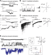
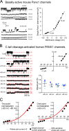
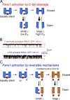
Pannexin 1 (Panx1) forms plasma membrane ion channels that are widely expressed throughout the body. Panx1 activation results in the release of nucleotides such as adenosine triphosphate and uridine triphosphate. Thus, these channels have been implicated in diverse physiological and pathological functions associated with purinergic signaling, such as apoptotic cell clearance, blood pressure regulation, neuropathic pain, and excitotoxicity. In light of this, substantial attention has been directed to understanding the mechanisms that regulate Panx1 channel expression and activation. Here we review accumulated evidence for the various activation mechanisms described for Panx1 channels and, where possible, the unitary channel properties associated with those forms of activation. We also emphasize current limitations in studying Panx1 channel function and propose potential directions to clarify the exciting and expanding roles of Panx1 channels.
© 2018 Chiu et al.
Figures


Similar articles
-
Physiological mechanisms for the modulation of pannexin 1 channel activity.J Physiol. 2012 Dec 15;590(24):6257-66. doi: 10.1113/jphysiol.2012.240911. Epub 2012 Oct 15. J Physiol. 2012. PMID: 23070703 Free PMC article. Review.
-
Intrinsic properties and regulation of Pannexin 1 channel.Channels (Austin). 2014;8(2):103-9. doi: 10.4161/chan.27545. Epub 2014 Jan 13. Channels (Austin). 2014. PMID: 24419036 Free PMC article. Review.
-
Emerging concepts regarding pannexin 1 in the vasculature.Biochem Soc Trans. 2015 Jun;43(3):495-501. doi: 10.1042/BST20150045. Biochem Soc Trans. 2015. PMID: 26009197 Free PMC article. Review.
-
Recent advances in the structure and activation mechanisms of metabolite-releasing Pannexin 1 channels.Biochem Soc Trans. 2023 Aug 31;51(4):1687-1699. doi: 10.1042/BST20230038. Biochem Soc Trans. 2023. PMID: 37622532 Review.
-
Mechanisms of pannexin1 channel gating and regulation.Biochim Biophys Acta Biomembr. 2018 Jan;1860(1):65-71. doi: 10.1016/j.bbamem.2017.07.009. Epub 2017 Jul 21. Biochim Biophys Acta Biomembr. 2018. PMID: 28735901 Review.
Cited by
-
Pannexin-1 Channels as Mediators of Neuroinflammation.Int J Mol Sci. 2021 May 14;22(10):5189. doi: 10.3390/ijms22105189. Int J Mol Sci. 2021. PMID: 34068881 Free PMC article. Review.
-
Pannexin 1 Regulates Dendritic Protrusion Dynamics in Immature Cortical Neurons.eNeuro. 2020 Aug 26;7(4):ENEURO.0079-20.2020. doi: 10.1523/ENEURO.0079-20.2020. Print 2020 Jul/Aug. eNeuro. 2020. PMID: 32737184 Free PMC article.
-
Pharmacology of pannexin channels.Curr Opin Pharmacol. 2023 Apr;69:102359. doi: 10.1016/j.coph.2023.102359. Epub 2023 Feb 28. Curr Opin Pharmacol. 2023. PMID: 36858833 Free PMC article. Review.
-
Structure-Based Design and Synthesis of Stapled 10Panx1 Analogues for Use in Cardiovascular Inflammatory Diseases.J Med Chem. 2023 Sep 28;66(18):13086-13102. doi: 10.1021/acs.jmedchem.3c01116. Epub 2023 Sep 13. J Med Chem. 2023. PMID: 37703077 Free PMC article.
-
ATP Release Channels.Int J Mol Sci. 2018 Mar 11;19(3):808. doi: 10.3390/ijms19030808. Int J Mol Sci. 2018. PMID: 29534490 Free PMC article. Review.
References
-
- Adamson S.E., Meher A.K., Chiu Y.H., Sandilos J.K., Oberholtzer N.P., Walker N.N., Hargett S.R., Seaman S.A., Peirce-Cottler S.M., Isakson B.E., et al. . 2015. Pannexin 1 is required for full activation of insulin-stimulated glucose uptake in adipocytes. Mol. Metab. 4:610–618. 10.1016/j.molmet.2015.06.009 - DOI - PMC - PubMed
-
- Ambrosi C., Gassmann O., Pranskevich J.N., Boassa D., Smock A., Wang J., Dahl G., Steinem C., and Sosinsky G.E.. 2010. Pannexin1 and Pannexin2 channels show quaternary similarities to connexons and different oligomerization numbers from each other. J. Biol. Chem. 285:24420–24431. 10.1074/jbc.M110.115444 - DOI - PMC - PubMed
-
- Anselmi F., Hernandez V.H., Crispino G., Seydel A., Ortolano S., Roper S.D., Kessaris N., Richardson W., Rickheit G., Filippov M.A., et al. . 2008. ATP release through connexin hemichannels and gap junction transfer of second messengers propagate Ca2+ signals across the inner ear. Proc. Natl. Acad. Sci. USA. 105:18770–18775. 10.1073/pnas.0800793105 - DOI - PMC - PubMed
Publication types
MeSH terms
Substances
Grants and funding
LinkOut - more resources
Full Text Sources
Other Literature Sources

