

The Complexity of Antibody Responses Elicited against the Respiratory Syncytial Virus Glycoproteins in Hospitalized Children Younger than 2 Years
- PMID: 29213258
- PMCID: PMC5702767
- DOI: 10.3389/fmicb.2017.02301
The Complexity of Antibody Responses Elicited against the Respiratory Syncytial Virus Glycoproteins in Hospitalized Children Younger than 2 Years
Abstract
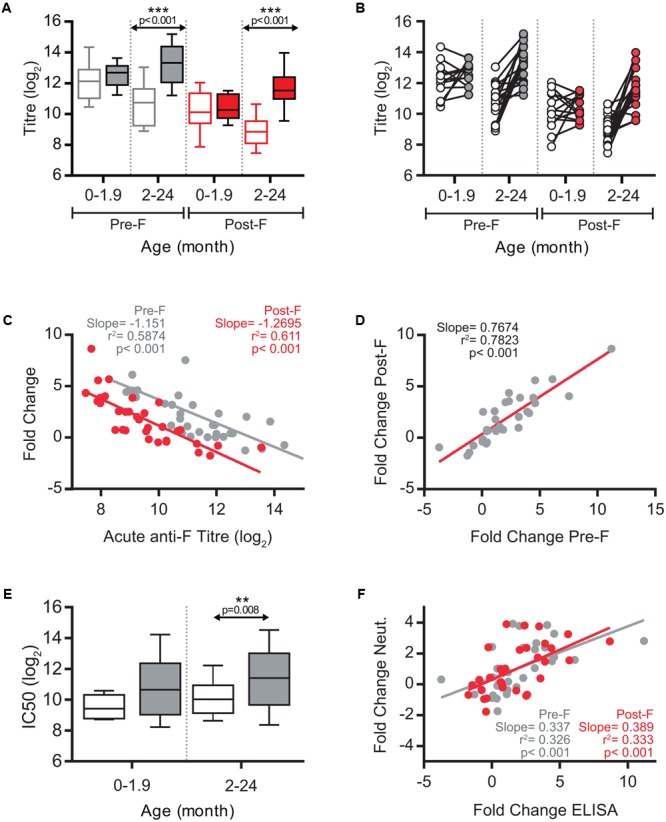
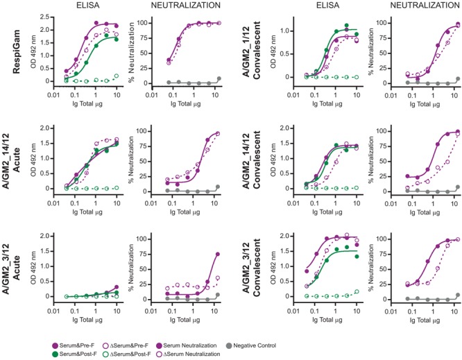
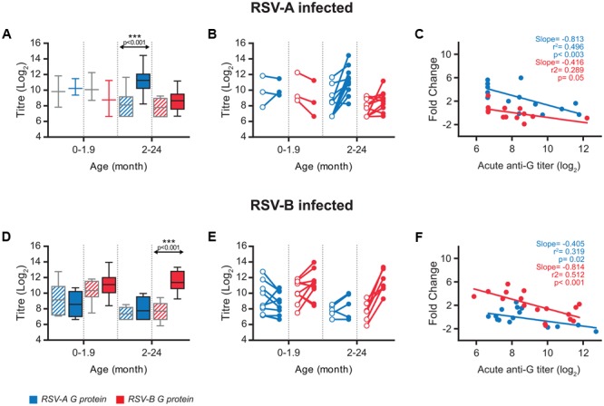
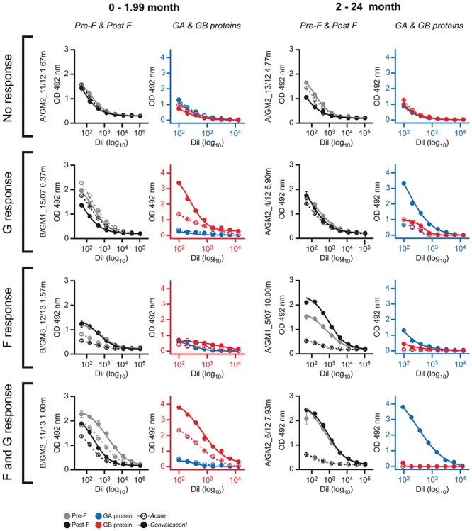
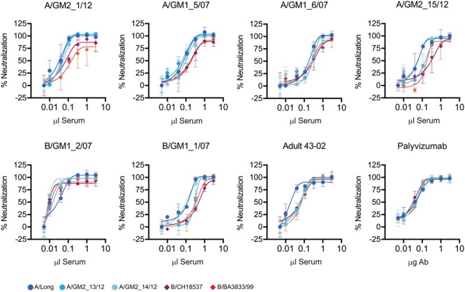
The influence of age and maternal antibodies on the antibody responses to human respiratory syncytial virus (hRSV) glycoproteins in very young children has been a matter of controversy. Both, immaturity of the immune system at very early age and suppression of the host immune response by high level of maternal antibodies have been claimed to limit the host antibody response to virus infection and to jeopardize the use of hRSV vaccines under development in that age group. Hence, the antibody responses to the two major hRSV glycoproteins (F and G) were evaluated in children younger than 2 years, hospitalized with laboratory confirmed hRSV bronchiolitis. A strong negative correlation was found between the titre of circulating ELISA antibodies directed against either prefusion or postfusion F in the acute phase, but not age, and their fold change at convalescence. These changes correlated also with the level of circulating neutralizing antibodies in sera. As reported in adults, most neutralizing antibodies in a subset of tested sera could not be depleted with postfusion F, suggesting that they were mostly directed against prefusion-specific epitopes. In contrast, a weak negative association was found for group-specific anti-G antibodies in the acute phase and their fold change at convalescence only after correcting for the antigenic group of the infecting virus. In addition, large discrepancies were observed in some individuals between the antibody responses specific for F and G glycoproteins. These results illustrate the complexity of the anti-hRSV antibody responses in children experiencing a primary severe infection and the influence of preexisting maternal antibodies on the host response, factors that should influence hRSV serological studies as well as vaccine development.
Keywords: antibody specificity; bronchiolitis; glycoproteins; immune responses; respiratory syncytial virus infections; viral.
Figures
Similar articles
-
Characterization of Epitope-Specific Anti-Respiratory Syncytial Virus (Anti-RSV) Antibody Responses after Natural Infection and after Vaccination with Formalin-Inactivated RSV.J Virol. 2016 Jun 10;90(13):5965-5977. doi: 10.1128/JVI.00235-16. Print 2016 Jul 1. J Virol. 2016. PMID: 27099320 Free PMC article.
-
Analysis of the binding of monoclonal and polyclonal antibodies to the glycoproteins of antigenic variants of human respiratory syncytial virus by surface plasmon resonance.J Immunol Methods. 2005 Feb;297(1-2):143-52. doi: 10.1016/j.jim.2004.12.017. J Immunol Methods. 2005. PMID: 15777938
-
Measurement of antibody against contemporary virus lineages of human respiratory syncytial virus sub-group A in infants and their mothers.J Clin Virol. 2004 May;30(1):73-80. doi: 10.1016/j.jcv.2003.09.013. J Clin Virol. 2004. PMID: 15072758
-
Human Respiratory Syncytial Virus: Role of Innate Immunity in Clearance and Disease Progression.Viral Immunol. 2016 Jan-Feb;29(1):11-26. doi: 10.1089/vim.2015.0098. Epub 2015 Dec 17. Viral Immunol. 2016. PMID: 26679242 Review.
-
CD8+ T cell immunity against human respiratory syncytial virus.Vaccine. 2014 Oct 21;32(46):6130-7. doi: 10.1016/j.vaccine.2014.08.063. Epub 2014 Sep 16. Vaccine. 2014. PMID: 25223272 Review.
Cited by
-
Development and comparison of immunologic assays to detect primary RSV infections in infants.Front Immunol. 2024 Jan 12;14:1332772. doi: 10.3389/fimmu.2023.1332772. eCollection 2023. Front Immunol. 2024. PMID: 38283339 Free PMC article.
-
During the COVID-19 pandemic where has respiratory syncytial virus gone?Pediatr Pulmonol. 2021 Oct;56(10):3106-3109. doi: 10.1002/ppul.25582. Epub 2021 Jul 26. Pediatr Pulmonol. 2021. PMID: 34273135 Free PMC article. Review.
-
Antibody Responses to Respiratory Syncytial Virus: A Cross-Sectional Serosurveillance Study in the Dutch Population Focusing on Infants Younger Than 2 Years.J Infect Dis. 2021 Jul 15;224(2):269-278. doi: 10.1093/infdis/jiaa483. J Infect Dis. 2021. PMID: 32964923 Free PMC article.
-
A multifaceted approach to RSV vaccination.Hum Vaccin Immunother. 2018 Jul 3;14(7):1734-1745. doi: 10.1080/21645515.2018.1472183. Epub 2018 Jun 19. Hum Vaccin Immunother. 2018. PMID: 29771625 Free PMC article. Review.
-
Transplacental Antibody Transfer of Respiratory Syncytial Virus Specific IgG in Non-Human Primate Mother-Infant Pairs.Pathogens. 2021 Nov 5;10(11):1441. doi: 10.3390/pathogens10111441. Pathogens. 2021. PMID: 34832599 Free PMC article.
References
LinkOut - more resources
Full Text Sources
Other Literature Sources
Research Materials