Review
. 2017 Dec;207(4):1231-1253.
doi: 10.1534/genetics.117.199885.

Regulation of Carbohydrate Energy Metabolism in Drosophila melanogaster

Affiliations
Review

Regulation of Carbohydrate Energy Metabolism in Drosophila melanogaster

Jaakko Mattila et al. Genetics. 2017 Dec.

Abstract

Carbohydrate metabolism is essential for cellular energy balance as well as for the biosynthesis of new cellular building blocks. As animal nutrient intake displays temporal fluctuations and each cell type within the animal possesses specific metabolic needs, elaborate regulatory systems are needed to coordinate carbohydrate metabolism in time and space. Carbohydrate metabolism is regulated locally through gene regulatory networks and signaling pathways, which receive inputs from nutrient sensors as well as other pathways, such as developmental signals. Superimposed on cell-intrinsic control, hormonal signaling mediates intertissue information to maintain organismal homeostasis. Misregulation of carbohydrate metabolism is causative for many human diseases, such as diabetes and cancer. Recent work in Drosophila melanogaster has uncovered new regulators of carbohydrate metabolism and introduced novel physiological roles for previously known pathways. Moreover, genetically tractable Drosophila models to study carbohydrate metabolism-related human diseases have provided new insight into the mechanisms of pathogenesis. Due to the high degree of conservation of relevant regulatory pathways, as well as vast possibilities for the analysis of gene-nutrient interactions and tissue-specific gene function, Drosophila is emerging as an important model system for research on carbohydrate metabolism.

Keywords: gene regulation; glucose; insulin; metabolism; nutrient sensing.


Figures

Figure 1
Figure 1
Intracellular sugar-responsive gene regulatory network. The main regulators of sugar-responsive gene expression are the heterodimeric bHLH-Zip transcription factors Mondo and Mlx. Mondo-Mlx has a direct role in regulating gene expression programs, which are essential in glucose and fatty acid metabolism. In addition, Mondo-Mlx activates the transcription of a second tier of transcriptional regulators, including Sugarbabe and Cabut as well as other regulatory proteins such as protein kinase SIK3. In parallel, glucose regulates indirectly, through the generation of fatty acids and NAD+, transcription factor HNF4 and deacetylase Sirt1, respectively. The transcriptome regulated by Mondo-Mlx and HNF4 are partially overlapping. However, how these factors interact is yet unknown. bHLH, basic helix-loop-helix; FA, fatty acid; HNF4, hepatocyte nuclear factor 4; Mlx, Max-like protein X; NAD+, nicotinamide adenine dinucleotide; NADPH, nicotinamide adenine dinucleotide phosphate; OXPHOS, oxidative phosphorylation; PPP, pentose phosphate pathway; SIK3, salt-inducible kinase 3; Sirt1, Sirtuin 1; TCA, tricarboxylic acid cycle.
Figure 2
Figure 2
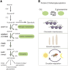
The role of the HBP and protein O-linked GlcNAc conjugation in the regulation of Drosophila physiology. Schematic presentation of the HBP (A) and the known processes regulated by protein O-linked GlcNAc conjugation (B). (A) HBP competes for F-6-P with PFK, the rate-limiting enzyme of glycolysis. In the first and rate-limiting step of HBP, GFAT conjugates an amine group from glutamine to the F-6-P yielding GlcN-6-P and glutamate. In the following step, GNPNAT conjugates the acetyl group from acetyl-CoA to yield GlcNAc-6-P, which is then isomerized to GlcNAc-1-P by PGM3. Finally, UDP is conjugated to the GlcNAc-1-P by UAP1 to yield UDP-GlcNAc, which is a substrate for macromolecule glycosylation and chitin biosynthesis. Hence, HBP integrates inputs from glucose (G-6-P), amino acid (glutamine), fatty acid (acetyl-CoA) and energy (UDP) metabolism, making it a sensor of cellular nutrient and energy metabolism. N-linked glycosylation, covalent attachment of an oligosaccharide to asparagine residues, is a mechanism of protein maturation and trafficking between cell compartments. The UDP-GlcNAc polymer, also known as chitin, is the key component of Drosophila exoskeleton. (B) O-linked GlcNAcylation is a transient protein post-translational modification mechanism, which targets threonine and serine residues. O-linked GlcNAcylation is mediated by the activities of OGT and OGA to conjugate and deconjugate glucosamine, respectively. O-linked GlcNAcylation can compete, enhance, or attenuate protein phosphorylation, making it an important mechanism to regulate protein activity. In Drosophila, the activity of OGT is known to be involved in maintenance of chromatin state, in regulating larval growth through insulin signaling, and in regulating the maintenance of circadian rhythm. F-6-P, fructose-6-phosphate; G-6-P, glucose-6-phosphate; GFAT1/2, glutamine fructose-6-phosphate amidotransferase 1/2; GlcN-6-P, glucosamine-6-phosphate; GlcNAc, N-acetylglucosamine; GlcNAc-1-P, N-acetyl-D-glucosamine-1-phosphate; GlcNAc-6-P, N-acetyl-D-glucosamine-6-phosphate; GNPNAT, glucosamine-phosphate N-acetyltransferase; HBP, hexosamine biosynthesis pathway; HXK, hexokinase; OGA, O-GlcNAcase; OGT, O-GlcNAc transferase; PFK, phosphofructokinase; PGI, phosphoglucose isomerase; PGM3, phosphoglucomutase 3; UAP1, UDP-N-acetylglucosamine pyrophosphorylase; UTP, uridine triphosphate.
Figure 3
Figure 3
The de novo synthesis of fatty acids and glycogenesis is coordinated in response to dietary sugars. The increase in cellular G-6-P levels leads to the orchestrated regulation of several metabolic processes important in the synthesis of fatty acids and glycogen and, as a result, clearance of intracellular sugars. The majority of G-6-P is channeled through glycolysis, resulting in elevated pyruvate and the production of acetyl-CoA. The process is coordinated with increased levels of CoA biosynthesis. Acetyl-CoA is utilized by the TCA cycle to produce intermediates of amino acid metabolism, ATP, NADH, and citrate. Citrate is further channeled to the fatty acid biosynthesis. The process of fatty acid synthesis is accompanied with the activity of the pentose phosphate pathway yielding the necessary reductive power in the form of NADPH. Parallel to the fatty acid synthesis, elevated levels of G-6-P shut down the process of lipid catabolism through lipolysis and generation of acetyl-CoA through β-oxidation. CoA, coenzyme A; G-6-P, glucose-6-phosphate; NADPH, nicotinamide adenine dinucleotide phosphate; TCA, tricarboxylic acid cycle.
Figure 4
Figure 4
Insulin-like peptide-glucagon circuit in Drosophila. Schematic presentation of larva (A) and adult fly (B), illustrating mechanisms that regulate the output of dILP and dAKH signaling in response to dietary carbohydrates. Dietary carbohydrates are digested in the midgut and glucose is taken up by the intestinal enterocytes. Glucose is converted into trehalose in the fat body and released into circulation. In the larva, trehalose has a biphasic effect to dAKH; low and high trehalose concentrations are shown to stimulate dAKH secretion. Whether such regulation also exists in adults is unknown. Larval IPCs are inherently insensitive to glucose, but carbohydrates regulate dILP secretion through remote mechanisms. These include dAKH from the CC as well as CCHamide-2, Dawdle, and Upd2 secreted from the fat body. Only Upd2 has been shown to function at the larval and adult stages. At the adult stage, glucose regulates IPCs directly by modulating the activity of KATP channels and cell depolarization leading to dILP secretion. The output of dAKH and dILP signaling is regulated through humoral factors, such as Activinβ, Imp-L2, dALS, Sdr, and NLaZ. Only NLaz has been shown to function at larval and Activinβ adult stage. Actβ, Activinβ; CC, corpora cardiaca; dAKH, Drosophila adipokinetic hormone; dALS, Drosophila acid-labile subunit; dILP, Drosophila insulin-like peptide; Imp-L2, imaginal morphogenesis protein-late 2; IPC, insulin producing cells; NLaz, Neural Lazarillo; Sdr, secreted decoy of InR; Upd2, unpaired 2.

Similar articles

Cited by

References

    1. Abruzzi K. C., Rodriguez J., Menet J. S., Desrochers J., Zadina A., et al. , 2011. Drosophila CLOCK target gene characterization: implications for circadian tissue-specific gene expression. Genes Dev. 25: 2374–2386. - PMC - PubMed
    1. Adamson A. W., Suchankova G., Rufo C., Nakamura M. T., Teran-Garcia M., et al. , 2006. Hepatocyte nuclear factor-4alpha contributes to carbohydrate-induced transcriptional activation of hepatic fatty acid synthase. Biochem. J. 399: 285–295. - PMC - PubMed
    1. Alejandro E. U., Bozadjieva N., Kumusoglu D., Abdulhamid S., Levine H., et al. , 2015. Disruption of O-linked N-Acetylglucosamine signaling induces ER stress and β cell failure. Cell Rep. 13: 2527–2538. - PMC - PubMed
    1. Alfa R. W., Kim S. K., 2016. Using Drosophila to discover mechanisms underlying type 2 diabetes. Dis. Model. Mech. 9: 365–376. - PMC - PubMed
    1. Alic N., Andrews T. D., Giannakou M. E., Papatheodorou I., Slack C., et al. , 2011. Genome-wide dFOXO targets and topology of the transcriptomic response to stress and insulin signalling. Mol. Syst. Biol. 7: 502. - PMC - PubMed

Publication types