

Induction of Apoptosis by Tithonia diversifolia in Human Hepatoma Cells
- PMID: 29200736
- PMCID: PMC5701414
- DOI: 10.4103/0973-1296.218113
Induction of Apoptosis by Tithonia diversifolia in Human Hepatoma Cells
Abstract
Background: Traditional Chinese herb Tithonia diversifolia, belonging to the Compositae family, has long been applied for the treatment of liver diseases. In recent years, many reports also indicated that it possesses hepato-protective, anti-inflammatory, and anti-cancer activities.
Objective: In this study, we evaluated whether T. diversifolia is an effective therapy for hepatocellular carcinoma (HCC).
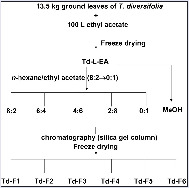
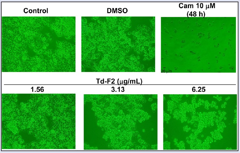
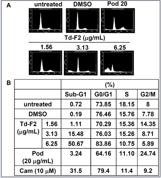
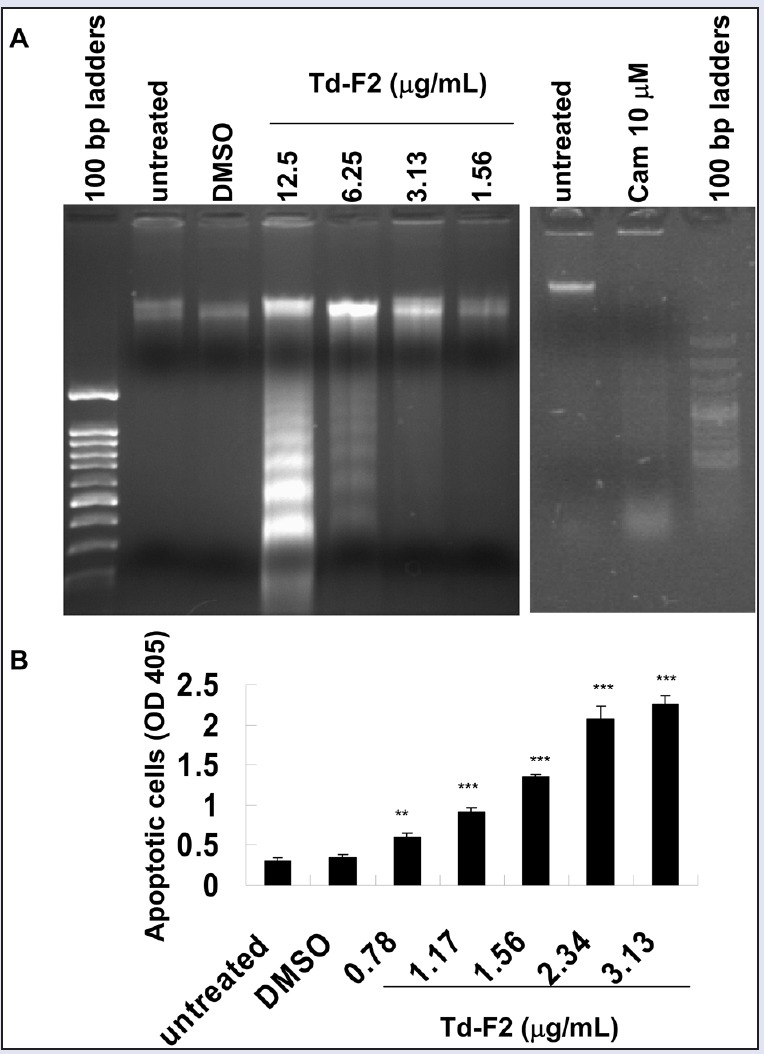
Materials and methods: Dry leaves of T. Diversifolia were first extracted in ethyl acetate, then further fractionated by different ratio of n-hexane-ethyl acetate (8:2→0:1) or methanol as fractions 1-6 (Td-F1 to Td-F6), respectively. We first showed that the ethyl acetate extracts of T. diversifolia leaves (Td-L-EA) exhibits growth inhibition on human hepatoma HepG2 cells. To further check the extracts-induced apoptosis, microscopic observation, fragmented chromosomal DNA electrophoresis, apoptotic DNA-detection ELISA assay, flow cytometry, and Western blot analysis were performed.
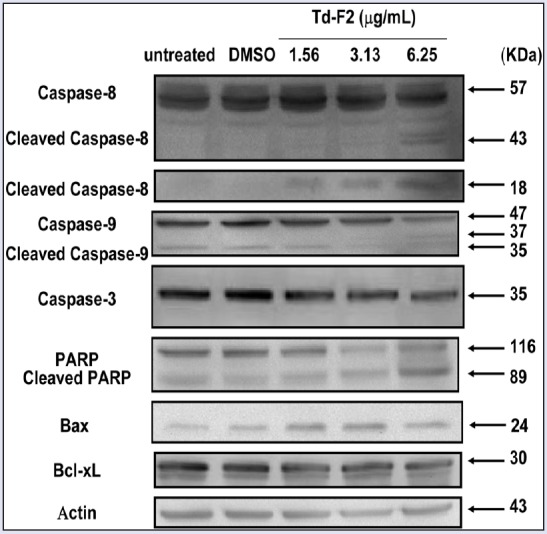
Results: After isolating the effective fractions from Td-L-EA, we found strong cytotoxic effects of fraction-2 (Td-F2). By further analyzing the mechanisms of cytotoxic activities using microscopic observation, fragmented chromosomal DNA electrophoresis, apoptotic DNA-detection ELISA assay, and flow cytometry, we found that induction of apoptosis such as DNA fragmentation increased the apoptosis rate and the apoptosis sub-G1 populations in Td-F2-treated HepG2 cells. In addition, we also confirmed Td-F2-induced degradation of caspase-8, caspase-9, caspase-3, and caspase-3 substrate PARP. Besides, Td-F2 also increased the Bcl-2 proapoptotic family protein Bax expression.
Conclusion: In short, our results clearly showed the induction of apoptosis by ethyl acetate extracts of T. diversifolia leaves in human hepatoma HepG2 cells, suggesting its potential application as an antitumor agent.
Summary: T. Diversifolia leaves were first extracted in ethyl acetate, then further fractionated by different ratio of n-hexane/ethyl acetate (8:2→0:1) or methanol.These extracts exhibit growth inhibition on human hepatoma (HCC) HepG2 cells.n-Hexane/ethyl acetate (6:4) extract (Td-F2) induces apoptosis of HCC. Abbreviations used:T. diversifolia, Tithonia diversifolia; HCC, Hepatocellular carcinoma DMEM, Dulbecco's modified Eagle's medium; DMSO, dimethyl sulfoxide; HRP, horseradish peroxidase; MTT, 3-(4,5-dimethylthiazol-2-yl)-2,5-diphenyl tetrazolium bromide; OD, optical density; SDS-PAGE, sodium dodecylsulfate-polyacrylamide gel electrophoresis; PARP, Poly (ADP-ribose) polymerase; PBS, phosphate buffered saline; PI, propidium iodide.
Keywords: Tithonia diversifolia; apoptosis; caspase; hepatocellular carcinoma.
Conflict of interest statement
There are no conflicts of interest.
Figures

Similar articles
-
Miltirone induces cell death in hepatocellular carcinoma cell through GSDME-dependent pyroptosis.Acta Pharm Sin B. 2020 Aug;10(8):1397-1413. doi: 10.1016/j.apsb.2020.06.015. Epub 2020 Jul 2. Acta Pharm Sin B. 2020. PMID: 32963939 Free PMC article.
-
In vitro Anti-Proliferative Effect of Tephrosia purpurea on Human Hepatocellular Carcinoma Cells.Pharmacogn Mag. 2017 Jan;13(Suppl 1):S16-S21. doi: 10.4103/0973-1296.203981. Epub 2017 Apr 7. Pharmacogn Mag. 2017. PMID: 28479720 Free PMC article.
-
Flavonoids Derived from Abelmoschus esculentus Attenuates UV-B Induced Cell Damage in Human Dermal Fibroblasts Through Nrf2-ARE Pathway.Pharmacogn Mag. 2016 May;12(Suppl 2):S129-38. doi: 10.4103/0973-1296.182175. Epub 2016 May 11. Pharmacogn Mag. 2016. PMID: 27279697 Free PMC article.
-
Anti-Cancer Effects of Queen Bee Acid (10-Hydroxy-2-Decenoic Acid) and Its Cellular Mechanisms against Human Hepatoma Cells.Molecules. 2023 Feb 19;28(4):1972. doi: 10.3390/molecules28041972. Molecules. 2023. PMID: 36838959 Free PMC article.
-
Characterization of the Phenolic Compound, Gallic Acid from Sansevieria roxburghiana Schult and Schult. f. Rhizomes and Antioxidant and Cytotoxic Activities Evaluation.Pharmacogn Mag. 2017 Oct;13(Suppl 3):S693-S699. doi: 10.4103/pm.pm_497_16. Epub 2017 Oct 11. Pharmacogn Mag. 2017. PMID: 29142435 Free PMC article.
Cited by
-
Ethnobotanical Survey on Bitter Tea in Taiwan.Front Pharmacol. 2022 Feb 18;13:816029. doi: 10.3389/fphar.2022.816029. eCollection 2022. Front Pharmacol. 2022. PMID: 35250565 Free PMC article.
References
-
- Zhao G, Li X, Chen W, Xi Z, Sun L. Three new sesquiterpenes from Tithonia diversifolia and their anti-hyperglycemic activity. Fitoterapia. 2012;83:1590–7. - PubMed
-
- Kuroda M, Yokosuka A, Kobayashi R, Jitsuno M, Kando H, Nosaka K, et al. Sesquiterpenoids and flavonoids from the aerial parts of Tithonia diversifolia and their cytotoxic activity. Chem Pharm Bull (Tokyo) 2007;55:1240–4. - PubMed
-
- Lin HR. Sesquiterpene lactones from Tithonia diversifolia act as peroxisome proliferator-activated receptor agonists. Bioorg Med Chem Lett. 2012;22:2954–8. - PubMed
-
- Chagas-Paula DA, Oliveira RB, da Silva VC, Gobbo-Neto L, Gasparoto TH, Campanelli AP, et al. Chlorogenic acids from Tithonia diversifolia demonstrate better anti-inflammatory effect than indomethacin and its sesquiterpene lactones. J Ethnopharmacol. 2011;136:355–62. - PubMed
-
- Hebeda CB, Bolonheis SM, Nakasato A, Belinati K, Souza PD, Gouvea DR, et al. Effects of chlorogenic acid on neutrophil locomotion functions in response to inflammatory stimulus. J Ethnopharmacol. 2011;135:261–9. - PubMed
LinkOut - more resources
Full Text Sources
Other Literature Sources
Research Materials
Miscellaneous