. 2017 Nov 14:8:1533.
doi: 10.3389/fimmu.2017.01533. eCollection 2017.

Tumor Necrosis Factor-Alpha Targeting Can Protect against Arthritis with Low Sensitization to Infection

Affiliations

Tumor Necrosis Factor-Alpha Targeting Can Protect against Arthritis with Low Sensitization to Infection

Nadia Belmellat et al. Front Immunol. .

Abstract

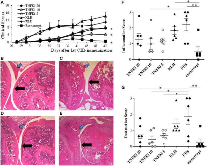
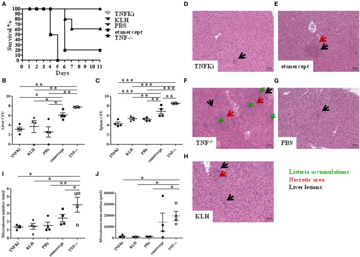
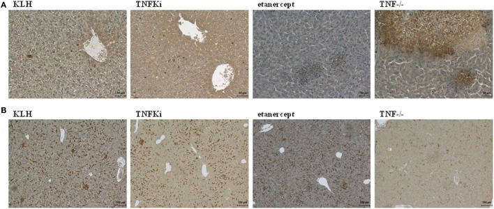
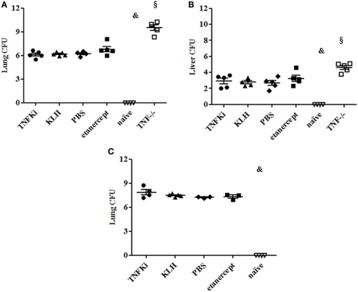
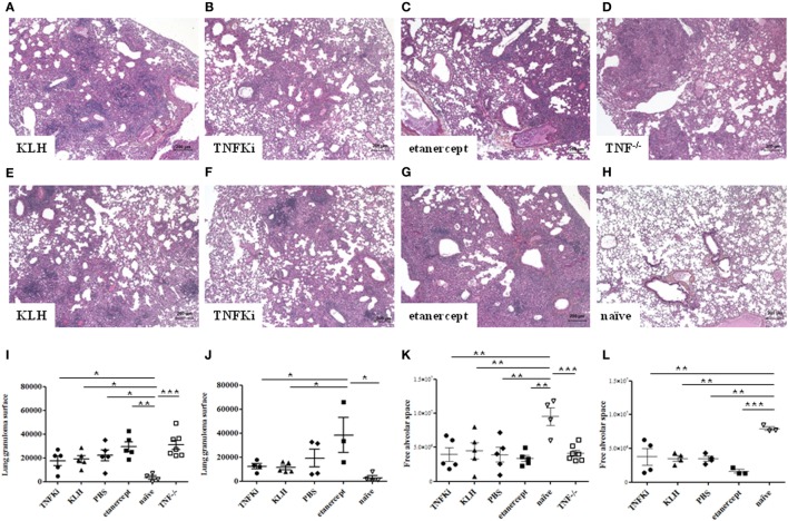
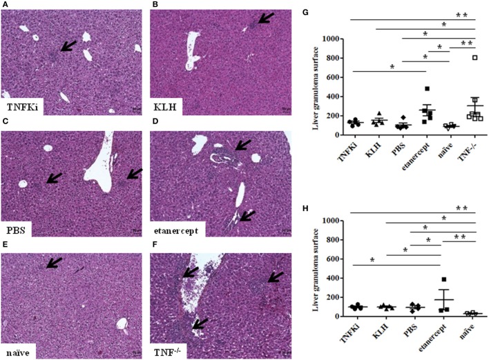
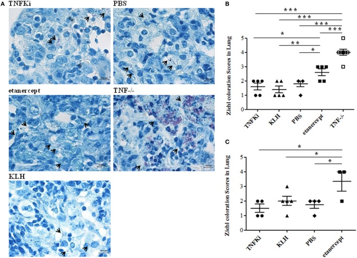
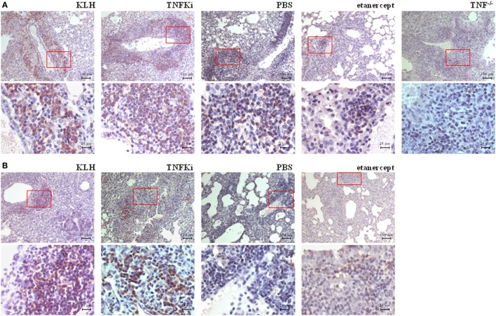
Tumor necrosis factor-alpha (TNF-α) blockade is an effective treatment for rheumatoid arthritis (RA) and other inflammatory diseases, but in patients, it is associated with reduced resistance to the infectious agents Mycobacterium tuberculosis and Listeria monocytogenes, among others. Our goal was to model infection and arthritis in mice and to compare etanercept, a currently used anti-TNF-α inhibitor, to an anti-TNF-α vaccine. We developed a murine surrogate of the TNF-α kinoid and produced an anti-murine TNF-α vaccine (TNFKi) composed of keyhole limpet hemocyanin conjugated to TNF-α, which resulted in anti-TNF-α antibody production in mice. We also used etanercept (a soluble receptor of TNF commonly used to treat RA) as a control of TNF neutralization. In a mouse model of collagen-induced arthritis, TNFKi protected against inflammation similar to etanercept. In a mouse model of acute L. monocytogenes infection, all TNFKi-treated mice showed cleared bacterial infection and survived, whereas etanercept-treated mice showed large liver granulomas and quickly died. Moreover, TNFKi mice infected with the virulent H37Rv M. tuberculosis showed resistance to infection, in contrast with etanercept-treated mice or controls. Depending on the TNF-α blockade strategy, treating arthritis with a TNF-α inhibitor could result in a different profile of infection suceptibility. Our TNFKi vaccine allowed for a better remaining host defense than did etanercept.

Keywords: host-defense; infection; rheumatoid arthritis; tumor necrosis factor; vaccine.


Figures

Figure 1
Figure 1
TNFKi protects mice against clinical and histological arthritis. DBA/1 mice were vaccinated three times with keyhole limpet hemocyanin (KLH) or differents doses of TNFKi (20, 10, 5 µg) at days −21, −7, 7 before and during collagen-induced arthritis (CIA) induced by two injections of CIIb (50 μg/mice; day 0 and 21). Treatment with etanercept (30 mg/kg, two times/week) or phosphate-buffered saline (PBS) began from day 22. (A) Clinical arthritis was measured by using the sum of arthritis scores (0–4) from four paws (n = 6/group). Data are mean ± SEM. *p < 0.05 vs. both PBS and KLH during the whole experiment by ANOVA. The area under the curve (AUC) of clinical scores for each animal are the raw value. (B–E) Histology of paws for treatment groups. (B) TNFKi-20, (C) TNFKi-10, (D) TNFKi-5 (20, 10, 5 µg, respectively), (E) KLH shows inflammatory synovitis (blue arrow) and articular surface (black arrow). (F) Histological inflammation score and (G) joint destruction in treatment groups. Horizontal bar is mean, outer bars are SEM, and whiskers are range. *p < 0.05 for TNFKi and **p < 0.01 for etanercept vs PBS (Newman–Keuls test with subsequent post hoc comparisons). CIA inhibition with TNFKi was observed in three independent experiments.
Figure 2
Figure 2
TNFKi vaccination does not alter survival, organ infiltration, and liver lesions during Listeria monocytogenes infection. Mice (n = 10 per group) were treated with TNFKi or keyhole limpet hemocyanin (KLH) (at days −44, −31, −17, −4) or given phosphate-buffered saline (PBS) or 30 mg/kg etanercept (days −4, −2, 0, and 3). TNF−/− mice were a genetic control. All mice were infected with 104 colony forming units (CFU) Listeria at day 0. 4 mice per group were euthanized for bacterial burden and histological analysis at day 4. Survival was evaluated for 11 days postinfection with the remaining mice (A). All TNF−/− mice (n = 6) died at 5 days postinfection. Four etanercept-treated mice died on day 6 (n = 5) and two mice treated with KLH (n = 5). All mice treated with TNFKi (n = 6) and PBS (n = 6) survived during this period. Survival of TNF−/− and etanercept groups was significantly reduced as compared with PBS, TNFKi, and KLH mice (Logrank test for trend, p < 0.0001). Colony-forming units (CFUs) evaluated in liver (B) and spleen (C) 4 days after infection in mice (*p < 0.05; **p < 0.01, ***p < 0.001, ANOVA). Liver lesions were studied 4 days after infection with Listeria after staining sections with hematoxylin-eosin. Liver sections (D–H) showing size and number of lesions (black arrow), with necrotic areas (red arrow) and Listeria accumulation (green arrow). Quantification of hepatic lesion number (I) and surface area (j) in slides involved use of NDPview software (Hamamatsu, Japan) (*p < 0.05, **p < 0.01, ANOVA).
Figure 3
Figure 3
TNFKi vaccination does not alter neutrophils and macrophages infiltration of livers during Listeria monocytogenes infection. Immunohistochemical staining of neutrophils expressing Ly6G (A) and macrophages expressing F4/80 (B) after 4 days of infection [scale bar (A) 50 µm; scale bar (B) 100 µm].
Figure 4
Figure 4
Organ weights during early and established M. tuberculosis infection. C57Bl/6 mice were vaccinated with keyhole limpet hemocyanin (KLH) (n = 10) or 10 µg TNFKi (n = 10) at days −44, −31, −17, and −4 before infection. One group of mice received 30 mg/kg of etanercept (n = 10) twice a week from day 4 to day 52. Except for naïve mice (n = 5), all treated mice and TNF−/− mice (n = 7) were infected intranasally with 2,250 CFU of M. tuberculosis on day 0. Five mice per treatment (day 28) and all TNF−/− mice (day 25) were euthanized and lung, spleen, and liver were weighed. Survival was monitored until day 56 (4–5 mice/group) and organs were weighed at euthanasia. Relative organ weights were calculated as the ratio of absolute organ weights to body weights. Organ weights of mice during early infection (A–C) and established infection (D–F). Organ weights were expressed as % of body weight. Horizontal bar is mean, outer bars are SEM, and whiskers are range. *p < 0.05 vs TNFKi, KLH, phosphate-buffered saline (PBS), ***p < 0.001 vs naïve mice; Newman–Keuls test with post hoc comparisons. &p < 0.05 naïve vs TNFKi, KLH, PBS, and etanercept.
Figure 5
Figure 5
TNFKi did not increase bacterial loads in lung and liver of Mycobacterium tuberculosis-infected mice. Vaccination schedule described previously. During early (A,B) and established infection (C), a part of lungs and liver were weighted and used after lysis for bacterial culture and colony formation units (CFU) evaluation. Results are expressed as Log10 of CFU count per organ. CFU number in organs of mice during early infection (A,B) and lung during established (C) infection. Horizontal bar is mean, outer bars are SEM, and whiskers are range (§p < 0.001 TNF−/− vs all groups; Newman–Keuls test with subsequent post hoc comparisons).
Figure 6
Figure 6
Limited effects of TNFKi on lung granuloma formation during Mycobacterium tuberculosis infection. Granulomas were assessed during early (A–D) and established (E–H) infection. Lung sections were stained with hematoxylin-eosin (n = 3–7 per group). Granulomas in lungs during early infection (A–D) and established infection (E–H). Quantification of lung granuloma surface area during early (I) and established infection (J) and alveolar space of lungs during early (K) and established infection (L). Horizontal bar is mean, outer bars are SEM, and whiskers are range (*p < 0.05, **p < 0.01, ***p < 0.001, ANOVA).
Figure 7
Figure 7
TNFKi did not induce larger granuloma formation in liver of Mycobacterium tuberculosis-infected mice. Histology of granulomas (arrows) in liver sections stained with hematoxylin-eosin (n = 3–7 per group) during early infection (A–F). Quantification of hepatic granuloma surface area during early (G) and established infection (H). Horizontal bar is mean, outer bars are SEM, and whiskers are range (*p < 0.05, **p < 0.01, ANOVA).
Figure 8
Figure 8
TNFKi did not increase bacterial load in lungs of Mycobacterium tuberculosis-infected mice. Ziehl–Neelsen coloration showing direct recognition of mycobacteria in lung sections. M. tuberculosis appeared as red bacilli (arrowheads). Examples of images at early phase of infection (A). Quantification of mycobacteria during the early (B) and established infection (C) after blinded observations of three fields per mice (n = 3–7 mice per group) (*p < 0.05 ANOVA). Scale bar, 10 µm.
Figure 9
Figure 9
TNFKi favored B-lymphocyte infiltration in lungs of Mycobacterium tuberculosis-infected mice. Immunohistochemical staining of B lymphocytes expressing CD45R. During early infection (A) and established infection (B), perivascular accumulation of B lymphocytes in lungs of mice. Representative images of 3–7 mice per group. Scale bar, 100 µm. 4× magnification of red boxes below original picture (scale bar, 25 µm).

Similar articles

Cited by

References

    1. Winsauer C, Kruglov AA, Chashchina AA, Drutskaya MS, Nedospasov SA. Cellular sources of pathogenic and protective TNF and experimental strategies based on utilization of TNF humanized mice. Cytokine Growth Factor Rev (2014) 25(2):115–23.10.1016/j.cytogfr.2013.12.005 - DOI - PubMed
    1. Minichiello E, Semerano L, Boissier MC. Time trends in the incidence, prevalence, and severity of rheumatoid arthritis: a systematic literature review. Joint Bone Spine (2016) 83(6):625–30.10.1016/j.jbspin.2016.07.007 - DOI - PubMed
    1. Singh JA, Christensen R, Wells GA, Suarez-Almazor ME, Buchbinder R, Lopez-Olivo MA, et al. A network meta-analysis of randomized controlled trials of biologics for rheumatoid arthritis: a Cochrane overview. CMAJ (2009) 181(11):787–96.10.1503/cmaj.091391 - DOI - PMC - PubMed
    1. Wallis RS. Infectious complications of tumor necrosis factor blockade. Curr Opin Infect Dis (2009) 22(4):403–9.10.1097/QCO.0b013e32832dda55 - DOI - PubMed
    1. Keane J, Gershon S, Wise RP, Mirabile-Levens E, Kasznica J, Schwieterman WD, et al. Tuberculosis associated with infliximab, a tumor necrosis factor-α-neutralizing agent. N Engl J Med (2001) 345(15):1098–104.10.1056/NEJMoa011110 - DOI - PubMed

LinkOut - more resources