. 2018 Jan 12;293(2):662-676.
doi: 10.1074/jbc.M117.802900. Epub 2017 Nov 28.

IL-10 promoter transactivation by the viral K-RTA protein involves the host-cell transcription factors, specificity proteins 1 and 3

Affiliations

IL-10 promoter transactivation by the viral K-RTA protein involves the host-cell transcription factors, specificity proteins 1 and 3

Masanori Miyazawa et al. J Biol Chem. .

Abstract

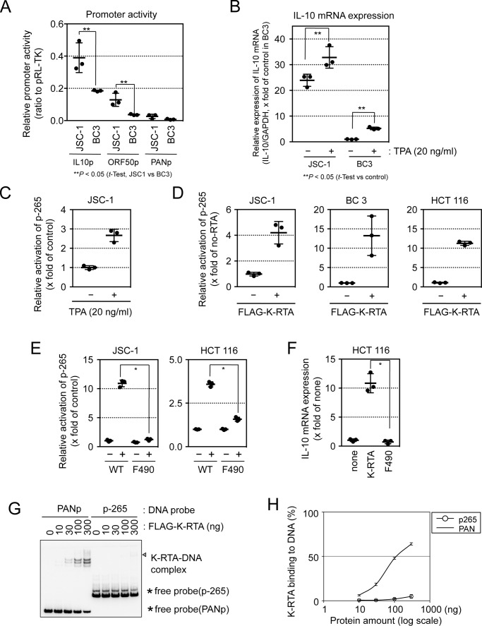
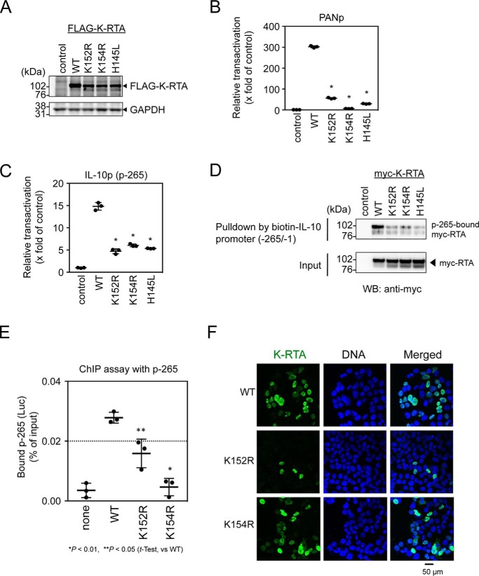
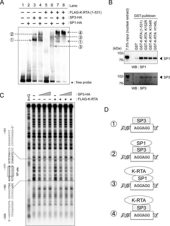
Kaposi's sarcoma-associated herpesvirus (KSHV)/human herpesvirus-8 (HHV-8) causes a persistent infection, presenting latent and lytic replication phases during its life cycle. KSHV-related diseases are associated with deregulated expression of inflammatory cytokines, including IL-6 and IL-10, but the mechanisms underlying this dysregulation are unclear. Herein, we report a molecular mechanism for KSHV-induced IL-10 gene expression. KSHV replication and transcription activator (K-RTA) is a molecular switch for the initiation of expression of viral lytic genes, and we describe, for the first time, that K-RTA significantly activates the promoter of the human IL-10 gene. Of note, mutations involving a basic region of K-RTA reduced the association of K-RTA with the IL-10 promoter. Moreover, the host-cell transcription factors, specificity proteins (SP) 1 and 3, play a pivotal cooperative role in K-RTA-mediated transactivation of the IL-10 promoter. K-RTA can interact with SP1 and SP3 directly in vitro, and electrophoresis mobility shift assays (EMSAs) revealed co-operative interaction involving K-RTA, SP1, and SP3 in binding to the IL-10 promoter. As DNase I footprinting assays indicated that K-RTA did not affect SP3 binding to the IL-10 promoter, SP3 can function to recruit K-RTA to the IL-10 promoter. These findings indicate that K-RTA can directly contribute to IL-10 up-regulation via a functional interplay with the cellular transcription factors SP1 and SP3.

Keywords: IL-10; SP3; herpesvirus; interleukin; specificity protein 1 (Sp1); transactivation; transcription; viral transcription.


Conflict of interest statement

The authors declare that they have no conflicts of interest with the contents of this article

Figures

Figure 1.
Figure 1.
K-RTA transactivates the human IL-10 promoter. A, KSHV-infected PEL cell line JSC-1 and BC3 cells were co-transfected with the reporter plasmids IL-10 p-1000, ORF50, or PANp and pRL-TK vector (internal control). Two days after transfection, a luciferase assay was performed. Results are presented as the mean ± S.D. (n = 3). Student's t test; **, p < 0.05. B, KSHV-infected PEL cell line JSC-1 and BC3 cells were treated with or without TPA (20 ng/ml) for 24 h, and human IL-10 mRNA expression was determined by RT-qPCR. GAPDH mRNA was used to normalize data. Results are presented as the mean ± S.D. (n = 3). Student's t test; **, p < 0.05. C, human IL-10 promoter reporter plasmid p-265 and internal control pRL-TK were transfected into JSC-1 cells. One day later, cells were treated or not with TPA (20 ng/ml) for 24 h. Luciferase activities were determined 2 days after transfection. Results are presented as the mean ± S.D. (n = 3). The activation was statistically significant by Student's t test; p < 0.01. D, three human cell lines, JSC-1, BC3, and HCT 116 cells, were co-transfected with FLAG–K-RTA, IL-10 promoter reporter plasmid p-265, and the internal control pRL-TK plasmid (ratio 1:1:1). Two days after transfection, luciferase activity was examined. Results are presented as the mean ± S.D. (n = 3). The activation was statistically significant by Student's t test; *, p < 0.01. E, human IL-10 promoter reporter plasmid p-265 and internal control pRL-TK were co-transfected into cells with either an empty plasmid (i.e. without K-RTA) or with a plasmid carrying the wild-type K-RTA (WT) or the transactivation domain-deleted K-RTA (F490). Luciferase activity was determined 2 days after transfection. Results are presented as the mean ± S.D. (n = 3). Student's t test; *, p < 0.01. F, HCT 116 cells were transiently transfected with either K-RTA WT or K-RTA F490 expression plasmids. Two days after transfection, mRNA was prepared, and human IL-10 gene expression was measured by RT-qPCR. GAPDH mRNA was used to normalize data. Results are presented as the mean ± S.D. (n = 3). Student's t test. *, p < 0.01. G and H, binding efficiencies of K-RTA to PAN (PANp) or IL-10 (−265/−1 from p-265) promoter DNA probes were analyzed by EMSA. Five-fmol biotin-labeled promoter probes were incubated with increasing amounts of FLAG–K-RTA protein (aa 1–531). The amount of FLAG–K-RTA increased from 0 to 300 ng. A complex of FLAG–K-RTA–DNA probes was examined by EMSA, indicated by the triangle (G). The asterisk indicates free probe DNA. The binding curves were calculated (H).
Figure 2.
Figure 2.
Effect of basic domain of K-RTA on IL-10 promoter activation. A, expression of basic domain mutated K-RTAs (K152R, K154R, and H145L) was analyzed in HCT 116 cells by Western blotting. FLAG-tagged K-RTAs were detected with anti-FLAG antibody. GAPDH is shown as loading control. B and C, HCT 116 cells were co-transfected with the mutant FLAG–K-RTA expression plasmids, promoter reporter plasmid, viral PANp (B) or IL-10 promoter p-265 (C), and internal control pRL-CMV (ratio 1:1:1). A luciferase assay for these mutant K-RTAs was performed. Results are presented as the mean ± S.D. (n = 3). Student's t test; *, p < 0.01 (WT versus mutant). D, loading of mutant K-RTA on IL-10 promoter p-265 was examined by ABCD assay. An extract from cells expressing mutant K-RTAs was subjected to a pulldown assay, using p-265 DNA probe, and p-265-bound K-RTA was detected by Western blotting. Two independent experiments showed similar results. E, occupancy of IL-10 promoter by FLAG–K-RTA was examined by ChIP assay. HEK293 cells were transfected with FLAG–K-RTA-expressing plasmid and p-265 reporter plasmid (ratio 1:1) and 2 days later were fixed with 3.7% formalin. Nuclei were sonicated, and FLAG–K-RTA-bound DNA was immunoprecipitated with anti-FLAG antibody. Eluted DNA was quantified by qPCR using primers targeting p-265. Results are presented as the mean ± S.D. (n = 3). Student's t test; *, p < 0.01, and **, p < 0.05 (WT versus mutant). F, subcellular locations of mutant K-RTAs (K152R and K154R) were examined by indirect immunofluorescent microscopic analysis. FLAG–K-RTAs (green) were detected by anti-FLAG antibody, and nuclei were stained with DAPI (blue). Representative images are shown. Scale bar, 50 μm.
Figure 3.
Figure 3.
Mutation analysis of IL-10 promoter. A, schematic representation of putative transcription factor-binding sites in the fragment from nucleotides −265 to −1 of the human IL-10 promoter. The binding elements for transcription factors are shown in dotted boxes, and mutation sites are shown in capital letters. B, HCT 116 cells were co-transfected with the FLAG–K-RTA–expressing plasmid, internal control pRL-CMV, and mutant p-265 plasmid (as illustrated in A) (ratio 1:1:1), and luciferase assays were performed 2 days later. Basal activity of wild-type p-265 reporter plasmid without K-RTA (in empty box) was used as relative control 1. Results are presented as the mean ± S.D. (n = 3). Student's t test; *, p < 0.01 (WT versus mutant p-265). C, SP-binding site-dependent K-RTA loading onto the IL-10 promoter was determined by ABCD assay. Nuclear extracts (100 μg) from myc-K-RTA–expressing HEK293 cells were incubated with biotin-labeled IL-10 promoter probe (from either wild type or mutated mSP p-265). Myc-K-RTA co-precipitating with biotinylated DNA probes was detected by Western blotting (upper panel). Signal intensity was quantified from triplicate Western blotting experiments and calculated as p-265-bound K-RTA/input K-RTA ratio (lower plot). Student's t test; **, p < 0.05 (WT versus mSP). D, WT or mutant IL-10 promoter occupancy by K-RTA was analyzed by ChIP assay. HCT 116 cells were co-transfected with FLAG–K-RTA–expressing plasmid and p-265 (either WT or mSP) reporter plasmid (ratio 1:1) and 2 days later were fixed with 3.7% formalin. Nuclei were sonicated, and FLAG–K-RTA-bound DNA was immunoprecipitated with anti-FLAG antibody. Eluted DNA was quantified by qPCR, using primers targeting p-265. Results are presented as the mean ± S.D. (n = 3). Student's t test; *, p < 0.01.
Figure 4.
Figure 4.
K-RTA–mediated IL-10 promoter activation involves SP1 and SP3. A, dose-dependent increase of K-RTA–mediated IL-10 promoter activation by SP1 and by SP3. HEK293 cells (1.5 × 10⁵ cells/well) were co-transfected with increasing amounts of SP1 or SP3 expression plasmid (0, 25, 50, 100, 200, and 400 ng), 100 ng of K-RTA–expressing plasmid, 100 ng of p-265 reporter plasmid, and 30 ng of pRL-CMV vector (internal control). Luciferase activity was measured 2 days later. Results are presented as the mean ± S.D. (n = 3). Student's t test; *, p < 0.01 (K-RTA only versus +SP1 or +SP3). Western blotting data for the expression of SP1-HA, SP3-HA, and FLAG–K-RTA are shown in the lower panels. B, knockdown of SP1 and SP3 by siRNA. HeLa cells were transfected with 5 nm SP1- or SP3-specific siRNA or with a scrambled siRNA. Knockdown of SP1 and SP3 proteins was confirmed by Western blotting at 72 h post-transfection. GAPDH was used as loading control. C, effect of SP1 and SP3 knockdown on K-RTA–mediated IL-10 promoter activation. HeLa cells were transfected with siRNA as described in C. Twenty four hours later, HeLa cells were co-transfected with the K-RTA–expressing plasmid, the reporter plasmid p-265, and the internal control pRL-CMV (ratio 1:1:1). Luciferase activity was measured after 2 days. Results are presented as the mean ± S.D. (n = 3). Student's t test. *, p < 0.01 (scramble versus SP1 or SP3).
Figure 5.
Figure 5.
Superior SP3 binding to IL-10 promoter in vitro. A, ABCD assay was employed to detect in vitro IL-10 promoter association of K-RTA. Increasing amounts of nuclear extracts (NE) from myc-K-RTA–expressing HEK293 cells were incubated with biotin-labeled DNA probes from either wild type (WT) or SP-binding site-mutated (mSP) IL-10 promoter (fragment from nucleotides −265 to −1). Myc-K-RTA and endogenous SP1 and SP3 co-precipitating with biotin-DNA probes were detected by Western blotting. Myc-K-RTA and SP3 co-precipitated with p-265 DNA probe in a nuclear extract dose-dependent manner. Two independent experiments showed similar results. B, SP1-HA and SP3-HA were expressed in HEK293 cells and immunopurified using anti-HA-agarose. Purified SP1-HA and SP3-HA were visualized by silver staining after SDS-PAGE. An eluted sample from an empty plasmid-transfected lysate was used as control; the molecular mass marker is shown (left lane, Marker). C and D, binding affinities of SP1-HA and SP3-HA to the IL-10 promoter were analyzed by EMSA. The biotin-labeled IL-10 promoter probes (fragment from nucleotides −265 to −1) were incubated with increasing amounts of SP1-HA (left lanes) and SP3-HA (right lanes). The amount of SP1-HA and SP3-HA increased from 0 to 15 ng. The triangle indicates SP3–DNA complex, and the asterisk indicates free DNA probe (C). EMSA experiments were performed three times, and the binding curves and the Kd values for SP3-HA on the IL-10 promoter probe were determined as in Fig. 1H (D). E and F, binding affinities of SP3-HA to wild type (WT p-265; left lanes) or SP3-binding site-mutated (mSP; right lanes) IL-10 promoter were analyzed by EMSA. The biotin-labeled IL-10 promoter probes (fragment from nucleotides −265 to −1) were incubated with increasing amounts of SP3-HA. The amount of SP3-HA increased from 0 to 15 ng. The triangle indicates SP3–DNA complex, and the asterisk indicates free DNA probe (E). EMSA experiments were performed three times, and the binding curve of WT is identical to that of SP3 in D (F).
Figure 6.
Figure 6.
Co-operative occupancy of IL-10 promoter by K-RTA, SP1, and SP3. A, immunopurified FLAG–K-RTA (aa 1–531), SP1-HA, and SP3-HA proteins from HEK293 cells were incubated with biotin-labeled IL-10 promoter probe (fragment from nucleotides −265 to −1) in the indicated combinations, and specific protein–DNA complexes were analyzed by EMSA. Gray arrows indicate protein–DNA complexes. Specific protein–DNA complexes are numbered as 1–4. Two independent experiments showed similar results. B, GST-pulldown experiments. Wild-type or mutant GST-K-RTA (aa 1–531) proteins were incubated with nuclear extracts, and the co-precipitated endogenous SP1 or SP3 was subsequently detected by Western blotting (WB). Two independent experiments showed similar results. C, DNase I footprinting assay with the IL-10 promoter DNA probe (−206/−72) labeled with [32P]dATP at the 3′ terminus. The 32P-labeled DNA probe was incubated with either immunopurified FLAG–K-RTA (0 or 2000 ng) and increasing amounts of SP3-HA (0, 55, 110, and 220 ng) An autoradiogram produced after gel electrophoresis of DNase I-digested samples is shown. A+G is DNA marker ladder by the Maxam-Gilbert A+G reaction. Boxes around predicted SP-binding site (−177/−172) and SP3-bound footprint are indicated. D, possible models of K-RTA recruitment to the IL-10 promoter. The protein–DNA complex of K-RTA, SP1, and SP3, as shown in A, may regulate IL-10 promoter transactivation. Specific protein–DNA complexes numbered as 1–4 correspond to those in A.

Similar articles

Cited by

References

    1. Chang Y., Cesarman E., Pessin M. S., Lee F., Culpepper J., Knowles D. M., and Moore P. S. (1994) Identification of herpesvirus-like DNA sequences in AIDS-associated Kaposi's sarcoma. Science 266, 1865–1869 10.1126/science.7997879 - DOI - PubMed
    1. Mesri E. A., Cesarman E., and Boshoff C. (2010) Kaposi's sarcoma and its associated herpesvirus. Nat. Rev. Cancer 10, 707–719 10.1038/nrc2888 - DOI - PMC - PubMed
    1. Boshoff C., and Weiss R. A. (1998) Kaposi's sarcoma-associated herpesvirus. Adv. Cancer Res. 75, 57–86 10.1016/S0065-230X(08)60739-3 - DOI - PubMed
    1. Boshoff C., and Chang Y. (2001) Kaposi's sarcoma-associated herpesvirus: a new DNA tumor virus. Annu. Rev. Med. 52, 453–470 10.1146/annurev.med.52.1.453 - DOI - PubMed
    1. Deng H., Liang Y., and Sun R. (2007) Regulation of KSHV lytic gene expression. Curr. Top. Microbiol. Immunol. 312, 157–183 - PubMed

Publication types

MeSH terms

LinkOut - more resources