. 2018 Jan 8;145(1):dev156588.
doi: 10.1242/dev.156588.

The skeletal phenotype of achondrogenesis type 1A is caused exclusively by cartilage defects

Affiliations

The skeletal phenotype of achondrogenesis type 1A is caused exclusively by cartilage defects

Ian M Bird et al. Development. .

Abstract

Inactivating mutations in the ubiquitously expressed membrane trafficking component GMAP-210 (encoded by Trip11) cause achondrogenesis type 1A (ACG1A). ACG1A is surprisingly tissue specific, mainly affecting cartilage development. Bone development is also abnormal, but as chondrogenesis and osteogenesis are closely coupled, this could be a secondary consequence of the cartilage defect. A possible explanation for the tissue specificity of ACG1A is that cartilage and bone are highly secretory tissues with a high use of the membrane trafficking machinery. The perinatal lethality of ACG1A prevents investigating this hypothesis. We therefore generated mice with conditional Trip11 knockout alleles and inactivated Trip11 in chondrocytes, osteoblasts, osteoclasts and pancreas acinar cells, all highly secretory cell types. We discovered that the ACG1A skeletal phenotype is solely due to absence of GMAP-210 in chondrocytes. Mice lacking GMAP-210 in osteoblasts, osteoclasts and acinar cells were normal. When we inactivated Trip11 in primary chondrocyte cultures, GMAP-210 deficiency affected trafficking of a subset of chondrocyte-expressed proteins rather than globally impairing membrane trafficking. Thus, GMAP-210 is essential for trafficking specific cargoes in chondrocytes but is dispensable in other highly secretory cells.

Keywords: Achondrogenesis type 1A; Cartilage; Conditional knockout; GMAP-210; Golgin; Proteomics.


Conflict of interest statement

Competing interestsThe authors declare no competing or financial interests.

Figures
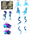
Fig. 1.
Fig. 1.
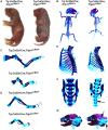
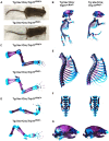
Mice homozygous for the recombined Trip11 conditional allele (Trip11) have a severe skeletal dysplasia. (A) E17.5 wild-type (Trip11+/+) and recombined (Trip11−/−) embryos. Note the short limbs, short snout, domed skull, protruding tongue and omphalocele in the recombined (i.e. knockout) embryo. (B-G) Skeletal preparations of the embryos shown in A. Compared with the Trip11+/+ embryo, the Trip11−/− embryo has: (B) a smaller ribcage; (C) decreased mineralization of the calvarium (arrow); (D) absent mineralization of the sternum (arrow); (E) absent mineralization of the vertebral bodies (arrow); (F) short forelimbs; and (G) short hindlimbs. n=3; one representative result is shown.
Fig. 2.
Fig. 2.
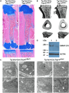
Swelling of ER cisternae and disruption of the Golgi stack structure in chondrocytes of mice homozygous for a recombined Trip11 conditional allele (Trip11). (A) Alcian Blue-stained sections through the humeri of E15.5 wild-type (Trip11+/+) and knockout (Trip11−/−) embryos. Note the delay in the formation of the primary ossification center in the knockout. Bottom panels: higher magnification of columnar chondrocytes. Note the swollen appearance of chondrocytes in the knockout. (B) Transmission electron microscopy pictures of epiphyseal chondrocytes from the humeri of E15.5 wild-type and knockout embryos (original magnifications: top, 2900×; bottom, 9300×). Note the increased size of ER cisternae in the knockout chondrocytes (top panel, arrow) and the disruption of the Golgi stack structure (bottom panels, arrow). (C) Western blot with a GMAP-210-specific antibody on the cell lysates of mouse embryonic fibroblasts extracted from E13.5 wild-type, Trip11−/+ and knockout embryos. Note the complete absence of GMAP-210 protein in the knockout (−/−) cell lysate. n=3; one representative result is shown.
Fig. 3.
Fig. 3.
Inactivation of Trip11 in chondrocytes recapitulates the skeletal dysplasia seen in germline knockout mice. (A) Control (Tg:Col2a1-Cre;Trip11cko/+) and chondrocyte knockout (Tg:Col2a1-Cre;Trip11cko/−) newborn pups. Note the short snout, domed skull and short limbs in the chondrocyte knockout. (B-G) Skeletal preparations of the pups shown in A. Compared with the control embryo, the embryo with the chondrocyte-specific deletion of Trip11 has: short (C) forelimbs and (D) hindlimbs; (E) a small ribcage; (F) delayed mineralization of the vertebral body (arrow); and (G) decreased mineralization of the skull (arrows). n=3; one representative result is shown.
Fig. 4.
Fig. 4.
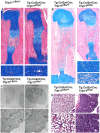
Specific inactivation of Trip11 in chondrocytes causes swelling of ER cisternae and disruption of the Golgi stack structure. (A) Alcian Blue-stained sections through the humeri of E15.5 control (Trip11cko/+) and chondrocyte knockout (Tg:Col2a1-Cre;Trip11cko/−) embryos. Bottom panels show higher magnification of columnar chondrocytes, indicating the delay in the formation of the primary ossification center and the swollen appearance of some chondrocytes in the humerus of the chondrocyte knockout (arrow). (B) Alcian Blue staining of sections through the humerus of P0 control (Tg:Col2a1-Cre;Trip11cko/+) and chondrocyte knockout (Tg:Col2a1-Cre;Trip11cko/−) pups. Lower right panels show higher magnification of columnar chondrocytes. Note the swollen appearance of chondrocytes in the humerus of the chondrocyte knockout. (C) Transmission electron microscopy pictures of epiphyseal chondrocytes from the humerus of P0 control (Tg:Col2a1-Cre;Trip11cko/+) and chondrocyte knockout (Tg:Col2a1-Cre;Trip11cko/−) pups (original magnifications: top, 2900×; bottom, 9300×). Note the increased size of ER cisternae in the chondrocyte knockout (ER) and the disruption of the Golgi stack structure (arrow). (D) Hematoxylin staining of sections through the lungs of P0 control (Tg:Col2a1-Cre;Trip11cko/+) and chondrocyte knockout (Tg:Col2a1-Cre;Trip11cko/−) pups. Higher magnification of the alveoli is shown in the bottom panels. Note the impaired alveolar development in the mutant lung. n=3; one representative result is shown.
Fig. 5.
Fig. 5.
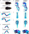
Specific inactivation of Trip11 in osteoblasts does not result in a skeletal dysplasia. (A) Five-month-old control (Tg:Bglap-Cre;Trip11cko/+;ROSA26mTmG/+) and osteoblast knockout (Tg:Bglap-Cre;Trip11cko/−;ROSA26mTmG/+) mice are indistinguishable. (B-G) Skeletal preparations of E17.5 control (Tg:Bglap-Cre;Trip11cko/+;ROSA26mTmG/+) and osteoblast knockout (Tg:Bglap-Cre;Trip11cko/−;ROSA26mTmG/+) embryos. (B) Whole skeletal preparations show no size difference between control and osteoblast knockouts. (C-G) Higher magnifications of the forelimbs (C), hindlimbs (D), sternums (E), lumbar vertebrae (F) and skulls (G) showing no differences between control and osteoblast knockout mice. n=3; one representative result is shown.
Fig. 6.
Fig. 6.
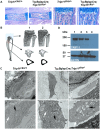
Specific inactivation of Trip11 in osteoblasts does not result in swelling of ER cisternae in their osteocyte descendants, but does disrupt Golgi stack structure. (A) Alcian Blue-stained sections through the humeri of P0 control (Trip11cko/+;ROSA26mTmG/+) and osteoblast knockout (Tg:Bglap-Cre;Trip11cko/−;ROSA26mTmG/+) littermates. Left: low magnification of the primary ossification centers. Right: higher magnification of the primary spongosia of the proximal growth plates. No differences between control and osteoblast knockout are observed. (B) µCT of the tibias of 6-week-old control (1: Trip11cko/+;ROSA26mTmG/+) and osteoblast knockout (2: Tg:Bglap-Cre;Trip11cko/−;ROSA26mTmG/+) mice shows the mice have similar trabecular and cortical bone architecture. (C) Transmission electron microscopy pictures of calvarial osteocytes from P5 control (Trip11cko/+;ROSA26mTmG/+) and osteoblast knockout (Tg:Bglap-Cre;Trip11cko/−;ROSA26mTmG/+) pups. Original magnifications: 2900× (top); 9300× (bottom). Note the absence of swelling of ER cisternae in control and mutant osteocytes (white arrows), but that Golgi cisternae (black arrows) are increased in size and not organized into stacks in the mutant. (D) Western blot analysis of lysates from GFP-sorted primary calvarial outgrowth cultures established from 5-day-old control (Tg:Bglap-Cre;Trip11cko/+;ROSA26mTmG/+) and osteoblast knockout (Tg:Bglap-Cre;Trip11cko/−;ROSA26mTmG/+) mice. (Lane 1) GFP-negative control cells; (lane 2) GFP-positive control cells; (lane 3) GFP-negative osteoblast knockout cells; and (lane 4) GFP-positive osteoblast knockout cells. Note the absence of GMAP-210 protein in GFP-positive osteoblast knockout cells. Re-probing of the western blot with an anti-actin antibody serves as a loading control. n=3; one representative result is shown.
Fig. 7.
Fig. 7.
Specific inactivation of Trip11 in the hematopoietic lineage does not affect viability or cause skeletal dysplasia. (A) Eight-week-old male control (Tg:Vav1-Cre;Trip11cko/+;ROSA26mTmG/+) and blood cell and osteoclast knockout (Tg:Vav1-Cre;Trip11cko/−;ROSA26mTmG/+) mice. No dwarfism is present in the blood cell and osteoclast knockout animal. (B-G) Skeletal preparations of P0 control (Tg:Vav1-Cre;Trip11cko/+;ROSA26mTmG/+) and blood cell and osteoclast knockout (Tg:Vav1-Cre;Trip11cko/−;ROSA26mTmG/+) newborns. (B) Whole skeletal preparations showing the mice are indistinguishable. (C-G) Higher magnifications of the: (C) forelimbs, (D) hindlimbs, (E) sternums, (F) lumbar vertebrae and (G) skulls showing no differences between control and blood cell and osteoclast knockout mice. n=3; one representative result is shown.
Fig. 8.
Fig. 8.
Inactivation of Trip11 in osteoclasts does not result in swelling of ER cisternae and does not disrupt Golgi stack structure. (A) Alcian Blue-stained sections through the humeri of P0 control (Tg:Vav1-Cre;Trip11cko/+;ROSA26mTmG/+) and blood cell and osteoclast knockout (Tg:Vav1-Cre;Trip11cko/−;ROSA26mTmG/+) pups. Bottom: higher magnification of the primary spongosia of the proximal growth plates. No differences between mutant and control can be observed. (B) µCT of the tibias of 8-week-old control (Tg:Vav1-Cre;Trip11cko/+;ROSA26mTmG/+) and mutant (Tg:Vav1-Cre;Trip11cko/−;ROSA26mTmG/+) mice. No differences in the density of trabeculae or in the thickness of the bone collar can be observed between mutant and control mice. (C) Transmission electron microscopy pictures of tibial osteoclasts from 8-week-old control, and blood cell and osteoclast knockout mice. Original magnifications: 2900× (left); 9300× (right) Note the presence of normal ER cisternae (arrowheads, top panels) and normal Golgi (arrowheads, bottom panels) in both mice. (D) Western blot analysis of lysates of isolated bone marrow hematopoietic precursor cells from 8-week-old control (lane 1), and blood cell and osteoclast knockout (lane 2) mice. Note the absence of GMAP-210 protein in the knockout mice. Re-probing of the western blot with an anti-actin antibody serves as a loading control. n=3; one representative result is shown.
Fig. 9.
Fig. 9.
Ex vivo inactivation of Trip11 in primary chondrocyte pellet cultures. (A) Western blot of cell lysates from 4-OH tamoxifen-treated primary chondrocyte pellet cultures using antibodies against perlecan (HSPG2) and GMAP-210. Lysates come from control (lane 1, Trip11cko/cko;ROSA26mTmG/+) and from two different induced Trip11 knockout (lanes 2 and 3) pellet cultures: (lane 2) Tg:CagCre/Esr1;Trip11cko/cko;ROSA26mTmG/+ and (lane 3) Tg:CagCre/Esr1;Trip11cko/−;ROSA26mTmG/+. Note the reduction in immunodetectable GMAP-210 and the increase in HSPG2 when Trip11 was inactivated. Re-probing of the western blot with an anti-actin antibody serves as a loading control. (B) Alcian Blue-stained sections through 4-OH tamoxifen-treated control (Tg:CagCre/Esr1;Trip11cko/+;ROSA26mTmG/+) and Trip11-inactivated (Tg:CagCre/Esr1;Trip11cko/−;ROSA26mTmG/+) primary chondrocyte pellet cultures. Both control and Trip11-inactivated cultures produce abundant extracellular matrix. (C) Representative electron microscopy images of 4-OH tamoxifen-treated control and Trip11-inactivated primary chondrocyte pellet cultures. Original magnification: 4800× (top). Note both chondrocytes have enlarged endoplasmic reticulum (ER) cisternae. Original magnification: 9300× (bottom). Note the Golgi apparatus (white arrows) is enlarged and more disorganized in the Trip11-inactivated chondrocytes. (D) Abundance of GMAP-210 and COL10A1 in lysates from 4-OH tamoxifen-treated control (red) and Trip11 inactivated (yellow) primary chondrocyte pellet cultures, as determined by tandem tag mass spectroscopy. Student's t-test adjusted P-value. Data are mean±s.d. TMT-RA, tandem mass tag relative abundance. (E) Graph depicting extracellular matrix proteins whose intracellular abundance increased or decreased in 4-OH tamoxifen-treated Trip11-inactivated (yellow; Tg:CagCre/Esr1;Trip11cko/−;ROSA26mTmG/+) versus control (red; (Trip11cko/cko;ROSA26mTmG/+) primary chondrocyte pellet cultures, as determined by tandem tag mass spectroscopy. CHADL, chondro-adherin like; HSPG2, perlecan; COL9A2, type 9 collagen α2 chain; ACAN, aggrecan; MATN4, matrillin 4; NID2, nidogen 2; NID1, nidogen 1; DCN, decorin. Data are mean±s.d. n values are in parentheses. Student's t-test adjusted P values and Benjamini-Hochberg FDR (B/H) (mutiple test) corrected P values are shown. GMAP-210, COL10A1 and CHADL are significantly different from control.

Similar articles

Cited by

References

    1. American Thoracic Society (2004). Mechanisms and limits of induced postnatal lung growth. Am. J. Respir. Crit. Care Med. 170, 319-343. 10.1164/rccm.200209-1062ST - DOI - PubMed
    1. Apodaca G. and Brown W. J. (2014). Membrane traffic research: challenges for the next decade. Front. Cell Dev. Biol. 2, 52 10.3389/fcell.2014.00052 - DOI - PMC - PubMed
    1. Balderhaar H. J. and Ungermann C. (2013). CORVET and HOPS tethering complexes - coordinators of endosome and lysosome fusion. J. Cell Sci. 126, 1307-1316. 10.1242/jcs.107805 - DOI - PubMed
    1. Benjamini Y. and Hochberg Y. (1995). Controlling the false discovery rate: a practical and powerful approach to multiple testing. J. R. Statist. Soc. B 57, 289-300.
    1. Bonifacino J. S. and Glick B. S. (2004). The mechanisms of vesicle budding and fusion. Cell 116, 153-166. 10.1016/S0092-8674(03)01079-1 - DOI - PubMed

Publication types

MeSH terms

Supplementary concepts