. 2018 Jan:147:119-127.
doi: 10.1016/j.bcp.2017.11.015. Epub 2017 Nov 23.

Src family kinase tyrosine phosphorylates Toll-like receptor 4 to dissociate MyD88 and Mal/Tirap, suppressing LPS-induced inflammatory responses

Affiliations

Src family kinase tyrosine phosphorylates Toll-like receptor 4 to dissociate MyD88 and Mal/Tirap, suppressing LPS-induced inflammatory responses

Jonathon Mitchell et al. Biochem Pharmacol. 2018 Jan.

Abstract

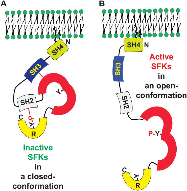
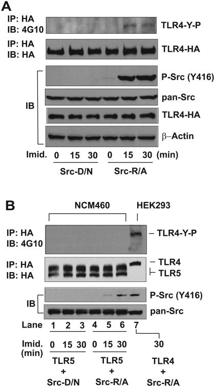
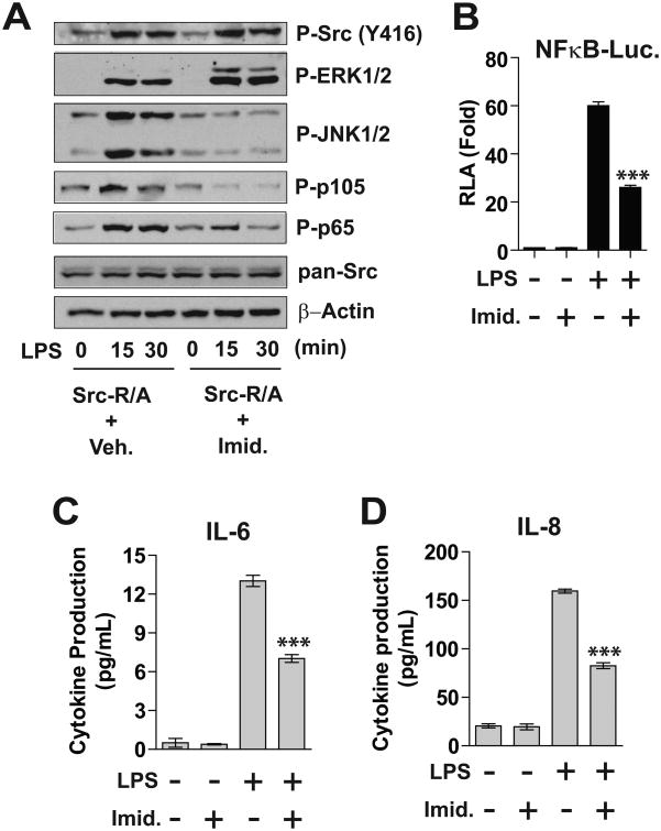
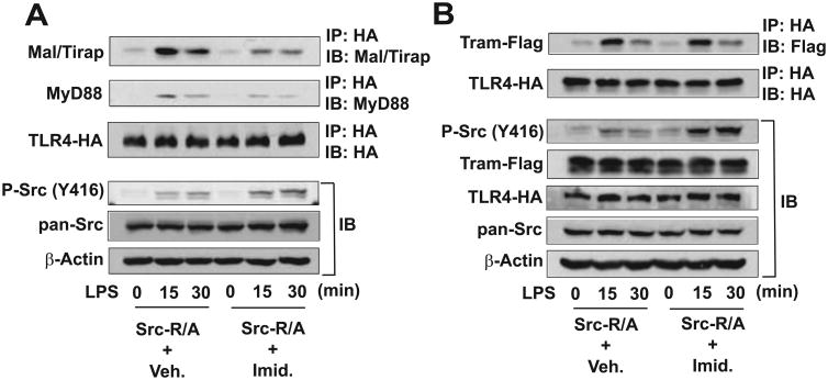
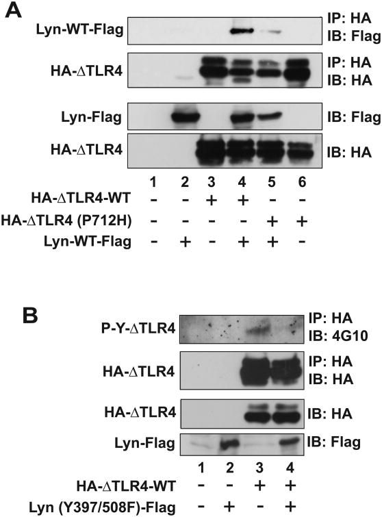
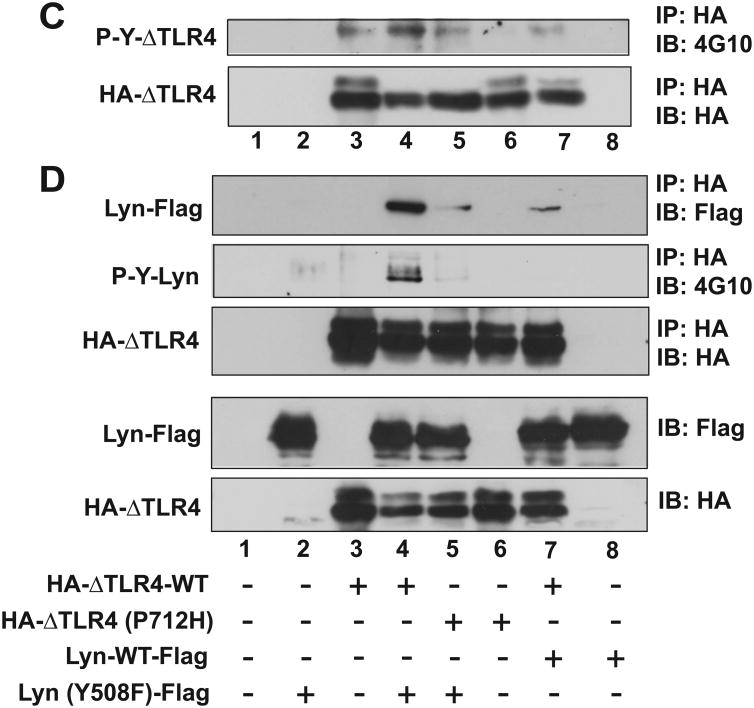
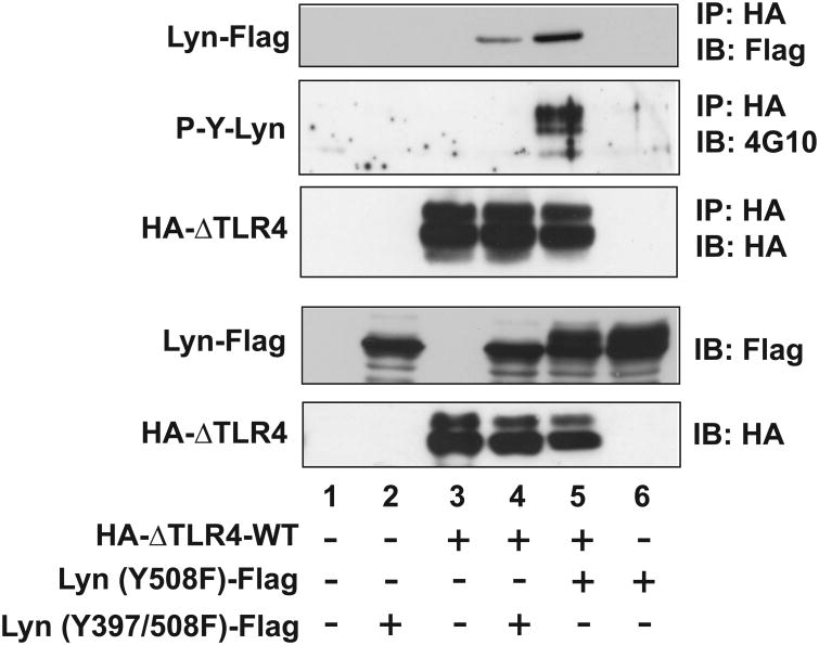
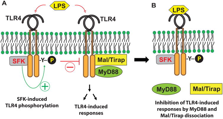
Src family kinases (SFKs) are a family of protein tyrosine kinases containing nine members: Src, Lyn, Fgr, Hck, Lck, Fyn, Blk, Yes, and Ylk. Although SFK activation is a major immediate signaling event in LPS/Toll-like receptor 4 (TLR4) signaling, its precise role has remained elusive due to various contradictory results obtained from a certain SFK member-deficient mice or cells. The observed inconsistencies may be due to the compensation or redundancy by other SFKs upon a SFK deficiency. The chemical rescuing approach was suggested to induce temporal and precise SFK activation in living cells, thereby limiting the chance of cellular adaption to a SFK-deficient condition. Using the rescuing approach, we demonstrate that restoring SFK activity not only induces tyrosine phosphorylation of TLR4, but also inhibits LPS-induced NFκB and JNK1/2 activation and consequently suppresses LPS-induced cytokine production. TLR4 normally recruits TIR domain-containing adaptors in response to LPS, however, temporally restored SFK activation disrupts the LPS-induced association of MyD88 and Mal/Tirap with TLR4. Additionally, using kinase-dead SFK-Lyn (Y397/508F) and constitutively active SFK-Lyn (Y508F), we found that the kinase-dead SFK inhibits TLR4 tyrosine phosphorylation with reduced binding affinity to TLR4, while the kinase-active SFK strongly binds to TLR4 and promotes TLR4 tyrosine phosphorylation, suggesting that SFK kinase activity is required for TLR4 tyrosine phosphorylation and TLR4-SFK interaction. Together, our results demonstrate that SFK activation induces TLR4 tyrosine phosphorylation, consequently dissociating MyD88 and Mal/Tirap from TLR4 and inhibiting LPS-induced inflammatory responses, suggesting a negative feedback loop regulated by SFK-induced tyrosine phosphorylation in TLR4.

Keywords: Chemical rescuing approach; Negative feedback; Sepsis; Src family kinase; Toll like receptor; Tyrosine phosphorylation.


Conflict of interest statement

Conflict of Interest: The authors declare that they have no conflicts of interest with the contents of this article.

Figures

Figure 1
Figure 1. Domain structure of SFKs and tyrosine phosphorylation-dependent SFK activation mechanism
(A and B) SFKs have an SH4 (Src homology 4) domain, which can be post-translationally modified with myristoylation and palmitoylation for plasma membrane targeting; an SH3 domain, which interacts with the SH1 domain; an SH1 (or kinase catalytic domain) in which tyrosine phosphorylation renders SFKs active; and an R (regulatory) domain in the C-terminal end in which phosphorylated tyrosine interacts with the SH2 domain. SH2 and SH3 participate in protein-protein interaction. Phosphorylation at the tyrosine residue in the C-terminus induces a closed conformation that is inactive (A). Dephosphorylation at the tyrosine residue in the C-terminus results in an open-conformation of SFKs, followed by autophosphorylation at the tyrosine residue in the kinase domain to become an active form of SFK (B).
Figure 2
Figure 2. Rescued SFK activity induces TLR4 tyrosine phosphorylation
(A and B) HEK293 cells (A) were stably transfected with HA-TLR4 WT (full length) construct together with SFK-Src R388A/Y527F (Src-R/A) or SFK-Src D386N/Y527F (Src-D/N) construct. Human colonic epithelial (NCM460) cells (B) were also stably transfected with HA-TLR5 construct and Src-D/N or Src-R/A construct. The cells were treated with fresh prepared imidazole (Imid., 10 mM) for the indicated time points, as we previously described (39). The cell lysates were subjected to immunoprecipitation (IP) assay with HA antibody, followed by immunoblot (IB) analysis with anti-phospho-tyrosine antibody (4G10) to examine the tyrosine phosphorylation of TLR4 (TLR4-Y-P). In parallel, the cell extracts were applied for IB analysis to determine imidazole-rescued SFK-Src activation represented by phospho-Src (Y416). Subsequently, total Src (pan-Src) and HA-TLR4 expression were evaluated. β-Actin was determined as a loading control. Presented are the representative from three independent experiments.
Figure 3
Figure 3. Restored SFK activity inhibits LPS-stimulated responses in SW480 and RAW264.7 cells
(A) Human colonic epithelial SW480 cells were stably transfected with SFK-Src-R/A construct, followed by LPS (0.5 μg/mL) and/or imidazole (10 mM) treatment for the indicated time points. Cell lysates were subjected to immunoblot analysis. (B) RAW264.7 cells were transiently co-transfected with NFκB-luciferase (Luc.) reporter and Src-R/A constructs, followed by LPS (20 ng/mL) and/or imidazole treatment for as indicated 6 hours. Cell lysates were used for measuring the luciferase activity (RLA, relative luciferase activity). (C and D) SW480-Src-R/A cells were treated with LPS (0.5 |ig/mL) and/or imidazole as indicated for 8 hours. The culture supernatants were collected to measure the cytokine production by ELISA performed in triplicate. All data presented are the representative from three independent experiments. The data in graphs were shown as mean + SEM. ***P<0.001 (Mann-Whitney U test).
Figure 4
Figure 4. Rescued SFK activation inhibits the recruitment of MyD88 and Mal/Tirap adaptors to TLR4
(A) HEK293 cells stably expressing SFK-Src (R/A) and HA-TLR4-WT were treated with a combination of LPS (100 ng/mL) and/or imidazole (10 mM) as indicated. Cell lysates were subjected to IP with HA antibody, followed by IB with antibody recognizing endogenous Mal/Tirap or MyD88. In parallel, the cell lysates were used for IB analysis to evaluate imidazole-induced SFK-Src activation [P-Src (Y416)] and total Src. (B) HEK293 cells expressing SFK-Src (R/A) and HA-TLR4-WT were transiently transfected with Tram-Flag encoding construct and treated with LPS and/or imidazole, followed by IP with HA and IB with Flag antibody. Cell lysates were also used for IB to evaluate SFK-Src activation and transfected gene expression. β-Actin was determined as a loading control. Presented are the representative from three independent experiments.
Figure 5
Figure 5. SFK kinase activity regulates TLR4 tyrosine phosphorylation and the interaction between TLR4 and SFK
HEK293 cells were transiently co-transfected with the plasmid constructs in an indicated combination, followed by IP assay with HA antibody and IB analysis with Flag antibody or anti-phospho-tyrosine antibody (4G10) to evaluate the tyrosine phosphorylation of TLR4 or SFK-Lyn. (A) SFK-Lyn wild-type interacts with the constitutively active TLR4 (ΔTLR4-WT), but not with the dominant negative TLR4 [ΔTLR4 (P712H)]. (B) The kinase-dead form of SFK-Lyn [Lyn (Y397/508F)] inhibits the tyrosine phosphorylation in the constitutively active TLR4. (C and D) The kinase-active form of SFK-Lyn [Lyn (Y508F)] enhances the tyrosine phosphorylation of ΔTLR4. Cell lysates of the transfected HEK293 cells were used for two sets of the experiments (C and D) of IP with HA, followed by IB with 4G10 antibody. The blots were stripped and reprobed with Flag antibody. The expression of transfected constructs was confirmed by IB analysis. The representative set was shown from three independent experiments.
Figure 6
Figure 6. SFK kinase activity promotes the interaction between TLR4 and SFK
HEK293 cells were transfected with a combination of the plasmid construct as indicated. Cell extracts were subjected to IP with HA antibody, followed by IB with 4G10 antibody. The blot was stripped and reprobed with Flag antibody. The expression of transfected constructs was confirmed by IB analysis. The representative set was presented from three independent experiments.
Figure 7
Figure 7. The activation of SFK by LPS results in the dissociation of MyD88 and Mal/Tirap from TLR4, leading to suppressed LPS-induced inflammatory responses
(A) LPS recognition by TLR4 immediately induces the recruitment of TIR-domain containing adaptor molecules, including MyD88 and Mal/Tirap, to the cytoplasmic region of TLR4, leading to immune and inflammatory responses. Simultaneously, LPS-stimulation elicits SFK binding to TLR4, which induces the tyrosine phosphorylation of TLR4. (B) The tyrosine phosphorylation of TLR4 by SFK dissociates MyD88 and Mal/Tirap from TLR4, leading to suppressed LPS-induced inflammatory responses.

Similar articles

Cited by

References

    1. Senis YA, Mazharian A, Mori J. Src family kinases: at the forefront of platelet activation. Blood. 2014;124:2013–2024. - PMC - PubMed
    1. Badolia R, Inamdar V, Manne BK, Dangelmaier C, Kunapuli SP. Gq pathway regulates proximal C-type lectin-like receptor 2 (CLEC-2) signaling in platelets. J Biol Chem 2017 - PMC - PubMed
    1. Wu Y, Span LM, Nygren P, Zhu H, Moore DT, Cheng H, Roder H, DeGrado WF, Bennett JS. The Tyrosine Kinase c-Src Specifically Binds to the Active Integrin alphaIIbbeta3 to Initiate Outside-in Signaling in Platelets. J Biol Chem. 2015;290:15825–15834. - PMC - PubMed
    1. Hamerman JA, Lanier LL. Inhibition of immune responses by ITAM-bearing receptors. Sci STKE. 2006;2006:re1. - PubMed
    1. Medvedev AE, Piao W, Shoenfelt J, Rhee SH, Chen H, Basu S, Wahl LM, Fenton MJ, Vogel SN. Role of TLR4 tyrosine phosphorylation in signal transduction and endotoxin tolerance. J Biol Chem. 2007;282:16042–16053. - PMC - PubMed

Publication types