. 2018 Jan 17;92(3):e01637-17.
doi: 10.1128/JVI.01637-17. Print 2018 Feb 1.

Tumor Necrosis Factor-Mediated Survival of CD169+ Cells Promotes Immune Activation during Vesicular Stomatitis Virus Infection

Affiliations

Tumor Necrosis Factor-Mediated Survival of CD169+ Cells Promotes Immune Activation during Vesicular Stomatitis Virus Infection

Prashant V Shinde et al. J Virol. .

Abstract

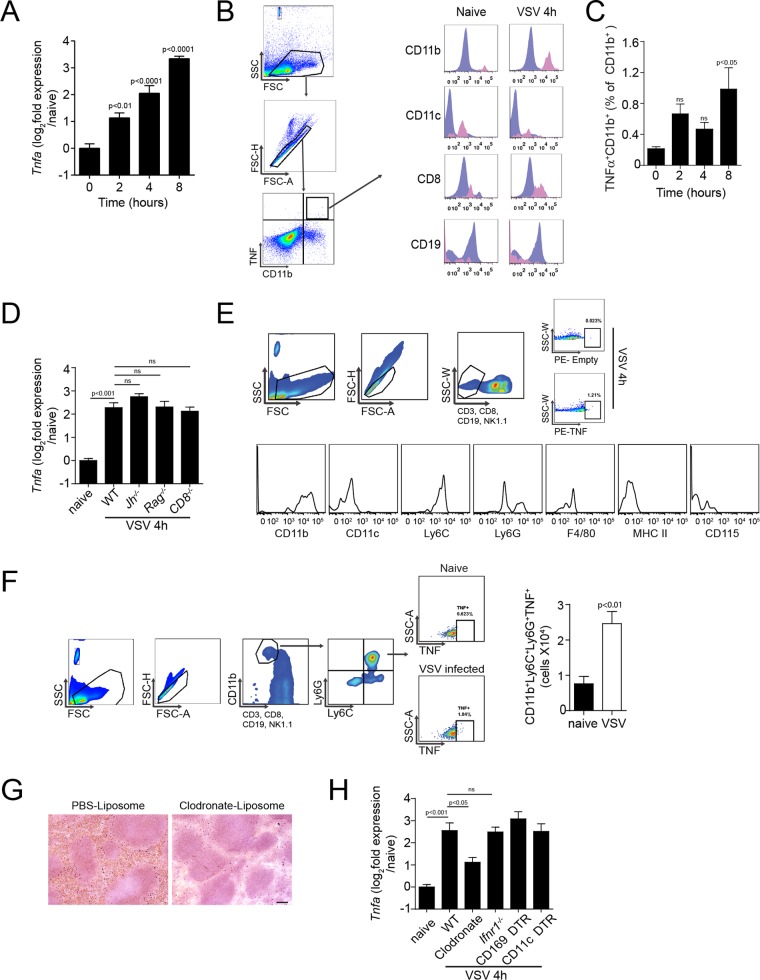
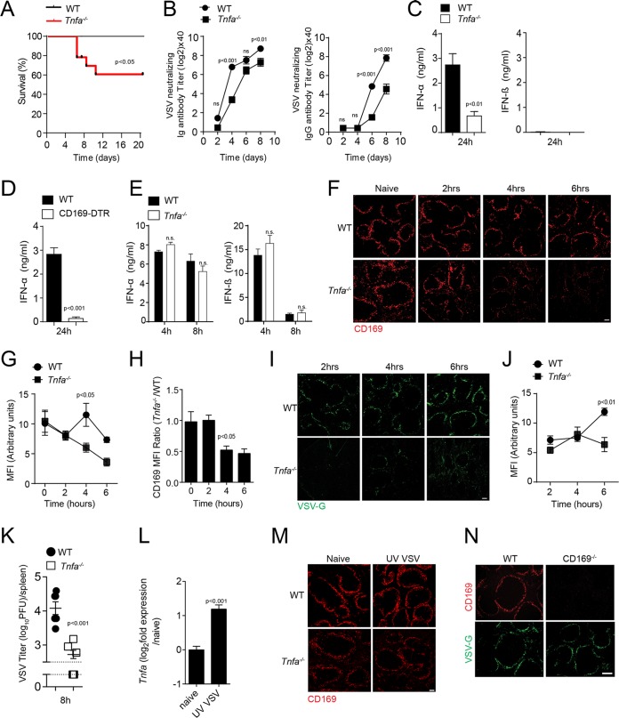
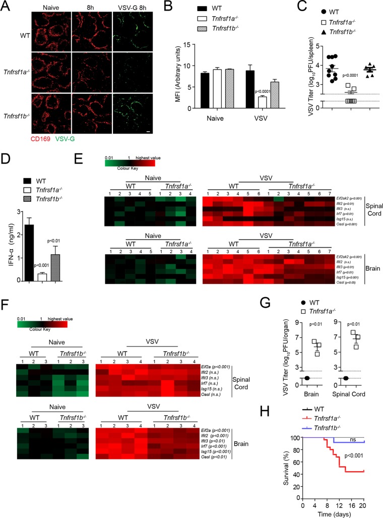
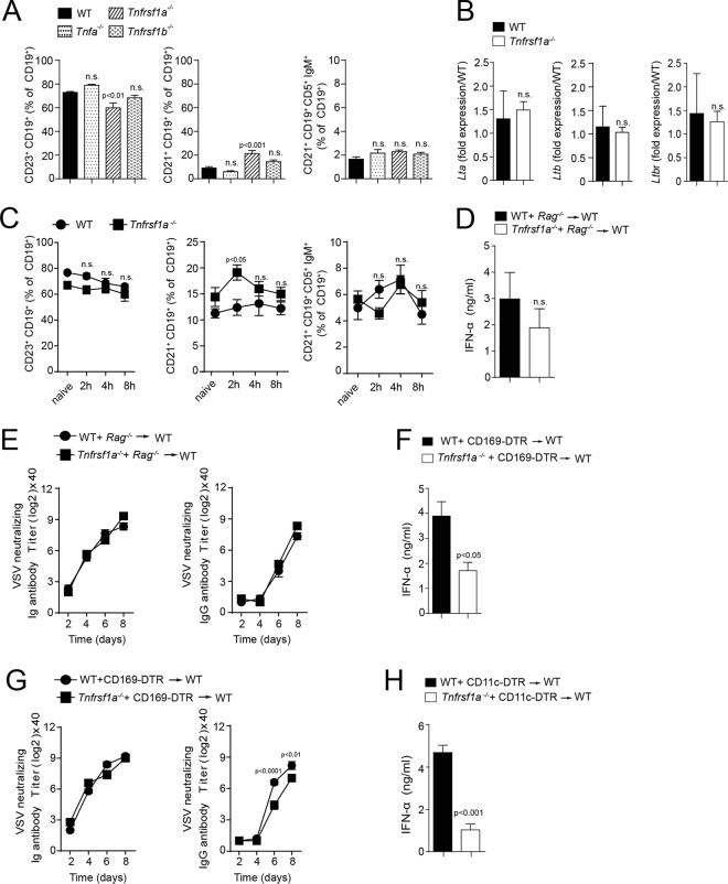
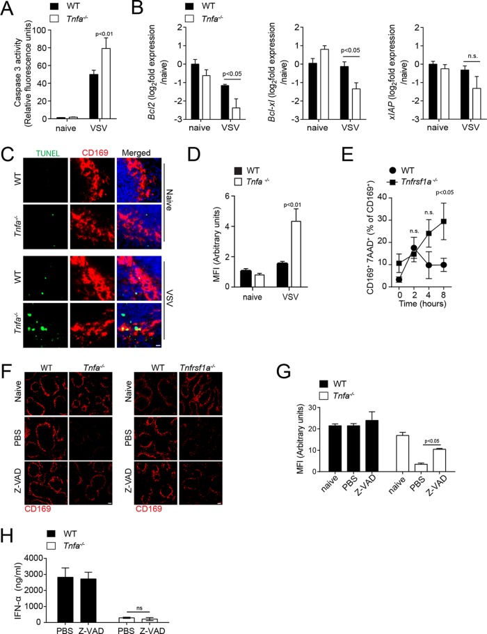
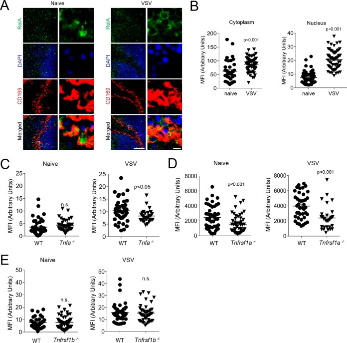
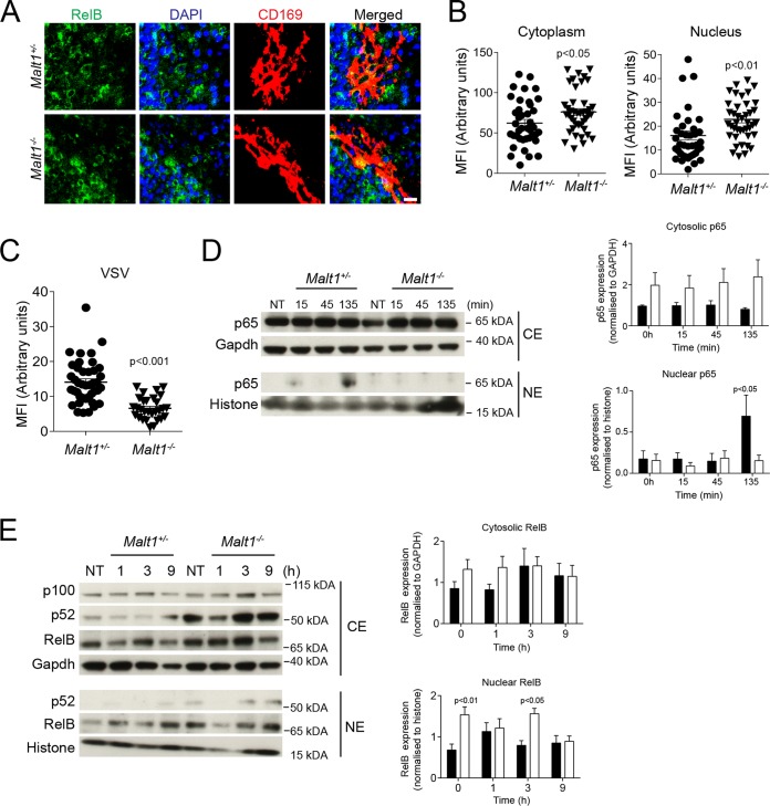
Innate immune activation is essential to mount an effective antiviral response and to prime adaptive immunity. Although a crucial role of CD169+ cells during vesicular stomatitis virus (VSV) infections is increasingly recognized, factors regulating CD169+ cells during viral infections remain unclear. Here, we show that tumor necrosis factor is produced by CD11b+ Ly6C+ Ly6G+ cells following infection with VSV. The absence of TNF or TNF receptor 1 (TNFR1) resulted in reduced numbers of CD169+ cells and in reduced type I interferon (IFN-I) production during VSV infection, with a severe disease outcome. Specifically, TNF triggered RelA translocation into the nuclei of CD169+ cells; this translocation was inhibited when the paracaspase MALT-1 was absent. Consequently, MALT1 deficiency resulted in reduced VSV replication, defective innate immune activation, and development of severe disease. These findings indicate that TNF mediates the maintenance of CD169+ cells and innate and adaptive immune activation during VSV infection.IMPORTANCE Over the last decade, strategically placed CD169+ metallophilic macrophages in the marginal zone of the murine spleen and lymph nodes (LN) have been shown to play a very important role in host defense against viral pathogens. CD169+ macrophages have been shown to activate innate and adaptive immunity via "enforced virus replication," a controlled amplification of virus particles. However, the factors regulating the CD169+ macrophages remain to be studied. In this paper, we show that after vesicular stomatitis virus infection, phagocytes produce tumor necrosis factor (TNF), which signals via TNFR1, and promote enforced virus replication in CD169+ macrophages. Consequently, lack of TNF or TNFR1 resulted in defective immune activation and VSV clearance.

Keywords: MALT1; NF-κB; TNF; innate immunity; interferon; interferons; tumor necrosis factor.


Figures

FIG 1
FIG 1
Vesicular stomatitis virus infection leads to infiltration of TNF-producing phagocytes. (A to F) WT mice were infected with 2 × 10⁸ PFU VSV. (A) TNF-α mRNA expression levels in WT spleen tissue were determined at the indicated time points after infection (n = 4 to 10). (B) Surface molecule expression of CD11b, CD11c, CD8, and CD19 on TNF+ cells 4 h after infection (purple gate, whole spleen; pink gate, TNF+ cells; one representative result out of 5 is shown). Numbers below the histograms indicate fluorescence intensities. FSC, forward scatter; SSC, side scatter. (C) Splenocytes from WT mice were stained for intracellular-TNF production. TNF+ CD11b+ cells were determined as percentages of total CD11b+ cells (n = 5). (D) TNF-α mRNA expression in the spleens of WT, Jh−/−, Rag−/−, and CD8−/− mice was determined 4 h after infection (n = 5 or 6). (E) Surface molecule expression of TNF-producing cells 4 h after infection. CD3 CD8 CD19 NK1.1 cells were further characterized for expression of CD11b, CD11c, Ly6C, Ly6G, F4/80, MHC-II, and CD115 on TNF+ cells (n = 6). The numbers in the boxes are percentages of the population positive for TNF expression. (F) CD3 CD8 CD19 NK1.1 CD11b+ Ly6C+ Ly6G+ TNF+ cells were quantified in spleen tissue 4 h after infection (n = 6). (G) Mice were injected with liposomes containing phosphate-buffered saline (PBS; PBS liposomes) or clodronate liposomes, and spleen tissue was harvested after 24 h. Sections of snap-frozen spleen tissue were stained with anti-F4/80 antibodies (n = 3). (H) TNF-α mRNA expression was determined in the spleens of WT, clodronate-treated WT, Ifnar−/−, DT-treated CD169-DTR, and CD11c-DTR mice 4 h after infection (n = 6). ns, not significant. The error bars indicate SEM.
FIG 2
FIG 2
Tumor necrosis factor is required for early innate immune activation via maintenance of CD169+ cells during viral infection. (A to D) Mice were infected with 105 PFU VSV. (A) Survival of WT and TNF-α-null (Tnfa−/−) mice was monitored for 20 days after infection (n = 9 to 12). (B) Titers of neutralizing total immunoglobulin (Ig) (left) and IgG (right) were determined in WT and Tnfa−/− mice at the indicated time points after infection (n = 7). (C) IFN-α and IFN-β concentrations were determined in the sera of WT and Tnfa−/− mice 24 h after infection (n = 6 to 9). (D) IFN-α levels were determined in sera from WT and CD169-DTR mice 24 h after infection (n = 6). (E) IFN-α and IFN-β concentrations were determined in the sera of WT and Tnfa−/− mice injected with 200 μg of poly(I·C) at the indicated time points (n = 3). (F) WT and Tnfa−/− mice were infected with 2 × 10⁸ PFU of VSV. Snap-frozen spleen sections were stained with anti-CD169 antibodies (clone MOMA-1) at the indicated time points (one representative result out of 6 mice is shown; scale bar = 100 μm). (G) The mean fluorescence intensity (MFI) of CD169 was quantified across spleen sections from naive and VSV-infected WT and Tnfa−/− mice using ImageJ (1 to 3 images per spleen from 3 or 4 mice were analyzed). (H) MFI from Tnfa−/− mice normalized to WT MFI. (I) Snap-frozen spleen sections from WT and Tnfa−/− mice were stained for VSV-G expression (clone Vi10) after infection with 2 × 10⁸ PFU VSV at the indicated time points (one representative result out of 6 mice is shown; scale bar = 100 μm). (J) MFI of VSV-G expression quantified across spleen sections from naive and VSV-infected WT and Tnfa−/− mice using ImageJ (1 to 3 images per spleen from 3 or 4 mice were analyzed). (K) WT and Tnfa−/− mice were infected with 105 PFU VSV. Viral titers were measured in the spleens of WT and Tnfa−/− mice 8 h after infection with VSV (n = 6). (L) Tnfa mRNA expression determined in spleen tissue of WT mice before and 4 h after injection with UV-inactivated VSV (n = 4). (M) Spleen tissue sections were stained with anti-CD169 antibodies in WT and Tnfa−/− mice 8 h after infection with 2 × 10⁸ PFU of UV-inactivated VSV (one representative result out of 3 is shown). (N) Sections from snap-frozen spleen tissue harvested from WT and CD169−/− mice were stained for CD169 and VSV-G 7 h after infection with 2 × 10⁸ PFU VSV (n = 3; scale bar = 100 μm). The error bars indicate SEM.
FIG 3
FIG 3
VSV replication is sustained via TNFR1 on CD169+ cells. (A) Spleen tissue sections from WT, Tnfrsf1a−/− (TNFR1), and Tnfrsf1b−/− (TNFR2) mice were stained with anti-CD169 and VSV-G antibodies 8 h after infection with 2 × 10⁸ PFU of VSV (one representative result out of 6 mice is shown; scale bar = 100 μm). (B) MFI of CD169 was quantified across spleen sections from WT, Tnfrsf1a−/−, and Tnfrsf1b−/− infected mice, using ImageJ (1 to 3 images per spleen from 3 or 4 mice were analyzed). (C to G) WT, Tnfrsf1a−/−, and Tnfrsf1b−/− mice were infected with 105 PFU VSV. (C) Viral titers were measured in spleen tissue 8 h after infection in WT, Tnfrsf1a−/−, and Tnfrsf1b−/− mice (n = 6 to 9). The gap and dotted lines represent the detection limit of the virus plaque assay. (D) IFN-α concentrations were determined in the sera of WT, Tnfrsf1a−/−, and Tnfrsf1b−/− mice 24 h after infection with VSV (n = 6 to 9). (E) WT and Tnfrsf1a−/− mice were infected with 105 PFU VSV. RNA expression levels of the indicated genes were determined in brains and spinal cords 24 h after infection (n = 4 to 7). The highest relative expression values (brain/spinal cord) were as follows: Eif2ak2, 13.72/7.98; Ifit2, 5.41/6.13; Ifit3, 35.99/34.15; Irf7, 68.80/54.55; Isg15, 42.54/51.23; Oasl1, 70.43/84.94. (F) WT and Tnfrsf1b−/− mice were infected with 105 PFU VSV. RNA expression levels of the indicated genes in brains and spinal cords were determined 24 h after infection (n = 3 or 4). The highest relative expression values (brain/spinal cord) were as follows: Eif2ak, 29.84/18.21; Ifit2, 7.99/10.24; Ifit3, 41.05/51.25; Irf7, 166.79/88.58; Isg15, 29.78/52.99; Oasl1, 75.60/114.39. (G) Viral titers were measured in brain and spinal cord tissue of WT and Tnfrsf1a−/− mice once Tnfrsf1a−/− mice exhibited hind limb paralysis (n = 3). (H) Survival of WT, Tnfrsf1a−/−, and Tnfrsf1b−/− mice was monitored over time after infection with VSV (n = 15 to 24). The error bars indicate SEM.
FIG 4
FIG 4
TNFR1 on CD169+ cells is essential for early IFN-I response. (A) Follicular B cells (CD19+ CD23+) (FB), marginal-zone B cells (CD19+ CD21+ CD23) (MZB), and regulatory B cells (CD19+ CD21+ CD5+ IgM+) (RB) were analyzed in naive WT and Tnfa−/−-, Tnfrsf1a−/−-, and Tnfrsf1b−/−-deficient mice (n = 6). (B) Lymphotoxin α (Ltα), Ltβ, and lymphotoxin β receptor (LtβR) gene expression was determined in spleen tissue from WT and Tnfrsf1a−/− mice by RT-PCR (n = 3). (C) Splenic B-cell populations (FB, MZB, and RB) were analyzed after infection with 2 × 10⁸ PFU of VSV in WT and Tnfrsf1a−/− mice at the indicated time points (n = 5). (D) IFN-α concentrations were determined 24 h after infection with 105 PFU VSV in the sera of lethally irradiated mice reconstituted with either WT-Rag−/− or Tnfrsf1a−/−-Rag−/− bone marrow at a ratio of 1:1 (n = 4). (E) Neutralizing total immunoglobulin (left) and IgG (right) antibody titers were determined in the sera of WT-Rag−/− or Tnfrsf1a−/−-Rag−/− reconstituted animals (n = 4). (F to H) Lethally irradiated WT mice were reconstituted with BM from WT or Tnfrsf1a−/− mice mixed with BM from CD169-DTR (F) and CD11c-DTR (H) mice at a 1:1 ratio. After 40 days, the mice were infected with 105 PFU of VSV. Before infection, the mice were treated with 2 doses of 100 ng DT via intraperitoneal injection. (F) IFN-α concentrations were determined 24 h after infection in the sera of WT–CD169-DTR and Tnfrsf1a−/−–CD169-DTR reconstituted animals (n = 4 or 5). (G) Neutralizing total immunoglobulin (left) and IgG (right) antibody titers were determined in the sera of WT–CD169-DTR and Tnfrsf1a−/−–CD169-DTR reconstituted animals after infection with 105 PFU VSV at the indicated time points (n = 4). (H) IFN-α concentrations were determined 24 h after infection in the sera of WT–CD11c-DTR and Tnfrsf1a−/−–CD11c-DTR reconstituted mice (n = 4 or 5). The error bars indicate SEM.
FIG 5
FIG 5
Tumor necrosis factor mediates survival of CD169+ cells via TNFR1. (A to E) Mice were infected with 2 × 10⁸ PFU VSV. (A) Caspase 3 activity was determined in spleen tissue harvested from WT and Tnfa−/− mice 6 h after infection with 2 × 10⁸ PFU VSV (n = 4 to 7). (B) Bcl2, Bclxl, and Xiap RNA expression was determined in spleen tissue from WT and Tnfrsf1a−/− mice 8 h after infection (n = 3). (C) Tissue sections from WT and Tnfa−/− mice were TUNEL stained 5 h after infection (one result representative of 3 or 4 mice is shown; scale bar = 10 μm). (D) MFI of TUNEL quantified across spleen sections from naive and VSV-infected WT and Tnfa−/− mice using ImageJ (1 or 2 images per spleen from 3 or 4 mice were analyzed). (E) At the indicated time points, the proportions of 7AAD+ cells among CD11b+ CD169+ cells were determined (n = 5) in WT and Tnfrsf1a−/− mice. (F) WT, Tnfa−/−, and Tnfrsf1a−/− mice were treated with Z-VAD and infected with 2 × 10⁸ PFU VSV. Spleen tissue sections were stained with anti-CD169 antibodies 8 h after infection (one result representative of 3 or 4 mice is shown; scale bar = 100 μm). (G) MFI of CD169 quantified across spleen sections from naive and VSV-infected WT and Tnfa−/− mice treated with Z-VAD, using ImageJ (1 to 3 images per spleen from 3 or 4 mice were analyzed). (H) IFN-α concentrations determined 24 h after infection in the sera of Z-VAD-treated WT and Tnfa−/− mice after infection with 105 PFU of VSV (n = 3). The error bars indicate SEM.
FIG 6
FIG 6
VSV infection leads to TNFR1-dependent canonical NF-κB activation in splenic CD169+ cells. (A to D) Sections of snap-frozen spleen tissue were harvested 4 h after infection with 2 × 10⁸ PFU VSV. (A) The sections were stained for RelA before and after infection (one representative result out of 3 is shown; scale bar = 100 μm). Enlarged images of the boxed areas in the merged images on the left side are shown on the right (scale bar = 5 μm). (B) MFI of cytoplasmic and respective nuclear RelA quantified in CD169+ cells from WT mice before and after VSV infection to evaluate nuclear translocation of RelA (n = 48 to 63). (C to E) Spleen sections from WT and Tnfa−/− (C), Tnfrsf1a−/− (D), and Tnfrsf1b−/− (E) mice were stained with anti-RelA antibodies 4 h after infection with 2 × 10⁸ PFU VSV. The MFI of RelA in the nuclei of CD169+ cells was determined with ImageJ software (n = 35 to 57). The error bars indicate SEM.
FIG 7
FIG 7
MALT1 regulates nuclear RelA expression after infection with vesicular stomatitis virus. (A) Sections from snap-frozen spleen tissue harvested from naive Malt1+/− and Malt1−/− mice were stained with anti-RelB antibodies (one representative result out of 3 is shown; scale bar = 10 μm). (B) MFI of cytoplasmic and nuclear RelB was quantified in CD169+ cells using ImageJ (n = 39 to 42). (C) Sections of snap-frozen spleen tissue from Malt1+/− and Malt1−/− mice were stained with anti-RelA antibodies 4 h after infection with 2 × 10⁸ PFU VSV. The MFI in the nuclei of CD169+ cells was quantified (n = 29 to 41). (D and E) Malt1+/− and Malt1−/− MEFs were stimulated with 100 ng/ml recombinant mouse tumor necrosis factor (rmTNF) at the indicated time points. Cytosolic extracts (CE) and nuclear extracts (NE) were harvested and probed for p65. Densitometry analysis of p65 and RelB was performed on the Western blot (WB) images from cytosolic and nuclear fractions at the indicated time points. Proteins were normalized to GAPDH or histone (n = 4). The error bars indicate SEM.
FIG 8
FIG 8
MALT1 promotes vesicular stomatitis virus replication in CD169+ cells and immune activation during viral infection. (A) Spleen sections from naive Malt1+/− and Malt1−/− mice were stained with anti-CD169 (one representative result out of 3 is shown; scale bar = 100 μm). (B) Sections of snap-frozen spleen tissue from Malt1+/− and Malt1−/− mice were analyzed 8 h after infection with 2 × 10⁸ PFU VSV and stained with anti-CD169 and anti-VSV-G) (one representative result out of 3 is shown; scale bar = 100 μm). (C) MFI of CD169 and VSV-G was quantified across spleen sections from VSV-infected Malt1+/− and Malt1−/− mice using ImageJ (n = 4). (D and E) Mice were infected with 105 PFU VSV. (D) Viral titers were measured in spleen tissue of Malt1+/− and Malt1−/− mice 8 h after infection (n = 6). (E) IFN-α concentrations were determined in the sera of Malt1+/− and Malt1−/− mice 24 h after infection (n = 6). (F) IFN-α concentrations were determined in the sera of Malt1+/− and Malt1−/− mice injected with 200 μg poly(I·C) at the indicated time points (n = 3 or 4). (G) Survival of Malt1+/− and Malt1−/− animals was monitored for 20 days after infection (n = 13 to 14). The error bars indicate SEM.

Similar articles

Cited by

References

    1. McNab F, Mayer-Barber K, Sher A, Wack A, O'Garra A. 2015. Type I interferons in infectious disease. Nat Rev Immunol 15:87–103. doi:10.1038/nri3787. - DOI - PMC - PubMed
    1. Chavez-Galan L, Olleros ML, Vesin D, Garcia I. 2015. Much more than M1 and M2 macrophages, There are also CD169(+) and TCR(+) macrophages. Front Immunol 6:263. doi:10.3389/fimmu.2015.00263. - DOI - PMC - PubMed
    1. Mebius RE, Kraal G. 2005. Structure and function of the spleen. Nat Rev Immunol 5:606–616. doi:10.1038/nri1669. - DOI - PubMed
    1. Snook T. 1964. Studies on the perifollicular region of the rat's spleen. Anat Rec 148:149–159. doi:10.1002/ar.1091480205. - DOI - PubMed
    1. Crocker PR, Gordon S. 1986. Properties and distribution of a lectin-like hemagglutinin differentially expressed by murine stromal tissue macrophages. J Exp Med 164:1862–1875. doi:10.1084/jem.164.6.1862. - DOI - PMC - PubMed

Publication types

MeSH terms

Substances