

Protective Effects of Estradiol and Dihydrotestosterone following Spinal Cord Injury
- PMID: 29132243
- PMCID: PMC5863086
- DOI: 10.1089/neu.2017.5329
Protective Effects of Estradiol and Dihydrotestosterone following Spinal Cord Injury
Abstract
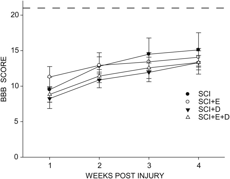
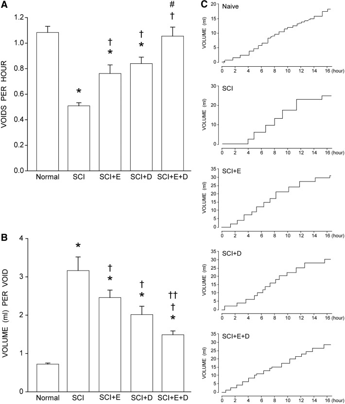
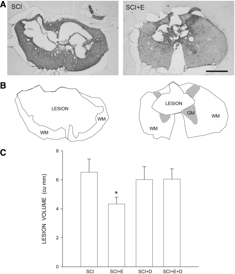
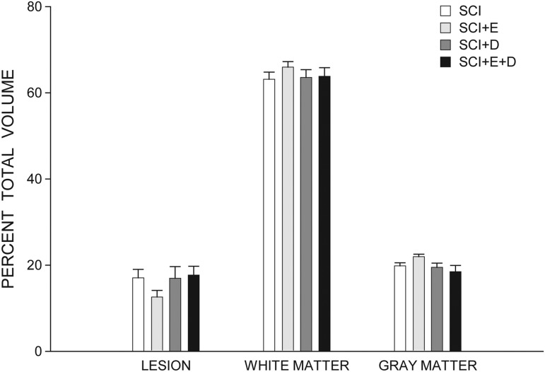
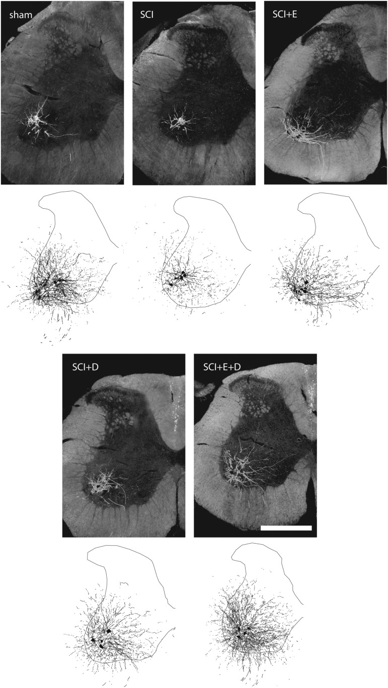
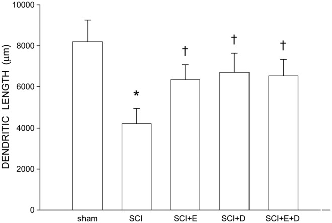
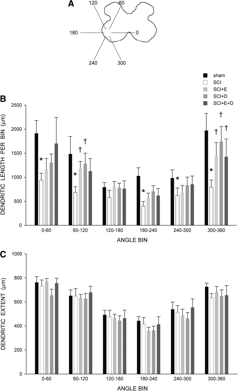
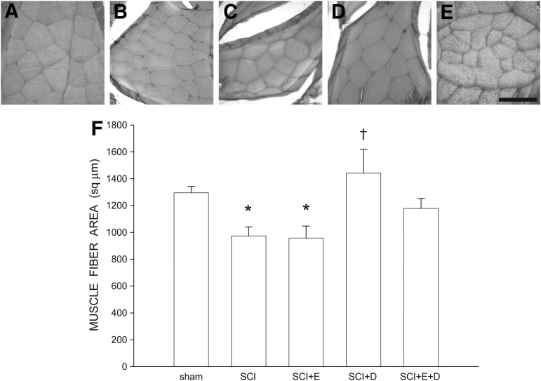
Spinal cord injury (SCI) results in lesions that destroy tissue and disrupt spinal tracts, producing deficits in locomotor and autonomic function. We previously demonstrated that motoneurons and the muscles they innervate show pronounced atrophy after SCI, and these changes are prevented by treatment with testosterone. Here, we assessed whether the testosterone active metabolites estradiol and dihydrotestosterone have similar protective effects after SCI. Young adult female rats received either sham or T9 spinal cord contusion injuries and were treated with estradiol, dihydrotestosterone, both, or nothing via Silastic capsules. Basso-Beattie-Bresnahan locomotor testing was performed weekly and voiding behavior was assessed at 3 weeks post-injury. Four weeks after SCI, lesion volume and tissue sparing, quadriceps muscle fiber cross-sectional area, and motoneuron dendritic morphology were assessed. Spontaneous locomotor behavior improved after SCI, but hormone treatments had no effect. Voiding behavior was disrupted after SCI, but was significantly improved by treatment with either estradiol or dihydrotestosterone; combined treatment was maximally effective. Treatment with estradiol reduced lesion volume, but dihydrotestosterone alone and estradiol combined with dihydrotestosterone were ineffective. SCI-induced decreases in motoneuron dendritic length were attenuated by all hormone treatments. SCI-induced reductions in muscle fiber cross-sectional areas were prevented by treatment with either dihydrotestosterone or estradiol combined with dihydrotestosterone, but estradiol treatment was ineffective. These findings suggest that deficits in micturition and regressive changes in motoneuron and muscle morphology seen after SCI are ameliorated by treatment with estradiol or dihydrotestosterone, further supporting a role for steroid hormones as neurotherapeutic agents in the injured nervous system.
Keywords: dendrites; morphology; neuroprotection; steroids.
Conflict of interest statement
No competing financial interests exist.
Figures
Similar articles
-
Neuroprotective effects of testosterone on motoneuron and muscle morphology following spinal cord injury.J Comp Neurol. 2012 Aug 15;520(12):2683-96. doi: 10.1002/cne.23066. J Comp Neurol. 2012. PMID: 22314886 Free PMC article.
-
Protective effects of gonadal hormones on spinal motoneurons following spinal cord injury.Neural Regen Res. 2018 Jun;13(6):971-976. doi: 10.4103/1673-5374.233434. Neural Regen Res. 2018. PMID: 29926818 Free PMC article. Review.
-
Neuroprotective effects of testosterone metabolites and dependency on receptor action on the morphology of somatic motoneurons following the death of neighboring motoneurons.Dev Neurobiol. 2017 Jun;77(6):691-707. doi: 10.1002/dneu.22445. Epub 2016 Oct 3. Dev Neurobiol. 2017. PMID: 27569375 Free PMC article.
-
Inhibition of Cytosolic Phospholipase A2 Has Neuroprotective Effects on Motoneuron and Muscle Atrophy after Spinal Cord Injury.J Neurotrauma. 2021 May 1;38(9):1327-1337. doi: 10.1089/neu.2014.3690. J Neurotrauma. 2021. PMID: 25386720 Free PMC article.
-
Evaluating Sex Steroid Hormone Neuroprotection in Spinal Cord Injury in Animal Models: Is It Promising in the Clinic?Biomedicines. 2024 Jul 4;12(7):1478. doi: 10.3390/biomedicines12071478. Biomedicines. 2024. PMID: 39062051 Free PMC article. Review.
Cited by
-
A comprehensive look at the psychoneuroimmunoendocrinology of spinal cord injury and its progression: mechanisms and clinical opportunities.Mil Med Res. 2023 Jun 9;10(1):26. doi: 10.1186/s40779-023-00461-z. Mil Med Res. 2023. PMID: 37291666 Free PMC article. Review.
-
Intravitreal Fluocinolone Acetonide May Decelerate Diabetic Retinal Neurodegeneration.Invest Ophthalmol Vis Sci. 2019 May 1;60(6):2134-2139. doi: 10.1167/iovs.18-24643. Invest Ophthalmol Vis Sci. 2019. PMID: 31100106 Free PMC article.
-
Neuroprotective Effects of Estradiol plus Lithium Chloride via Anti-Apoptosis and Neurogenesis Pathway in In Vitro and In Vivo Parkinson's Disease Models.Parkinsons Dis. 2021 Oct 22;2021:3064892. doi: 10.1155/2021/3064892. eCollection 2021. Parkinsons Dis. 2021. PMID: 34721835 Free PMC article.
-
Novel Therapeutic Effects in Rat Spinal Cord Injuries: Recovery of the Definitive and Early Spinal Cord Injury by the Administration of Pentadecapeptide BPC 157 Therapy.Curr Issues Mol Biol. 2022 Apr 27;44(5):1901-1927. doi: 10.3390/cimb44050130. Curr Issues Mol Biol. 2022. PMID: 35678659 Free PMC article.
-
Diminished enteric neuromuscular transmission in the distal colon following experimental spinal cord injury.Exp Neurol. 2020 Sep;331:113377. doi: 10.1016/j.expneurol.2020.113377. Epub 2020 Jun 8. Exp Neurol. 2020. PMID: 32526238 Free PMC article.
References
-
- National SCI Statistical Center. Available at: www.nscisc.uab.edu Accessed December1, 2017
-
- Liu D., Thangnipon W., and McAdoo D.J. (1991). Excitatory amino acids rise to toxic levels upon impact injury to the rat spinal cord. Brain Res. 547, 344–348 - PubMed
-
- Diaz-Ruiz A., Ibarra A., Perez-Severiano F., Guizar-Sahagun G., Grijalva I., and Rios C. (2002). Constitutive and inducible nitric oxide synthase activities after spinal cord contusion in rats. Neurosci. Lett. 319, 129–132 - PubMed
-
- Wang C.X., Olshowka J.A., and Wrathall J.R. (1997). Increase of interleukin-1beta mRNA and protein in the spinal cord following experimental traumatic injury in the rat. Brain Res. 759, 190–196 - PubMed
Publication types
MeSH terms
Substances
Grants and funding
LinkOut - more resources
Full Text Sources
Other Literature Sources
Medical
