. 2018 Jan 2;475(1):1-22.
doi: 10.1042/BCJ20170802.

Development of phospho-specific Rab protein antibodies to monitor in vivo activity of the LRRK2 Parkinson's disease kinase

Affiliations

Development of phospho-specific Rab protein antibodies to monitor in vivo activity of the LRRK2 Parkinson's disease kinase

Pawel Lis et al. Biochem J. .

Abstract

Mutations that activate the LRRK2 (leucine-rich repeat protein kinase 2) protein kinase predispose to Parkinson's disease, suggesting that LRRK2 inhibitors might have therapeutic benefit. Recent work has revealed that LRRK2 phosphorylates a subgroup of 14 Rab proteins, including Rab10, at a specific residue located at the centre of its effector-binding switch-II motif. In the present study, we analyse the selectivity and sensitivity of polyclonal and monoclonal phospho-specific antibodies raised against nine different LRRK2-phosphorylated Rab proteins (Rab3A/3B/3C/3D, Rab5A/5B/5C, Rab8A/8B, Rab10, Rab12, Rab29[T71], Rab29[S72], Rab35 and Rab43). We identify rabbit monoclonal phospho-specific antibodies (MJFF-pRAB10) that are exquisitely selective for LRRK2-phosphorylated Rab10, detecting endogenous phosphorylated Rab10 in all analysed cell lines and tissues, including human brain cingulate cortex. We demonstrate that the MJFF-pRAB10 antibodies can be deployed to assess enhanced Rab10 phosphorylation resulting from pathogenic (R1441C/G or G2019S) LRRK2 knock-in mutations as well as the impact of LRRK2 inhibitor treatment. We also identify rabbit monoclonal antibodies displaying broad specificity (MJFF-pRAB8) that can be utilised to assess LRRK2-controlled phosphorylation of a range of endogenous Rab proteins, including Rab8A, Rab10 and Rab35. The antibodies described in the present study will help with the assessment of LRRK2 activity and examination of which Rab proteins are phosphorylated in vivo These antibodies could also be used to assess the impact of LRRK2 inhibitors in future clinical trials.

Keywords: Parkinson's disease; Rab GTPases; Rab10; antibodies; leucine-rich repeat kinase; protein kinase.


Conflict of interest statement

M.-Y.C. is an employee of Abcam, a global antibody company that may benefit financially through future sales of antibodies resulting from this publication. The other authors of the present paper declare no conflict of interest.

Figures

Figure 1.
Figure 1.. Sequence alignment encompassing the phosphorylation sites of the 14 Rab proteins phosphorylated by LRRK2.
Phosphorylation sites marked in red. Sequence shown is human, but is identical in most other mammals including mouse.
Figure 2.
Figure 2.. Cross-reactivity of the polyclonal sheep and rabbit phospho-Rab protein antibodies.
HEK293 cell extracts overexpressing LRRK2[Y1699C] and the indicated HA-tagged Rab protein were treated ±150 nM MLi-2 for 90 min and then lysed. The cell lysates were immunoblotted with the indicated sheep (A) and rabbit (B) affinity-purified phospho-specific polyclonal antibodies (all at 1 µg/ml antibody — for only sheep polyclonal antibodies, we also included 10 µg/ml dephospho-peptide antigen). Ten micrograms of whole cell lysate were loaded per lane for the sheep polyclonal antibodies (A) and 1 µg of cell lysate per lane for the rabbit polyclonal antibodies (B). Immunoblots developed with ECL.
Figure 3.
Figure 3.. Detection of endogenous LRRK2-mediated Rab protein phosphorylation with polyclonal antibodies.
(A) The indicated littermate wild-type and knock-in or knock-out MEF cells were treated ±100 nM MLi-2 for 90 min and lysed. The cell lysates (10 µg) were immunoblotted with the indicated antibodies and the membranes developed using the Odyssey CLx Western Blot imaging system (all at 1 µg/ml antibody). Similar results were obtained in two independent experiments. (B) Quantitation of immunoblots was undertaken by analysing pRab8/tubulin (Bi) or pRab10/tubulin (Bii). (C) As in (A) except that Rab12 was immunoprecipitated using sheep polyclonal total Rab12 antibody from 5 mg of each of the indicated cell extracts. Ninety percent of the immunoprecipitates was immunoblotted with the phospho-Rab12 pSer106 antibody and 1% immunoblotted with the total Rab12 antibody (all at 1 µg/ml antibody and for the phospho-Rab12 pSer106 antibody, 10 µg/ml of dephospho-peptide was also included). Immunoblot (A) developed with LI-COR, and immunoblot (C) developed with ECL.
Figure 4.
Figure 4.. Characterisation of the selectivity and potency of MJFF-pRab10 rabbit monoclonal antibodies.
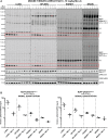
(A) HEK293 cell extracts overexpressing LRRK2[Y1699C] and the indicated HA-tagged Rab protein were treated ±150 nM MLi-2 for 90 min and then lysed. The cell lysates (0.1 µg) were immunoblotted with the indicated MJFF-pRab10 hybridoma clones (all at 0.5 µg/ml antibody). (B) The indicated wild-type and knock-out A549 cells were treated ±100 nM MLi-2 for 90 min and then lysed. The cell lysates (20 µg) were immunoblotted with the indicated MJFF-pRab10 hybridoma clones as well as shown total antibodies (all at 0.5 µg/ml antibody). (C) The indicated amounts of recombinant LRRK2-phosphorylated Rab10 were subjected to immunoblotting with the specified MJFF-pRab10 hybridoma clones as well as the total RAB10 antibody purchased from Cell Signaling Technology (CST) (all at 0.5 µg/ml antibody). (D) LRRK2[R1441G] MEFSs were treated ±100 nM MLi-2 for 90 min and then lysed. The cell lysates (5 µg) were immunoblotted with the indicated MJFF-pRab10 hybridoma clones (all at 0.5 µg/ml antibody). (E) The indicated littermate wild-type and knock-in or knock-out MEF cells were treated ±100 nM MLi-2 for 90 min and lysed. The cell lysates (5 µg) were immunoblotted with the indicated antibodies and the membranes developed using the Odyssey CLx scan Western Blot imaging system (all at 1 µg/ml antibody). Quantitation of immunoblots was undertaken by analysing pRab10/tubulin (Ei). Immunoblots were developed with LI-COR.
Figure 5.
Figure 5.. Characterisation of MJFF-pRab10 rabbit monoclonal antibodies in wild-type and LRRK2[R1441G] knock-in mouse tissue extracts.
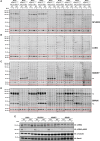
Wild-type and LRRK2[R1441G] knock-in mice were administered with MLi-2 (3 mg/kg) by subcutaneous injection. After 60 min, animals were killed and the spleen (A), lung (B), kidney (C) and brain (D) extracts were generated and immunoblotted with the indicated MJFF-pRab10 hybridoma clones (all at 0.5 µg/ml antibody). Thirty micrograms of the extract were immunoblotted for the spleen, lung, kidney and 40 µg for brain. (E) The denoted extracts were immunoblotted with the indicated total and phospho-Ser935 LRRK2 antibodies. The regions of the nitrocellulose membrane containing phosphorylated Rab10 are highlighted in a red box, *NS, major non-specific bands. Immunoblots were developed with LI-COR.
Figure 6.
Figure 6.. Characterisation of MJFF-pRab10clone-1 and MJFF-pRab10clone-2 rabbit monoclonal antibodies in LRRK2[R1441C] knock-in mouse tissue extracts.
LRRK2[R1441C] knock-in mice were administered with MLi-2 (10 mg/kg) by subcutaneous injection. After 60 min, animals were killed, and the lung, spleen, kidney and brain extracts were generated and immunoblotted with the indicated MJFF-pRab10 hybridoma clones (at 1 µg/ml antibody). Forty micrograms of the extract were immunoblotted for the spleen, lung, kidney and brain (A). Quantitation of immunoblots was undertaken by analysing pRab10/GAPDH (B). The regions of the nitrocellulose membrane containing phosphorylated Rab10 are highlighted in a red box. *NS, major non-specific bands. Immunoblots were developed with LI-COR.
Figure 7.
Figure 7.. Phosphorylation of Rab10 in control and idiopathic Parkinson's disease patients' cingulate cortex.
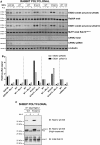
(A) Cingulate cortex samples derived from Control (Ctrl) and idiopathic Parkinson's disease (IPD) donors were obtained from the Queen Square Brain Bank (UCL). Extracts from each of these were generated and 20 µg subjected to immunoblot analysis using the indicated antibodies. Donors marked with an asterisk are over 90 years of age. The sample marked with an arrow (IPD donor 1) displays the highest levels of Rab10 phosphorylation. (B) As in (A) except that samples were subjected to Phos-tag polyacrylamide gel electrophoresis prior to immunoblot analysis employing a total Rab10 antibody. Cell lysates prepared from LRRK2[R1441C] knock-in MEFs were used as a positive control (+) for Rab10 phosphorylation and run in the last lane. The open circles on the Phos-tag gel correspond to the position that the phosphorylated Rab protein migrates and the closed circle the position that the dephosphorylated Rab protein migrates. (C) Control (donors 1 and 2) and IPD samples (donors 1 and 2) were subjected to immunoblot analysis using the MJFF-pRab10clone-1–3 monoclonal antibodies. The arrow indicates the band corresponding to the phospho-Rab10 signal. Immunoblots were developed with LI-COR.
Figure 8.
Figure 8.. Characterisation of the selectivity and potency of MJFF-pRab8 rabbit monoclonal antibodies.
(A) HEK293 cell extracts overexpressing LRRK2[Y1699C] and the indicated HA-tagged Rab protein were treated ±150 nM MLi-2 for 90 min and then lysed. The cell lysates (0.1 µg) were immunoblotted with the indicated MJFF-pRab8 hybridoma clones (all at 0.5 µg/ml antibody). (B) The indicated wild-type and knock-out A549 cells were treated ±100 nM MLi-2 for 90 min and then lysed. The cell lysates (20 µg) were immunoblotted with the specified MJFF-pRab8 hybridoma clones and the denoted total antibodies (all at 0.5 µg/ml antibody). (C) The shown amounts of recombinant LRRK2-phosphorylated Rab8A were subjected to immunoblotting with the indicated MJFF-pRab8 hybridoma clones as well as the total RAB8A antibody purchased from Cell Signaling Technology (CST) (all at 0.5 µg/ml antibody). Immunoblots were developed with LI-COR.
Figure 9.
Figure 9.. The pan-selective MJFF-pRab8 antibodies immunoprecipiate various LRRK2-phosphorylated Rab proteins.
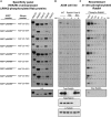
Littermate wild-type and LRRK2[R1441C] knock-in MEFs were treated ±100 nM MLi-2 for 90 min and lysed. The cell lysates (5 mg) were subjected to immunoprecipitation using the indicated MJFF-pRab8 monoclonal antibodies (20 µg) or rabbit polyclonal antibody (E8263) (50 µg). Forty percent of the immunoprecipitates was immunoblotted with total Rab43 antibody, 40% with total Rab35 antibody, 10% with Rab8A antibody and 10% with total Rab10 antibody (all at 1 µg/ml antibody). The specificities of the total Rab8A, Rab10 and Rab35 have been previously validated using appropriate A549 CRISPR/CAS9 knock-out cell extracts [12]. Immunoblots were developed with ECL.
Figure 10.
Figure 10.. Comparison of the phospho-immunoblotting and Phos-tag protocols to study LRRK2-mediated phosphorylation of Rab proteins.
(A) HEK293 cell extracts overexpressing LRRK2[Y1699C] and the indicated HA-Rab8A (A) or HA-Rab10 (B) were treated ±150 nM MLi-2 for 90 min and then lysed. The indicated amounts of cell lysates were subjected to either conventional polyacrylamide gel electrophoresis (upper panel) or Phos-tag polyacrylamide gel electrophoresis (lower panel). Proteins on both gels were transferred to nitrocellulose and subjected to identical immunoblot analysis with indicated antibodies (all at 0.5 µg/ml antibody). The open circles on the Phos-tag gel correspond to the position that phosphorylated Rab protein migrates, and the closed circle to the position that the dephosphorylated Rab protein migrates. Immunoblots were developed with ECL.

Comment in

Similar articles

Cited by

References

    1. Zimprich A., Biskup S., Leitner P., Lichtner P., Farrer M., Lincoln S. et al. (2004) Mutations in LRRK2 cause autosomal-dominant parkinsonism with pleomorphic pathology. Neuron 44, 601–607 10.1016/j.neuron.2004.11.005 - DOI - PubMed
    1. Paisán-Ruı´z C., Jain S., Evans E.W., Gilks W.P., Simón J., van der Brug M. et al. (2004) Cloning of the gene containing mutations that cause PARK8-linked Parkinson's disease. Neuron 44, 595–600 10.1016/j.neuron.2004.10.023 - DOI - PubMed
    1. Simón-Sánchez J., Schulte C., Bras J.M., Sharma M., Gibbs J.R., Berg D. et al. (2009) Genome-wide association study reveals genetic risk underlying Parkinson's disease. Nat. Genet. 41, 1308–1312 10.1038/ng.487 - DOI - PMC - PubMed
    1. Cookson M.R. (2017) Mechanisms of mutant LRRK2 neurodegeneration. Adv. Neurobiol. 14(suppl. 1), 227–239 10.1007/978-3-319-49969-7_12 - DOI - PubMed
    1. Ozelius L.J., Senthil G., Saunders-Pullman R., Ohmann E., Deligtisch A., Tagliati M. et al. (2006) LRRK2 g2019s as a cause of Parkinson's disease in Ashkenazi Jews. N. Engl. J. Med. 354, 424–425 10.1056/NEJMc055509 - DOI - PubMed

Publication types

MeSH terms

Substances