. 2017 Dec:113:494-504.
doi: 10.1016/j.freeradbiomed.2017.10.380. Epub 2017 Oct 28.

Respiratory syncytial virus induces NRF2 degradation through a promyelocytic leukemia protein - ring finger protein 4 dependent pathway

Affiliations

Respiratory syncytial virus induces NRF2 degradation through a promyelocytic leukemia protein - ring finger protein 4 dependent pathway

Narayana Komaravelli et al. Free Radic Biol Med. 2017 Dec.

Abstract

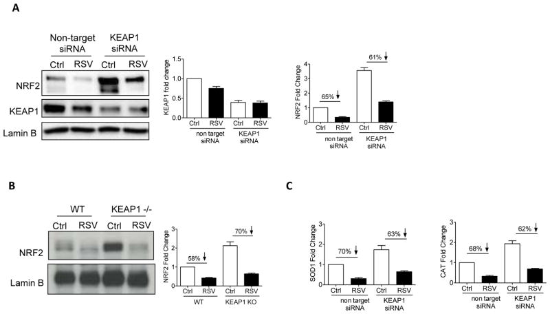
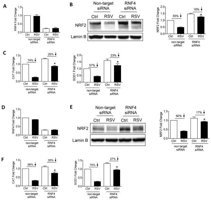
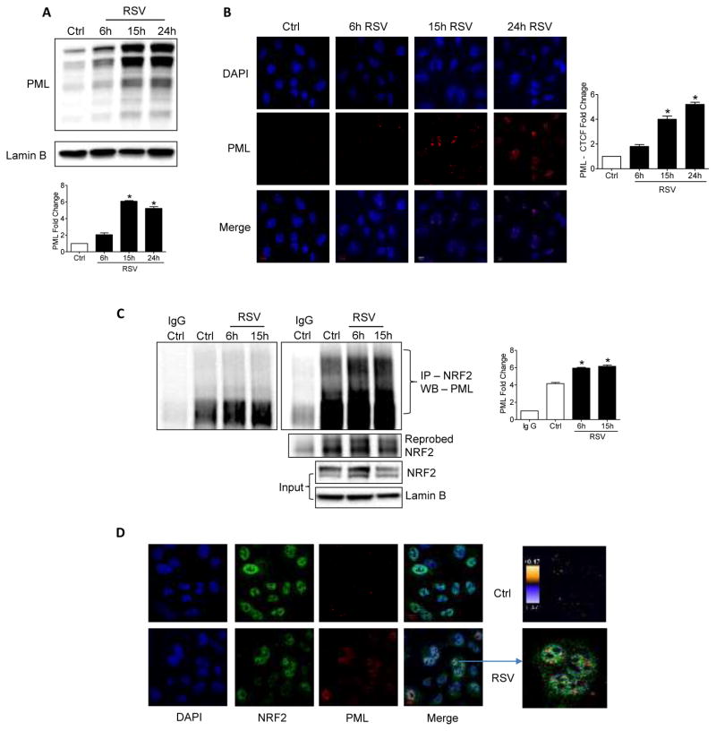
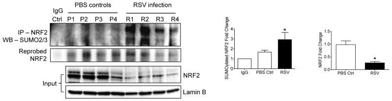
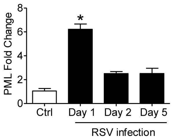
Respiratory syncytial virus (RSV) is the most important cause of viral acute respiratory tract infections and hospitalizations in children, for which no vaccine or specific treatments are available. RSV causes airway mucosa inflammation and cellular oxidative damage by triggering production of reactive oxygen species and by inhibiting at the same time expression of antioxidant enzymes, via degradation of the transcription factor NF-E2-related factor 2 (NRF2). RSV infection induces NRF2 deacetylation, ubiquitination, and degradation through a proteasome-dependent pathway. Although degradation via KEAP1 is the most common mechanism, silencing KEAP1 expression did not rescue NRF2 levels during RSV infection. We found that RSV-induced NRF2 degradation occurs in an SUMO-specific E3 ubiquitin ligase - RING finger protein 4 (RNF4)-dependent manner. NRF2 is progressively SUMOylated in RSV infection and either blocking SUMOylation or silencing RNF4 expression rescued both NRF2 nuclear levels and transcriptional activity. RNF4 associates with promyelocytic leukemia - nuclear bodies (PML-NBs). RSV infection induces the expression of PML and PML-NBs formation in an interferon (INF)-dependent manner and also induces NRF2 - PMN-NBs association. Inhibition of PML-NB formation by blocking IFN pathway or silencing PML expression resulted in a significant reduction of RSV-associated NRF2 degradation and increased antioxidant enzyme expression, identifying the RNF4-PML pathway as a key regulator of antioxidant defenses in the course of viral infection.

Keywords: Antioxidant enzymes; KEAP1; NRF2; PML nuclear bodies; RNF4; Respiratory syncytial virus; SUMOylation; Ubiquitination.


Conflict of interest statement

DECLARATION OF INTEREST

Conflicts of interest: none

Figures

Figure 1
Figure 1
(A) Nuclear protein prepared from SAE cells transfected with nontarget siRNA or KEAP1 siRNA, uninfected or infected with RSV for 18 h, were subjected to Western blot analysis with anti-NRF2 antibody. Membranes were stripped and reprobed with anti-KEAP1 and anti-Lamin B antibodies for loading control. The blots are representative of three independent experiments. Densitometric analysis of NRF2 band intensity is shown after normalization to Lamin B. The groups were analyzed by one-way ANOVA followed by Tukey’s post hoc test. Data are shown as mean ± SEM. (B) Nuclear protein prepared from wild type (WT) or KEAP1 knockout mouse embryonic fibroblasts, uninfected or infected with RSV for 24 h, were subjected to Western blot analysis with anti-NRF2 antibody. Membranes were stripped and reprobed with anti-Lamin B antibody for loading control. The blots are representative of three independent experiments. Densitometric analysis of NRF2 band intensity is shown after normalization to Lamin B. The groups were analyzed by one-way ANOVA followed by Tukey’s post hoc test. Data are shown as mean ± SEM. (C) SAE cells transfected with nontarget siRNA or KEAP1 siRNA, uninfected or infected with RSV for 18 h, were harvested to prepare total RNA. KEAP1 (left panel), catalase (middle panel) and SOD1 (right panel) gene expression were quantified by RT-PCR. Data are representative of three independent experiments. The groups were analyzed by one-way ANOVA followed by Tukey’s post hoc test. Data are shown as mean ± SEM.
Figure 2
Figure 2
A549 cells (A to C) and SAE cells (D to F) transfected with non-target siRNA or RNF4 siRNA, uninfected or infected with RSV for 18 h, were harvested to prepare total RNA or nuclear proteins. RNF4 (A and D), catalase and SOD1 (C and F) gene expression were quantified by RT-PCR, while NRF2 levels were analyzed by Western blot analysis (B and D). Membranes were stripped and reprobed with anti-Lamin B antibody for loading control. Densitometric analysis of NRF2 band intensity is shown after normalization to Lamin B. Data are representative of three independent experiments. The groups were analyzed by one-way ANOVA followed by Tukey’s post hoc test. Data are shown as mean ± SEM. *P < 0.05 relative to nontarget transfected, infected cells.
Figure 3
Figure 3
(A) Nuclear proteins of A549 cells infected with RSV for 3, 6 and 15 h were immunoprecipitated using anti-NRF2 antibody and subjected to Western blot using anti-SUMO2/3 antibody. Membranes were stripped and reprobed with anti-NRF2 antibody to determine the level of immunoprecipitated NRF2. Lower panel shows NRF2 Western blot of input proteins and Lamin B as loading control. The blots are representative of three independent experiments. The groups were analyzed by one-way ANOVA followed by Tukey’s post hoc test. Data are shown as mean ± SEM. *P < 0.05 relative to uninfected control cells. Nuclear extracts from A549 cells (B) or SAE cells (D) uninfected or infected with RSV for 18h in the presence or absence of 10 μM Anacardic acid were subjected to Western blot analysis using anti-NRF2 antibody. Membranes were stripped and reprobed with anti-Lamin B antibody for loading control. The blots are representative of three independent experiments. Densitometric analysis of NRF2 band intensity is shown after normalization to Lamin B. The groups were analyzed by one-way ANOVA followed by Tukey’s post hoc test. Data are shown as mean ± SEM. *P < 0.05 relative to untreated, RSV infected cells. (C) A549 cells or (E) SAE cells uninfected or infected with RSV for 18 h in the presence or absence of 10 μM Anacardic acid were harvested to prepare total RNA. Catalase and SOD1 gene expression were quantified by RT-PCR. Data are representative of three independent experiments. The groups were analyzed by one-way ANOVA followed by Tukey’s post hoc test. Data are shown as mean ± SEM. *P < 0.05 relative to untreated, RSV infected cells. (F) Nuclear protein prepared from A549 cells transfected with nontarget siRNA or SUMO2/3 siRNA, uninfected or infected with RSV for 18 h, were immunoprecipitated using anti-NRF2 antibody and immune complexes were analyzed by Western blots using anti-SUMO2/3 and anti-ubiquitin antibodies. Membranes were stripped and reprobed with anti-NRF2 antibody to determine the level of immunoprecipitated NRF2. Lower panel shows NRF2 Western blot of input proteins and Lamin B as internal control. Blots are representative of three independent experiments. Densitometric analysis of SUMOylated NRF2 and input NRF2 band intensities are shown. The groups were analyzed by one-way ANOVA followed by Tukey’s post hoc test. Data are shown as mean ± SEM. *P < 0.05 relative to untreated RSV infected cells.
Figure 4
Figure 4
(A) A549 cells were infected with RSV for 6, 15 and 24 h and nuclear protein were subjected to Western blot using anti-PML antibody. Membranes were stripped and reprobed with Lamin B for loading control. Densitometric analysis of PML band intensity is shown after normalization to Lamin B. The blots are representative of three independent experiments. The groups were analyzed by one-way ANOVA followed by Tukey’s post hoc test. Data are shown as mean ± SEM. *P < 0.05 relative to uninfected control cells. (B) Immunofluorescence and confocal microscopy analysis of uninfected or RSV infected A549 cells (6–24h p.i.). Nuclei (DAPI), PML (red) and merged images are shown. Scale bar, 10μm. Image analysis was done by ImageJ software. The plotted graph represents total cell fluorescence (CTCF) signal intensity from 50 cells of six different fields. Data are shown as mean ± SEM. *P < 0.05 relative to uninfected control cells. (C) A549 cells were infected with RSV for 6 h and 15 h and nuclear protein were immunoprecipitated using anti-NRF2 antibody and subjected to Western blot using anti-PML antibodies. Two panels of different exposure times are shown for better visibility of higher and lower molecular weight PML bands. Membranes were stripped and reprobed with anti-NRF2 antibody to determine the level of immunoprecipitated NRF2. Lower panel shows NRF2 Western blot of input proteins and Lamin B as loading control. The blots are representative of three independent experiments. The groups were analyzed by one-way ANOVA followed by Tukey’s post hoc test. Data are shown as mean ± SEM. *P < 0.05 relative to uninfected control cells. (D) Immunofluorescence and confocal microscopy analysis of uninfected or RSV infected A549 cells (15h p.i.). Nuclei (DAPI), NRF2 (green), PML (red) and merged images are shown. Scale bar, 10μm. Enlarged image of cells showing co-localization of NRF2 and PML, as well as PDM image of the same cells, are shown.
Figure 5
Figure 5
(A) Immunofluorescence and confocal microscopy analysis of A549 cells uninfected or infected with RSV for 18 h in the presence or absence of 5 μM Cerdulatinib. Nuclei (DAPI), PML (red) and merged images are shown. Scale bar, 10μm. Image analysis was done by ImageJ software. The plotted graph represents total cell fluorescence (CTCF) signal intensity from 50 cells of six different fields. Data are shown as mean ± SEM. *P < 0.05 relative to untreated, RSV infected cells. (B) Nuclear protein prepared from A549 cells uninfected or infected with RSV for 18 h in the presence or absence of 5 μM Cerdulatinib were subjected to Western blot analysis using anti-PML and anti-NRF2 antibody. Membranes were stripped and reprobed with anti-Lamin B antibody for loading control. The blots are representative of three independent experiments. Densitometric analysis of PML and NRF2 band intensities were shown after normalization to Lamin B. The groups were analyzed by one-way ANOVA followed by Tukey’s post hoc test. Data are shown as mean ± SEM. *P < 0.05 relative to untreated, RSV infected cells. (C) A549 cells transfected with nontarget siRNA or PML siRNA, uninfected or infected with RSV for 18 h, were harvested to prepare nuclear protein and subjected to Western blot analysis with anti-NRF2 and -PML antibody. Membranes were stripped and reprobed with anti-Lamin B antibody for loading control. The blots are representative of three independent experiments. Densitometric analysis of PML and NRF2 band intensities are shown after normalization to Lamin B. The groups were analyzed by one-way ANOVA followed by Tukey’s post hoc test. Data are shown as mean ± SEM. *P < 0.05 relative to PML siRNA transfected cells to nontarget siRNA transfected cells. (D) A549 cells uninfected or infected with RSV for 18h in the presence or absence of 5 μM Cerdulatinib were harvested to prepare total RNA. Catalase (left panel) and SOD1 (right panel) gene expression were quantified by real-time PCR. Data are representative of three independent experiments. The groups were analyzed by one-way ANOVA followed by Tukey’s post hoc test. Data are shown as mean ± SEM. *P < 0.05 relative to untreated, RSV infected cells. (E) A549 cells transfected with nontarget siRNA or siRNAs for PML, uninfected or infected with RSV for 18 h, were harvested to prepare total RNA. Catalase (left panel) and SOD1 (right panel) gene expression were quantified by real-time PCR. Data are representative of three independent experiments. The groups were analyzed by one-way ANOVA followed by Tukey’s post hoc test. Data are shown as mean ± SEM. *P < 0.05 relative to PML siRNA transfected cells to nontarget transfected cells. (F) Nuclear protein prepared from A549 cells uninfected or infected with RSV for 12 h in the presence or absence of 5 μM Cerdulatinib were immunoprecipitated using anti-NRF2 antibody and immune complexes were analyzed by Western blots using anti-SUMO2/3 and anti-ubiquitin antibodies. Membranes were stripped and reprobed with anti-NRF2 antibody to determine the level of immunoprecipitated NRF2. Lower panel shows NRF2 Western blot of input proteins and Lamin B as internal control. Blots are representative of three independent experiments. Densitometric analysis of SUMOylated NRF2 and ubiquitinated NRF2 band intensities are shown. The groups were analyzed by one-way ANOVA followed by Tukey’s post hoc test. Data are shown as mean ± SEM. *P < 0.05 relative to untreated RSV infected cells.
Figure 6
Figure 6
Nuclear protein isolated from lungs of mice that were either PBS inoculated (P1–P4) or infected with RSV (R1–R4) for 24h were immunoprecipitated using anti-NRF2 antibody and subjected to Western blot using anti-SUMO2/3 antibody. Membrane was stripped and reprobed with anti-NRF2 antibody. Lower panel shows NRF2 Western blot for input of the IP. Lamin B was used as loading control. The blots are representative of three independent experiments. Densitometric analysis of SUMOylated NRF2 and input NRF2 is shown after normalization to Lamin B. The groups were analyzed by one-way ANOVA followed by Tukey’s post hoc test. Data are shown as mean ± SEM. *P < 0.05 relative to PBS inoculated mice.
Figure 7
Figure 7
Total RNA isolated from lungs of mice that were either PBS-inoculated or infected with RSV for 1, 2 or 5 days. PML gene expression was quantified by RT-PCR. Data are representative of three independent experiments. The groups were analyzed by one-way ANOVA followed by Tukey’s post hoc test. Data are shown as mean ± SEM. *P < 0.05 relative to PBS inoculated mice.

Similar articles

Cited by

References

    1. Hall CB, Weinberg GA, Iwane MK, Blumkin AK, Edwards KM, Staat MA, et al. The burden of respiratory syncytial virus infection in young children. N Engl J Med. 2009;360:588–98. - PMC - PubMed
    1. Hall CB. Respiratory syncytial virus and parainfluenza virus. N Engl J Med. 2001;344:1917–28. - PubMed
    1. Falsey AR, Hennessey PA, Formica MA, Cox C, Walsh EE. Respiratory syncytial virus infection in elderly and high-risk adults. N Engl J Med. 2005;352:1749–59. - PubMed
    1. Nair H, Nokes DJ, Gessner BD, Dherani M, Madhi SA, Singleton RJ, et al. Global burden of acute lower respiratory infections due to respiratory syncytial virus in young children: a systematic review and meta-analysis. Lancet. 2010;375:1545–55. - PMC - PubMed
    1. Burr ML, Butland BK, King S, Vaughan-Williams E. Changes in asthma prevalence: two surveys 15 years apart. Arch Dis Child. 1989;64:1452–6. - PMC - PubMed

Publication types

MeSH terms