

The Spatial Organization of the Intranuclear Structures of Human Brain Dopaminergic Neurons
- PMID: 29104779
- PMCID: PMC5662277
The Spatial Organization of the Intranuclear Structures of Human Brain Dopaminergic Neurons
Abstract
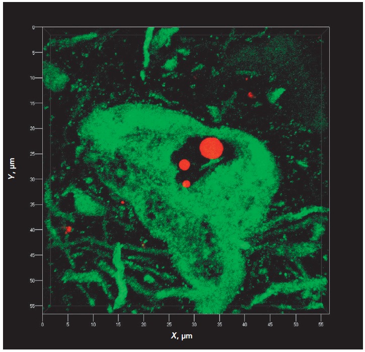
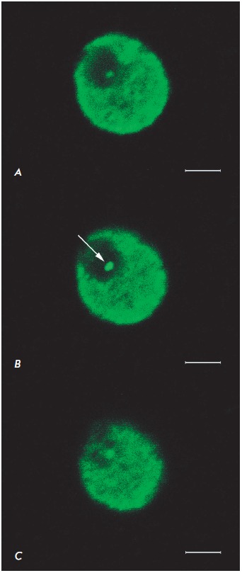
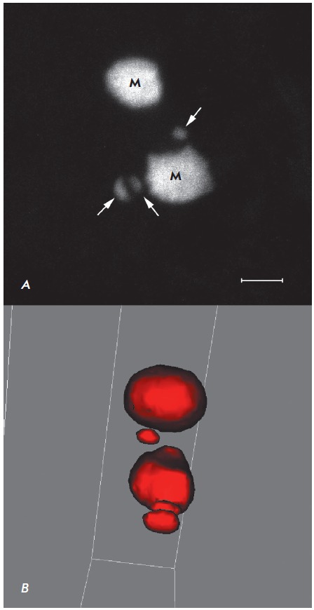
We studied the intranuclear localization of protein nucleophosmin (B23) and ubiquitin in the dopaminergic neurons of human substantia nigra (n = 6, age of 25-87 years) using immunohistochemistry and confocal laser microscopy. Intranuclear ubiquitin-immunopositive bodies that morphologically correspond to Marinesco bodies were found to be present in substantia nigra dopaminergic (tyrosine hydroxylase-immunopositive) neurons but absent in non-dopaminergic neurons. The number of bodies varied from 0 to 6 per cell nucleus. Nucleophosmin (B23) was found in the neuronal nucleolus, with the nucleolus size being constant in the nigral neurons of each individual brain. All the observed neurons had only one large nucleolus with intense nucleophosmin immunoreactivity and a lightly stained region (1-2 μm in diameter) that apparently represents the giant fibrillar center (GFC). An intensely immunostained nucleophosmin-containing granule was often observed at the GFC periphery. Double labeling demonstrated that nucleophosmin-immunoreactive nucleolus and ubiquitin-immunoreactive Marinesco bodies can occur both closely to and remotely from each other. Three-dimensional reconstruction indicates that rounded Marinesco bodies are polymorphic and often have a complex shape, with some flattening and concavities, which may be associated with contact not only with the nucleolus, but also, presumably, with other intranuclear structures free of ubiquitin or nucleophosmin. Ubiquitin-immunoreactive structures with a relatively small size (up to 1 μm in length) and various clastosome-like shapes (Lafarga et al., 2002) often occur near Marinesco bodies. There were no cases of detection of ubiquitin in the nucleoli of dopaminergic neurons and nucleophosmin/B23 in typical Marinesco bodies. The obtained information may be helpful in unraveling the molecular mechanisms of the selective vulnerability of substantia nigra dopaminergic neurons to damaging factors.
Keywords: Marinesco body; brain; dopaminergic neurons; human; nucleolus; nucleophosmin; substantia nigra; ubiquitin.
Figures


Similar articles
-
[INTRANUCLEAR UBIQUITIN-IMMUNOPOSITIVE STRUCTURES OF THE HUMAN SUBSTANTIA NIGRA NEURONS].Tsitologiia. 2015;57(11):780-7. Tsitologiia. 2015. PMID: 27012092 Russian.
-
[DISTRIBUTION OF THE MARINESCO BODIES IN THE NEURONS OF HUMAN BRAIN SUBSTANTIA NIGRA].Morfologiia. 2015;148(5):28-31. Morfologiia. 2015. PMID: 26987214 Russian.
-
Intranuclear rodlets in the substantia nigra: interactions with marinesco bodies, ubiquitin, and promyelocytic leukemia protein.J Neuropathol Exp Neurol. 2004 Nov;63(11):1200-7. doi: 10.1093/jnen/63.11.1200. J Neuropathol Exp Neurol. 2004. PMID: 15581187
-
Promyelocytic leukemia protein is redistributed during the formation of intranuclear inclusions independent of polyglutamine expansion: an immunohistochemical study on Marinesco bodies.J Neuropathol Exp Neurol. 2002 Nov;61(11):984-91. doi: 10.1093/jnen/61.11.984. J Neuropathol Exp Neurol. 2002. PMID: 12430715
-
Substantia nigra Marinesco bodies are associated with decreased striatal expression of dopaminergic markers.J Neuropathol Exp Neurol. 2004 Apr;63(4):329-37. doi: 10.1093/jnen/63.4.329. J Neuropathol Exp Neurol. 2004. PMID: 15099023
Cited by
-
Neuronal Senescence in the Aged Brain.Aging Dis. 2023 Oct 1;14(5):1618-1632. doi: 10.14336/AD.2023.0214. Aging Dis. 2023. PMID: 37196117 Free PMC article. Review.
References
-
- Gavrilov A.A., Razin S.V.. Mol Biol (Mosk). 2015;49(1):26–45. - PubMed
-
- Kettner M., Willwohl D., Hubbard G.B., Rüb U., Dick E.J. Jr., Cox A.B., Trottier Y., Auburger G., Braak H., Schultz C.. Exp. Neurol. 2002;176:117–121. - PubMed
-
- Grigorev I.P., Korzhevskii D.E., Medical Academic Journal. 2015;15(2):28–34.
-
- Okuwaki M.. J. Biochem. 2008;143(4):441–448. - PubMed
-
- Colombo E., Alcalay M., Pelicci P.G.. Oncogene. 2011;30(23):2595–2609. - PubMed
LinkOut - more resources
Full Text Sources