

A novel mtDNA repair fusion protein attenuates maladaptive remodeling and preserves cardiac function in heart failure
- PMID: 29101177
- PMCID: PMC5867654
- DOI: 10.1152/ajpheart.00515.2017
A novel mtDNA repair fusion protein attenuates maladaptive remodeling and preserves cardiac function in heart failure
Abstract
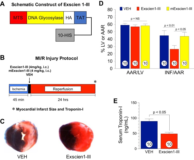
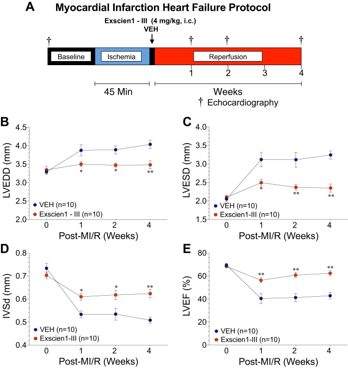
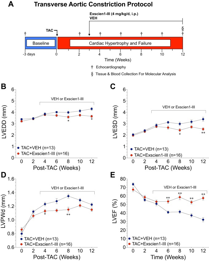
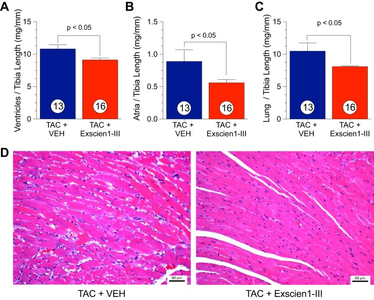
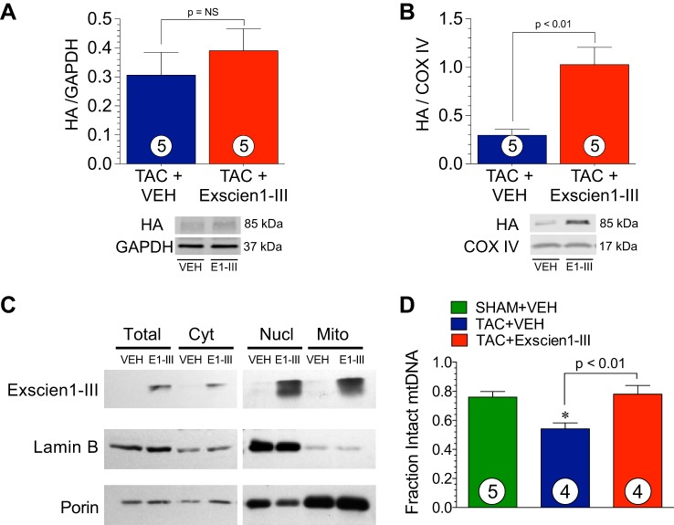
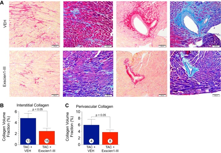
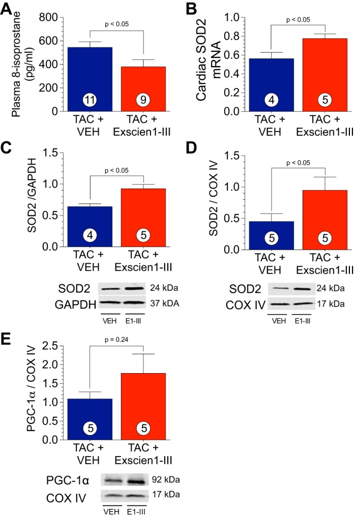
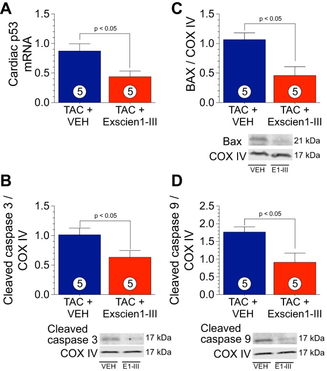
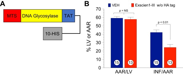
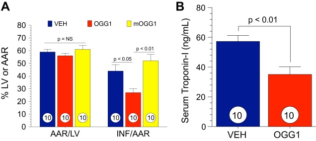
Oxidative stress results in mtDNA damage and contributes to myocardial cell death. mtDNA repair enzymes are crucial for mtDNA repair and cell survival. We investigated a novel, mitochondria-targeted fusion protein (Exscien1-III) containing endonuclease III in myocardial ischemia-reperfusion injury and transverse aortic constriction (TAC)-induced heart failure. Male C57/BL6J mice (10-12 wk) were subjected to 45 min of myocardial ischemia and either 24 h or 4 wk of reperfusion. Exscien1-III (4 mg/kg ip) or vehicle was administered at the time of reperfusion. Male C57/BL6J mice were subjected to TAC, and Exscien1-III (4 mg/kg i.p) or vehicle was administered daily starting at 3 wk post-TAC and continued for 12 wk. Echocardiography was performed to assess left ventricular (LV) structure and function. Exscien1-III reduced myocardial infarct size ( P < 0.01) at 24 h of reperfusion and preserved LV ejection fraction at 4 wk postmyocardial ischemia. Exscien1-III attenuated TAC-induced LV dilation and dysfunction at 6-12 wk post-TAC ( P < 0.05). Exscien1-III reduced ( P < 0.05) cardiac hypertrophy and maladaptive remodeling after TAC. Assessment of cardiac mitochondria showed that Exscien1-III localized to mitochondria and increased mitochondrial antioxidant and reduced apoptotic markers. In conclusion, our results indicate that administration of Exscien1-III provides significant protection against myocardial ischemia and preserves myocardial structure and LV performance in the setting of heart failure. NEW & NOTEWORTHY Oxidative stress-induced mitochondrial DNA damage is a prominent feature in the pathogenesis of cardiovascular diseases. In the present study, we demonstrate the efficacy of a novel, mitochondria-targeted fusion protein that traffics endonuclease III specifically for mitochondrial DNA repair in two well-characterized murine models of cardiac injury and failure.
Keywords: DNA repair; hypertrophic cardiomyopathy; mitochondria-targeted peptide; mitochondrial injury; myocardial infarction.
Figures
Comment in
-
"Mighty-chondrial" DNA repair for mitigation of cardiac injury: focus on "A novel mtDNA repair fusion protein attenuates maladaptive remodeling and preserves cardiac function in heart failure".Am J Physiol Heart Circ Physiol. 2018 Feb 1;314(2):H268-H269. doi: 10.1152/ajpheart.00661.2017. Epub 2017 Nov 16. Am J Physiol Heart Circ Physiol. 2018. PMID: 29146615 Free PMC article. No abstract available.
Similar articles
-
Water-soluble alkaloids extracted from Aconiti Radix lateralis praeparata protect against chronic heart failure in rats via a calcium signaling pathway.Biomed Pharmacother. 2021 Mar;135:111184. doi: 10.1016/j.biopha.2020.111184. Epub 2021 Jan 5. Biomed Pharmacother. 2021. PMID: 33418305
-
Adiponectin determines farnesoid X receptor agonism-mediated cardioprotection against post-infarction remodelling and dysfunction.Cardiovasc Res. 2018 Aug 1;114(10):1335-1349. doi: 10.1093/cvr/cvy093. Cardiovasc Res. 2018. PMID: 29668847
-
USP20 deletion promotes eccentric cardiac remodeling in response to pressure overload and increases mortality.Am J Physiol Heart Circ Physiol. 2024 Nov 1;327(5):H1257-H1271. doi: 10.1152/ajpheart.00329.2024. Epub 2024 Oct 4. Am J Physiol Heart Circ Physiol. 2024. PMID: 39365672
-
Myocardial reverse remodeling: how far can we rewind?Am J Physiol Heart Circ Physiol. 2016 Jun 1;310(11):H1402-22. doi: 10.1152/ajpheart.00696.2015. Epub 2016 Mar 18. Am J Physiol Heart Circ Physiol. 2016. PMID: 26993225 Review.
-
Peripheral Blood Mitochondrial DNA and Myocardial Function.Adv Exp Med Biol. 2017;982:347-358. doi: 10.1007/978-3-319-55330-6_19. Adv Exp Med Biol. 2017. PMID: 28551797 Review.
Cited by
-
Novel Molecular Targets Participating in Myocardial Ischemia-Reperfusion Injury and Cardioprotection.Cardiol Res Pract. 2019 May 28;2019:6935147. doi: 10.1155/2019/6935147. eCollection 2019. Cardiol Res Pract. 2019. PMID: 31275641 Free PMC article. Review.
-
Energy substrate metabolism and oxidative stress in metabolic cardiomyopathy.J Mol Med (Berl). 2022 Dec;100(12):1721-1739. doi: 10.1007/s00109-022-02269-1. Epub 2022 Nov 17. J Mol Med (Berl). 2022. PMID: 36396746 Review.
-
Mitochondria and cardiovascular diseases-from pathophysiology to treatment.Ann Transl Med. 2018 Jun;6(12):256. doi: 10.21037/atm.2018.06.21. Ann Transl Med. 2018. PMID: 30069458 Free PMC article. Review.
-
Calpain-mediated protein targets in cardiac mitochondria following ischemia-reperfusion.Sci Rep. 2022 Jan 7;12(1):138. doi: 10.1038/s41598-021-03947-9. Sci Rep. 2022. PMID: 34997008 Free PMC article.
-
"Mighty-chondrial" DNA repair for mitigation of cardiac injury: focus on "A novel mtDNA repair fusion protein attenuates maladaptive remodeling and preserves cardiac function in heart failure".Am J Physiol Heart Circ Physiol. 2018 Feb 1;314(2):H268-H269. doi: 10.1152/ajpheart.00661.2017. Epub 2017 Nov 16. Am J Physiol Heart Circ Physiol. 2018. PMID: 29146615 Free PMC article. No abstract available.
References
-
- Bhushan S, Kondo K, Polhemus DJ, Otsuka H, Nicholson CK, Tao YX, Huang H, Georgiopoulou VV, Murohara T, Calvert JW, Butler J, Lefer DJ. Nitrite therapy improves left ventricular function during heart failure via restoration of nitric oxide-mediated cytoprotective signaling. Circ Res 114: 1281–1291, 2014. doi:10.1161/CIRCRESAHA.114.301475. - DOI - PMC - PubMed
-
- Bradley JM, Islam KN, Polhemus DJ, Donnarumma E, Brewster LP, Tao YX, Goodchild TT, Lefer DJ. Sustained release nitrite therapy results in myocardial protection in a porcine model of metabolic syndrome with peripheral vascular disease. Am J Physiol Heart Circ Physiol 309: H305–H317, 2015. doi:10.1152/ajpheart.00163.2015. - DOI - PMC - PubMed
Publication types
MeSH terms
Substances
Grants and funding
LinkOut - more resources
Full Text Sources
Other Literature Sources
Medical