. 2017 Nov 3;14(1):213.
doi: 10.1186/s12985-017-0875-5.

Investigation of antiviral state mediated by interferon-inducible transmembrane protein 1 induced by H9N2 virus and inactivated viral particle in human endothelial cells

Affiliations

Investigation of antiviral state mediated by interferon-inducible transmembrane protein 1 induced by H9N2 virus and inactivated viral particle in human endothelial cells

Bo Feng et al. Virol J. .

Abstract

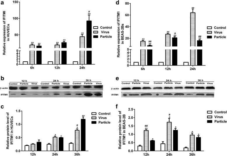
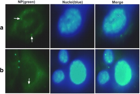
Background: Endothelial cells are believed to play an important role in response to virus infection. Our previous microarray analysis showed that H9N2 virus infection and inactivated viral particle inoculation increased the expression of interferon-inducible transmembrane protein 1 (IFITM1) in human umbilical vein endothelial cells (HUVECs). In present study, we deeply investigated the expression patterns of IFITM1 and IFITM1-mediated antiviral response induced by H9N2 virus infection and inactivated viral particle inoculation in HUVECs. Epithelial cells that are considered target cells of the influenza virus were selected as a reference control.

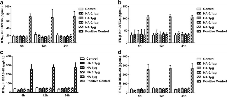
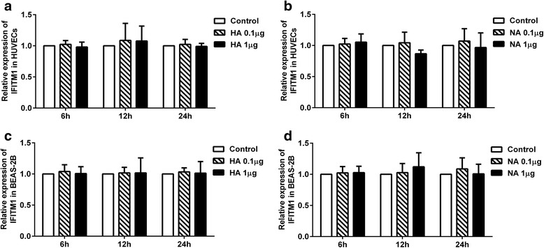
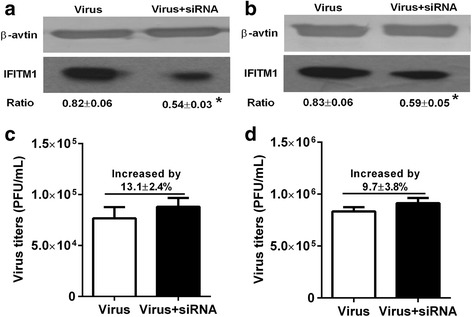
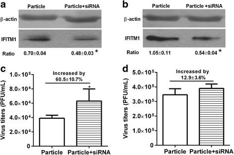
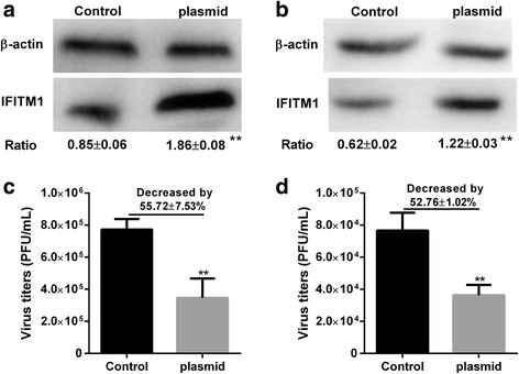
Methods: First, we quantified the expression levels of IFITM1 in HUVECs induced by H9N2 virus infection or viral particle inoculation using quantitative real-time PCR and western blot. Second, we observed whether hemagglutinin or neuraminidase affected IFITM1 expression in HUVECs. Finally, we investigated the effect of induced-IFITM1 on the antiviral state in HUVECs by siRNA and activation plasmid transfection.

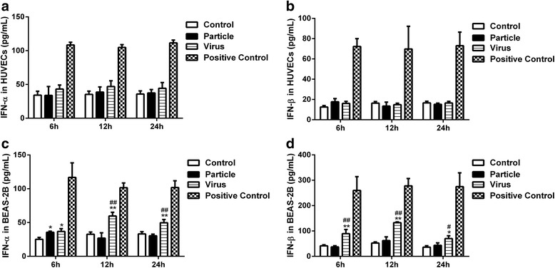
Results: Both H9N2 virus infection and viral particle inoculation increased the expression of IFITM1 without elevating the levels of interferon-ɑ/β in HUVECs. HA or NA protein binding alone is not sufficient to increase the levels of IFITM1 and interferon-ɑ/β in HUVECs. IFITM1 induced by viral particle inoculation significantly decreased the virus titers in culture supernatants of HUVECs.

Conclusions: Our results showed that inactivated viral particle inoculation increased the expression of IFITM1 at mRNA and protein levels. Moreover, the induction of IFITM1 expression mediated the antiviral state in HUVECs.

Keywords: Antiviral state; H9N2 influenza virus; Human endothelial cells; Human epithelial cells; IFITM1; Inactivated viral particle.


Conflict of interest statement

Ethics approval and consent to participate

Not applicable

Consent for publication

Not applicable

Competing interests

The authors declare that they have no competing interests.

Publisher’s Note

Springer Nature remains neutral with regard to jurisdictional claims in published maps and institutional affiliations.

Figures

Fig. 1
Fig. 1
Quantitation of IFITM1 induced by H9N2 virus or inactivated viral particle in HUVECs and BEAS-2Bs. HUVECs and BEAS-2Bs were treated with H9N2 virus (i.e., Virus) or viral particle (i.e., Particle) at MOI of 5. Cells used for RT-PCR and western blot analysis were collected at different time points. a The expression of IFITM1 at mRNA level in HUVECs. b, c The expression of IFITM1 at protein level in HUVECs, and the relative protein level to β-actin. d The expression of IFITM1 at mRNA level in BEAS-2Bs. e, f The expression of IFITM1 at protein level in BEAS-2Bs, and the relative protein level to β-actin. * means particle group and virus group compared with control group (*, P < 0.05. **, P < 0.01, ANOVA). # means particle group compared with H9N2 virus group (#, P < 0.05. ##, P < 0.01, ANOVA)
Fig. 2
Fig. 2
Production of interferon-α/β by HUVECs and BEAS-2Bs inoculated with H9N2 virus or viral particle. HUVECs and BEAS-2Bs were inoculated with H9N2 virus or viral particle at a MOI of 5, and then supernatants were collected at 6, 12, 24 h. Levels of interferon-α/β were detected using ELISA kits. Values represent the means from three independent experiments plus standard deviations. a, b The concentration of interferon-α/β in culture supernatants of HUVECs. c, d The concentration of interferon-α/β in culture supernatants of BEAS-2Bs. *means particle group and virus group compared with control group (*, P < 0.05. **, P < 0.01, ANOVA). # means particle group compared with H9N2 virus group (#, P < 0.05. ##, P < 0.01, ANOVA)
Fig. 3
Fig. 3
Evaluation of interferon-α/β in HUVECs and BEAS-2B incubated with hemagglutinin (HA) and neuraminidase (NA). HUVECs and BEAS-2Bs were incubated with HA or NA at concentrations of 0.1, 1 μg/mL, supernatants were collected at 6, 12, 24 h. Levels of interferon-α/β were determined using ELISA kits. Values represent the means from three independent experiments plus standard deviations. a, b The concentration of interferon-α/β in culture supernatants of HUVECs. c, d The concentration of interferon-α/β in culture supernatants of BEAS-2Bs
Fig. 4
Fig. 4
Expression of IFITM1 at mRNA level in HUVECs and BEAS-2Bs treated with hemagglutinin (HA) and neuraminidase (NA). HUVECs and BEAS-2Bs were incubated with HA or NA at concentrations of 0.1, 1 μg/mL. Cells used for RT-PCR analysis were collected at 6, 12, 24 h. a The mRNA levels of IFITM1 induced by HA in HUVECs. b The mRNA levels of IFITM1 induced by NA in HUVECs. c The mRNA levels of IFITM1 induced by HA in BEAS-2Bs. d The mRNA levels of IFITM1 induced by NA in BEAS-2Bs
Fig. 5
Fig. 5
Localization of inactivated viral particles in HUVECs and BEAS-2Bs. HUVECs and BEAS-2Bs were inoculated with viral particles at a MOI of 5 for 8 h, the distribution of viral particles were visualized by immunofluorescence. Cells in each group were double-stained with an anti-nucleoprotein antibody (NP, green) and 4′, 6-diamidino-2-phenylindole (DAPI, blue). a Localization of viral particles in HUVECs. b Localization of viral particles in BEAS-2Bs
Fig. 6
Fig. 6
Antiviral activity of IFITM1 induced by H9N2 virus infection in HUVECs and BEAS-2Bs. HUVECs and BEAS-2Bs were infected with H9N2 virus at MOI of 5 and incubated for 1 h, then cells were transfected with control siRNA or IFITM1 specific siRNA for 36 h. Virus titer in each group was detected using plaque assay at 36 h postinfection. The effect of IFIMT1 specific siRNA on IFITM1 expression was detected using western blot. a IFITM1 protein level after transfected with siRNA in HUVECs. b IFITM1 protein level after transfected with siRNA in BEAS-2Bs. c Virus titers in HUVECs transfected with control siRNA or IFITM1 specific siRNA. Compared to virus group (control siRNA), the virus titer in virus + siRNA group (IFITM1 specific siRNA) was increased by 13.1 ± 2.4% (P > 0.05, t-test). d Virus titers in BEAS-2Bs transfected with control siRNA or IFITM1 specific siRNA. Compared to virus group (control siRNA), the virus titer in virus + siRNA group (specific siRNA) was increased by 9.7% ± 3.8% (P > 0.05, t-test)
Fig. 7
Fig. 7
Antiviral activity of IFITM1 induced by viral particle inoculation in HUVECs and BEAS-2Bs. HUVECs and BEAS-2Bs were inoculated with viral particle at MOI of 5 and incubated for 1 h, then cells were transfected with control siRNA or IFITM1 specific siRNA for 36 h before infected with H9N2 virus at MOI of 5. Virus titer of each group was detected by plaque assay at 36 h postinfection. The effect of IFIMT1 specific siRNA on IFITM1 expression was detected using western blot. a IFITM1 protein level after transfected with siRNA in HUVECs. b IFITM1 protein level after transfected with siRNA in BEAS-2Bs. c Virus titers in HUVECs transfected with control siRNA or IFITM1 specific siRNA. Compared to particle group (control siRNA), the virus titer in particle + siRNA group (IFITM1 specific siRNA) was increased by 60.5 ± 10.7% (P < 0.05, t-test). d Virus titers in BEAS-2Bs transfected with control siRNA or IFITM1 specific siRNA. Compared to particle group (control siRNA), the virus titer in particle + siRNA group (specific siRNA) was increased by 12.9 ± 3.6% (P > 0.05, t-test). * means particle group compared with particle + siRNA group (*, P < 0.05, t-test)
Fig. 8
Fig. 8
Overexpression of IFITM1 significantly reduced virus titers in HUVECs and BEAS-2Bs. HUVECs and BEAS-2Bs were transfected with control plasmid (Control) or IFITM1 CRISPR activation plasmid (plasmid) for 36 h, then cells were infected with H9N2 virus at MOI of 5. Virus titer of each group was detected by plaque assay at 36 h postinfection. The overexpression of IFIMT1 was detected by western blot. a IFITM1 protein level after transfected with plasmid in BEAS-2Bs. b IFITM1 protein level after transfected with plasmid in HUVECs. c Virus titers in BEAS-2Bs transfected with control plasmid or IFITM1 CRISPR activation plasmid. Compared to control group (control plasmid), the virus titer in plasmid group (IFITM1 CRISPR activation plasmid) was decreased by 55.72.5 ± 7.53% (P < 0.01, t-test). d Virus titers in HUVECs transfected with control plasmid or IFITM1 CRISPR activation plasmid. Compared to control group (control plasmid), the virus titer in plasmid group (IFITM1 CRISPR activation plasmid) was decreased by 52.76 ± 1.02% (P < 0.01, t-test). * means control group compared with plasmid group (**, P < 0.01, t-test)

Similar articles

Cited by

References

    1. Guo YJ, Krauss S, Senne DA, Mo IP, Lo KS, Xiong XP, Norwood M, Shortridge KF, Webster RG, Guan Y. Characterization of the pathogenicity of members of the newly established H9N2 influenza virus lineages in Asia. Virology. 2000;267:279–288. doi: 10.1006/viro.1999.0115. - DOI - PubMed
    1. Lee CW, Song CS, Lee YJ, Mo IP, Garcia M, Suarez DL, Kim SJ. Sequence analysis of the hemagglutinin gene of H9N2 Korean avian influenza viruses and assessment of the pathogenic potential of isolate MS96. Avian Dis. 2000;44:527–535. doi: 10.2307/1593091. - DOI - PubMed
    1. Butt KM, Smith GJ, Chen H, Zhang LJ, Leung YH, Xu KM, Lim W, Webster RG, Yuen KY, Peiris JS, Guan Y. Human infection with an avian H9N2 influenza a virus in Hong Kong in 2003. J Clin Microbiol. 2005;43:5760–5767. doi: 10.1128/JCM.43.11.5760-5767.2005. - DOI - PMC - PubMed
    1. Cong YL, Pu J, Liu QF, Wang S, Zhang GZ, Zhang XL, Fan WX, Brown EG, Liu JH. Antigenic and genetic characterization of H9N2 swine influenza viruses in China. J Gen Virol. 2007;88:2035–2041. doi: 10.1099/vir.0.82783-0. - DOI - PubMed
    1. Cong YL, Wang CF, Yan CM, Peng JS, Jiang ZL, Liu JH. Swine infection with H9N2 influenza viruses in China in 2004. Virus Genes. 2008;36:461–469. doi: 10.1007/s11262-008-0227-z. - DOI - PubMed

MeSH terms

LinkOut - more resources