. 2017 Nov 1;8(1):1246.
doi: 10.1038/s41467-017-01255-3.

Endosomal phosphatidylserine is critical for the YAP signalling pathway in proliferating cells

Affiliations

Endosomal phosphatidylserine is critical for the YAP signalling pathway in proliferating cells

Tatsuyuki Matsudaira et al. Nat Commun. .

Abstract

Yes-associated protein (YAP) is a recently discovered growth-promoting transcription coactivator that has been shown to regulate the malignancy of various cancers. How YAP is regulated is not fully understood. Here, we show that one of the factors regulating YAP is phosphatidylserine (PS) in recycling endosomes (REs). We use proximity biotinylation to find proteins proximal to PS. Among these proteins are YAP and multiple proteins related to YAP signalling. Knockdown of ATP8A1 (an RE PS-flippase) or evectin-2 (an RE-resident protein) and masking of PS in the cytoplasmic leaflet of membranes, all suppress nuclear localization of YAP and YAP-dependent transcription. ATP8A1 knockdown increases the phosphorylated (activated) form of Lats1 that phosphorylates and inactivates YAP, whereas evectin-2 knockdown reduces the ubiquitination and increased the level of Lats1. The proliferation of YAP-dependent metastatic cancer cells is suppressed by knockdown of ATP8A1 or evectin-2. These results suggest a link between a membrane phospholipid and cell proliferation.


Conflict of interest statement

The authors declare no competing financial interests.

Figures

Fig. 1
Fig. 1
Identification of proteins proximal to PS in live cells. a Schematic illustration of biotinylation of proteins proximal to PS with GFP-BirA*-2xPH. b COS-1 cells stably expressing GFP-BirA*-2xPH were incubated with or without 50 µM biotin for 24 h. The cells were then fixed, permeabilized, and stained for TfnR and biotin with Alexa594-streptavidin. c, d Lysates from cells in b were mixed with streptavidin-coupled magnetic beads. The proteins pulled down by the beads were blotted with streptavidin-HRP in c, or the indicated antibodies in d. Nuclei were stained with DAPI. Scale bars, 10 μm
Fig. 2
Fig. 2
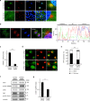
A PS-flippase at REs contributes to the nuclear localization of YAP. a COS-1 cells at low density or high density were fixed, permeabilized, and stained for YAP and TfnR. Magnified images of the boxed areas around the perinuclear REs are shown in the right column. b COS-1 cells at low density were fixed, permeabilized, and stained for YAP and phosphorylated YAP (S127). A fluorescence intensity profile along the dotted line in the magnified image is shown in the right panel. c qRT-PCR analysis of ATP8A1 mRNA in COS-1 cells treated with ATP8A1 siRNA for 48 h. GAPDH was used as an internal control. d COS-1 cells were treated with control or ATP8A1 siRNA for 48 h. The cells were then fixed, permeabilized, and stained for YAP. e Subcellular localization of YAP in cells in d was examined. f Lysates from cells in d were immunoblotted for the indicated proteins. α-tubulin was used as a loading control. g qRT-PCR analysis of CTGF mRNA from cells in d. GAPDH was used as an internal control. Data are mean ± s.d. from two (for e, n > 40 cells) or three (for c, g) independent experiments. Statistical significance was determined with two-tailed Student’s t-test (for c, g) or two-sided Fisher’s exact test (for e); **P < 0.01, ***P < 0.001. Nuclei were stained with DAPI. Scale bars, 10 μm
Fig. 3
Fig. 3
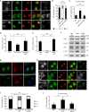
Cellular PS levels contribute to the nuclear localization of YAP. a GFP, GFP-2xPH (evectin-2, WT), or GFP-2xPH (K20E) was expressed in COS-1 cells for 24 h. The cells were then fixed, permeabilized, and stained for YAP and p-YAP. b Subcellular localization of YAP in cells in a was examined. c The fluorescence intensity of p-YAP in a was quantified and normalized to that of GFP-transfected cells. d Transcriptional coactivity of YAP was examined by the TEAD reporter system. GFP, GFP-2xPH, or GFP-2xPH (K20E) was co-expressed with 8xGTIIC TEAD reporter and pRL-TK (as internal control). e PSA-3 cells were cultured for 72 h with three different conditions (FBS + 10 μM Etn, dFBS, or dFBS + 40 μM Etn). qRT-PCR analysis of CTGF mRNA in these cells was then performed. GAPDH was used as an internal control. f Lysates from cells in e were immunoblotted for the indicated proteins. α-tubulin was used as a loading control. g COS-1 cells were cotransfected with GFP-2xPH and Myc-yPSD1ΔN (WT or S463A). After 24 h, the cells were fixed and stained for Myc. h COS-1 cells were transfected with mKate2, Myc-yPSD1ΔN (WT), or Myc-yPSD1ΔN (S463A). After 24 h, the cells were fixed and costained for YAP, p-YAP, and Myc. i Subcellular localization of YAP in cells in h was examined. j The fluorescence intensity of p-YAP in h was quantified and normalized to that of mKate2-transfected cells. Data are mean ± s.d. from two (for b, c, n > 30 cells), three (for e), three (for i, j, n > 85 cells), or six (for d) independent experiments. Statistical significance was determined with one-way analysis of variance followed by Tukey–Kramer post hoc test (for c, d, e, j) or Kruskal–Wallis test followed by Steel–Dwass post hoc test (for b, i); *P < 0.05, ***P < 0.001, NS not significant. The nuclei were stained with DAPI. Scale bars, 10 μm
Fig. 4
Fig. 4
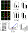
Evectin-2 contributes to the nuclear localization of YAP. a COS-1 cells were treated with control siRNA, evectin-2 siRNA#1, or evectin-2 siRNA#2 for 48 h. The cells were then fixed, permeabilized, and stained for YAP. b Subcellular localization of YAP in cells in a was examined. c qRT-PCR analysis of CTGF mRNA from cells in a. GAPDH was used as an internal control. d COS-1 cells were treated with control, Rab11, or VAMP3 siRNA for 48 h. The cells were then fixed, permeabilized, and stained for YAP. e Subcellular localization of YAP in cells in d was examined. f qRT-PCR analysis of CTGF mRNA in MDA-MB-231 cells depleted of evectin-2. GAPDH was used as an internal control. g MDA-MB-231 cells were treated with evectin-2 siRNA for 72 h. Cells were lysed and immunoblotted for the indicated proteins. α-tubulin was used as a loading control. h MDA-MB-231 cells were treated with siRNA for evectin-2, ATP8A1, or YAP/TAZ for 48 h and then trypsinized. A total of 5 × 104 cells were replated and further treated with the same siRNA 24 h after replating. The cells were counted every 24 h after replating. i MDA-MB-231 cells were treated with siRNA for evectin-2, evectin-2/Lats1, or evectin-2/Lats1/Lats2 for 48 h and then trypsinized. A total of 5 × 104 cells were replated and further treated with the same siRNA 24 h after replating. The cells were counted 96 h after replating. Data are mean ± s.d. from two (for b, e, n > 40 cells) or three (for c, f, h, i) independent experiments. Statistical significance was determined with two-tailed Student’s t test (for f), one-way analysis of variance followed by Tukey–Kramer post hoc test (for c, h, i), or Kruskal–Wallis test followed by Steel–Dwass post hoc test (for b, e); **P < 0.01, ***P < 0.001, NS not significant. The nuclei were stained with DAPI. Scale bars, 10 μm
Fig. 5
Fig. 5
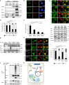
Binding of evectin-2 and Nedd4 E3 ligases is required for the nuclear localization of YAP. a FLAG-tagged evectin-2 was expressed in COS-1 cells for 24 h. Cell lysates with 1% Triton X-100 were immunoprecipitated with anti-FLAG antibody. The lysate (0.8%) and the immunoprecipitated fraction (10%) were then blotted for FLAG, Itch, WWP1, or WWP2. β-actin was used as a loading control. b Indicated proteins were expressed in COS-1 cells. The cells were then fixed, permeabilized, and stained for Myc. c COS-1 cells were treated with control, Itch, WWP1, or WWP2 siRNA for 48 h. The cells were then fixed, permeabilized, and stained for YAP. d Subcellular localization of YAP in cells in c was examined. e COS-1 cells were treated with the indicated siRNAs for 48 h. qRT-PCR analysis of CTGF mRNA was performed. GAPDH was used as an internal control. f The lysate of COS-1 cells in e was immunoblotted for the indicated proteins. α-tubulin was used as a loading control. A closed arrowhead indicates WWP1. g FLAG-tagged evectin-2 protein (WT or mutants) was expressed in COS-1 cells for 24 h. Cell lysates with 1% Triton X-100 were immunoprecipitated with anti-FLAG antibody. The lysate (0.8%) and the immunoprecipitated fraction (30%) were then blotted for FLAG, Itch, WWP1, or WWP2. α-tubulin was used as a loading control. h COS-1 cells that express siRNA-resistant Myc-evectin-2 WT or PPPA mutant in a cumate-dependent fashion were established. These cells were first treated with control or evectin-2 siRNA for 24 h, and incubated with 30 μM cumate for another 24 h. The cells were then fixed, permeabilized, and stained for YAP and Myc. Arrows indicate cells that express Myc-evt-2 WT or PPPA. i Subcellular localization of YAP in cells in h was examined. j FLAG-WWP2 and Myc-evectin-2 were expressed in HEK293T cells for 24 h. Cell lysates with 1% Triton X-100 were immunoprecipitated with anti-FLAG antibody. The immunoprecipitates were then blotted for ubiquitin (Ub), FLAG, or Myc. α-tubulin was used as a loading control. k A model of the regulation of YAP on RE membranes in proliferating cells. Data are mean ± s.d. from two (for d, n > 40 cells; for i, n > 30 cells) or three (for e) independent experiments. Statistical significance was determined with one-way analysis of variance followed by Tukey–Kramer post hoc test (for e) or Kruskal–Wallis test followed by Steel–Dwass post hoc test (for d, i); **P < 0.01, ***P < 0.001, NS not significant. The nuclei were stained with DAPI. Scale bars, 10 μm

Similar articles

Cited by

References

    1. Di Paolo G, De Camilli P. Phosphoinositides in cell regulation and membrane dynamics. Nature. 2006;443:651–657. doi: 10.1038/nature05185. - DOI - PubMed
    1. Behnia R, Munro S. Organelle identity and the signposts for membrane traffic. Nature. 2005;438:597–604. doi: 10.1038/nature04397. - DOI - PubMed
    1. van Meer G, Voelker DR, Feigenson GW. Membrane lipids: where they are and how they behave. Nat. Rev. Mol. Cell Biol. 2008;9:112–124. doi: 10.1038/nrm2330. - DOI - PMC - PubMed
    1. Devaux PF. Protein involvement in transmembrane lipid asymmetry. Annu. Rev. Biophys. Biomol. Struct. 1992;21:417–439. doi: 10.1146/annurev.bb.21.060192.002221. - DOI - PubMed
    1. Leventis PA, Grinstein S. The distribution and function of phosphatidylserine in cellular membranes. Annu. Rev. Biophys. 2010;39:407–427. doi: 10.1146/annurev.biophys.093008.131234. - DOI - PubMed

Publication types

MeSH terms

LinkOut - more resources