

Microstructure of the Distal Radius and Its Relevance to Distal Radius Fractures
- PMID: 29085733
- PMCID: PMC5658220
- DOI: 10.1055/s-0037-1602849
Microstructure of the Distal Radius and Its Relevance to Distal Radius Fractures
Erratum in
-
Erratum: Microstructure of the Distal Radius and Its Relevance to Distal Radius Fractures.J Wrist Surg. 2017 Nov;6(4):e1-e2. doi: 10.1055/s-0037-1603805. Epub 2017 Jun 16. J Wrist Surg. 2017. PMID: 29119975 Free PMC article.
Abstract
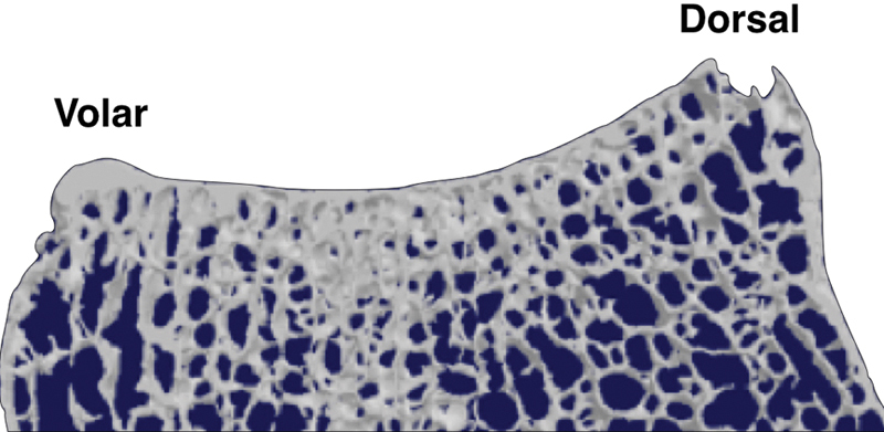
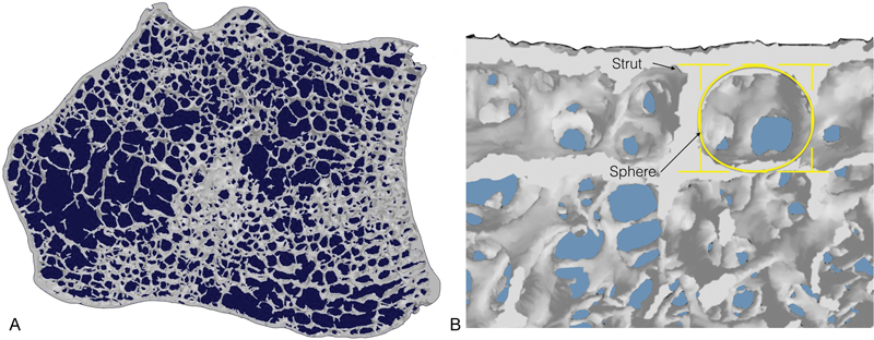
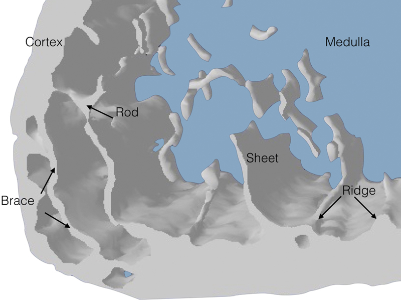
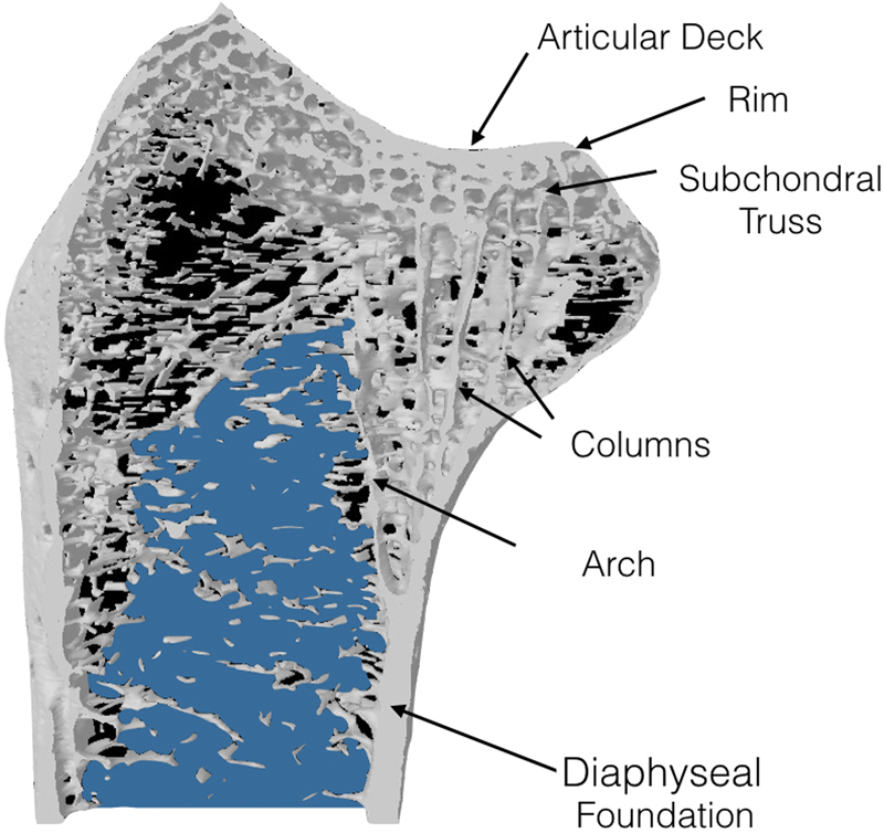
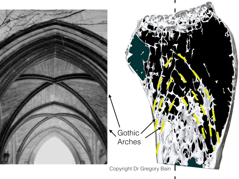
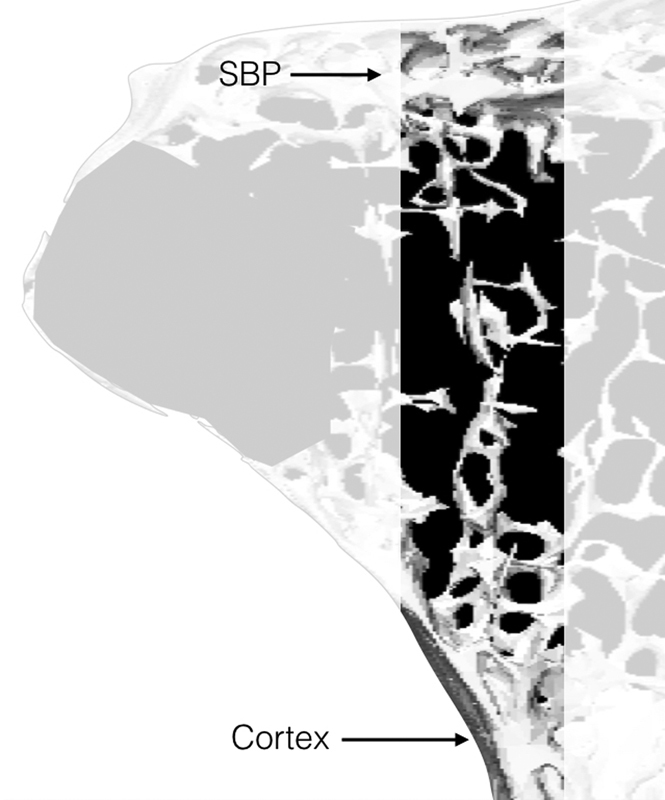
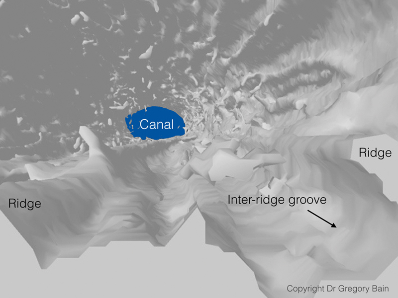
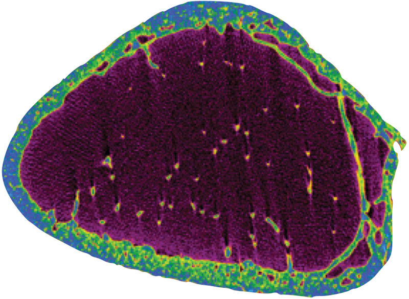
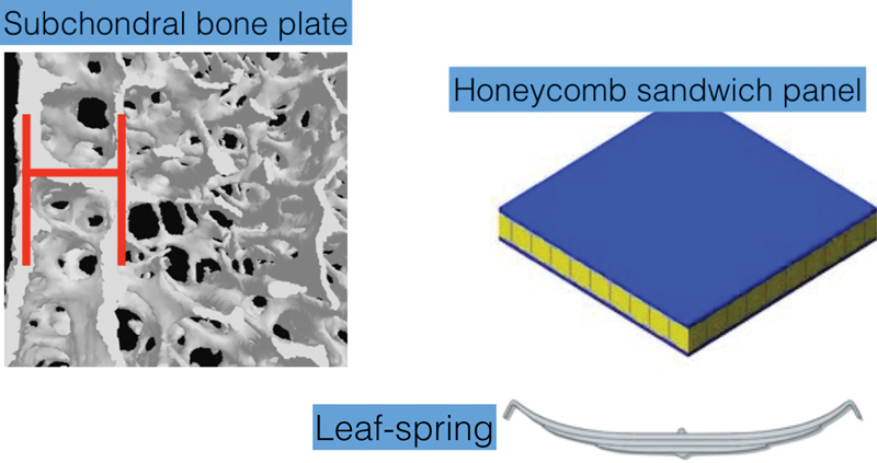
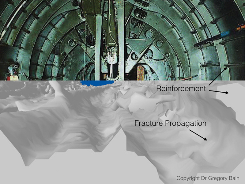
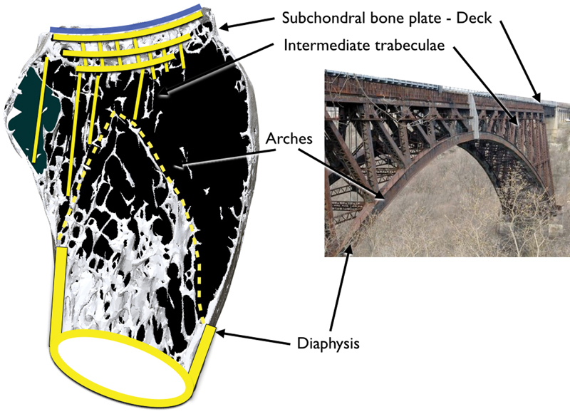
Background There is a paucity of information on the microstructure of the distal radius, and how this relates to its morphology and function. Purpose This study aims to assess the microanatomical structure of the distal radius, and relate this to its morphology, function, and modes of failure. Methods Six dry adult skeletal distal radii were examined with microcomputed tomography scan and analyzed with specialist computer software. From 3D and 2D images, the subchondral, cortical, and medullary trabecular were assessed and interpreted based on the overall morphology of the radius. Results The expanded distal radial metaphysis provides a wide articular surface for distributing the articular load. The extrinsic wrist ligaments are positioned around the articular perimeter, except on the dorsal radial corner. The subchondral bone plate is a 2 mm multilaminar lattice structure, which is thicker below the areas of the maximal articular load. There are spherical voids distally, which become ovoid proximally, which assist in absorbing articular impact. It does not have Haversian canals. From the volar aspect of the lunate facet, there are thick trabecular columns that insert into the volar cortex of the radius at the metaphyseal-diaphyseal junction. For the remainder of the subchondral bone plate, there is an intermediate trabecular network, which transmits the load to the intermediate trabeculae and then to the trabecular arches. The arches pass proximally and coalesce with the ridges of the diaphyseal cortex. Conclusion The distal radius morphology is similar to an arch bridge. The subchondral bone plate resembles the smooth deck of the bridge that interacts with the mobile load. The load is transmitted to the rim, intermediate struts, and arches. The metaphyseal arches allow the joint loading forces to be transmitted proximally and laterally, providing compression at all levels and avoiding tension. The arches have a natural ability to absorb the impact which protects the articular surface. The distal radius absorbs and transmits the articular impact to the medullary cortex and intermediate trabeculae. The medullary arches are positioned to transmit the load from the intermediate trabeculae to the diaphysis. Clinical Relevance The microstructure of the distal radius is likely to be important for physiological loading of the radius. The subchondral bone plate is a unique structure that is different to the cancellous and cortical bone. All three bone types have different functions. The unique morphology and microstructure of the distal radius allow it to transmit load and protect the articular cartilage.
Keywords: distal radius; fracture; micro-CT; microarchitecture; ultrastructure.
Conflict of interest statement
Figures


Similar articles
-
Deterioration of trabecular plate-rod and cortical microarchitecture and reduced bone stiffness at distal radius and tibia in postmenopausal women with vertebral fractures.Bone. 2016 Jul;88:39-46. doi: 10.1016/j.bone.2016.04.003. Epub 2016 Apr 12. Bone. 2016. PMID: 27083398 Free PMC article.
-
Cartilage and subchondral bone distributions of the distal radius: a 3-dimensional analysis using cadavers.Osteoarthritis Cartilage. 2020 Dec;28(12):1572-1580. doi: 10.1016/j.joca.2020.08.008. Epub 2020 Aug 26. Osteoarthritis Cartilage. 2020. PMID: 32860992
-
Characterization of trabecular bone microstructure in premenopausal women with distal radius fractures.Osteoporos Int. 2018 Feb;29(2):409-419. doi: 10.1007/s00198-017-4293-8. Epub 2017 Nov 3. Osteoporos Int. 2018. PMID: 29101409
-
Use of a distraction plate for distal radial fractures with metaphyseal and diaphyseal comminution. Surgical technique.J Bone Joint Surg Am. 2006 Mar;88 Suppl 1 Pt 1:29-36. doi: 10.2106/JBJS.E.01094. J Bone Joint Surg Am. 2006. PMID: 16510798 Review.
-
Sustentaculum Lunatum: Appreciation of the Palmar Lunate Facet in Management of Complex Intra-Articular Fractures of the Distal Radius.Am J Orthop (Belle Mead NJ). 2015 Sep;44(9):E303-7. Am J Orthop (Belle Mead NJ). 2015. PMID: 26372756 Review.
Cited by
-
Relationship between distal radius fracture severity and 25-hydroxyvitamin-D level among perimenopausal and postmenopausal women.Bone Jt Open. 2022 Mar;3(3):261-267. doi: 10.1302/2633-1462.33.BJO-2022-0004.R1. Bone Jt Open. 2022. PMID: 35311581 Free PMC article.
-
The Volar Cortical Hinge: An Independent Risk Factor for Distal Radius Fracture Displacement.J Wrist Surg. 2023 Aug 21;13(3):222-229. doi: 10.1055/s-0043-1771376. eCollection 2024 Jun. J Wrist Surg. 2023. PMID: 38808183 Free PMC article.
-
Posttraumatic Arthritis After Combined Plating of Distal Radius Fractures AO Type C: A 7-Year Follow-up of 97 Cases.Hand (N Y). 2022 Dec;17(1_suppl):50S-59S. doi: 10.1177/15589447211028991. Epub 2021 Sep 7. Hand (N Y). 2022. PMID: 34490825 Free PMC article.
-
Management of complex wrist fractures with volar and dorsal locked (double-locked) K-lock.OTA Int. 2023 Sep 22;6(4):e286. doi: 10.1097/OI9.0000000000000286. eCollection 2023 Dec. OTA Int. 2023. PMID: 37744996 Free PMC article.
-
Comparison of corticocancellous bone graft from the anterolateral metaphysis of the distal radius versus iliac crest for the treatment of unstable scaphoid nonunion with humpback deformity.BMC Musculoskelet Disord. 2024 Jan 2;25(1):20. doi: 10.1186/s12891-023-07134-x. BMC Musculoskelet Disord. 2024. PMID: 38167040 Free PMC article.
References
-
- Marble H C. Baltimore, MD: Williams & Wilkins; 1966. History of hand surgery; pp. 1–10.
-
- Chappard C. Microarchitecture assessment of human trabecular bone: description of methods [in French] 2012;28(12):1111–1115. - PubMed
-
- Singh M, Nagrath A R, Maini P S. Changes in trabecular pattern of the upper end of the femur as an index of osteoporosis. J Bone Joint Surg Am. 1970;52(03):457–467. - PubMed
-
- Currey J. Princeton, NJ: Princeton University Press; 2002. Bones: Structure and Mechanics.
-
- Currey J D. Bone architecture and fracture. Curr Osteoporos Rep. 2005;3(02):52–56. - PubMed
LinkOut - more resources
Full Text Sources
Other Literature Sources