. 2017 Oct 30;13(10):e1006700.
doi: 10.1371/journal.ppat.1006700. eCollection 2017 Oct.

Salmonella exploits the host endolysosomal tethering factor HOPS complex to promote its intravacuolar replication

Affiliations

Salmonella exploits the host endolysosomal tethering factor HOPS complex to promote its intravacuolar replication

Aastha Sindhwani et al. PLoS Pathog. .

Abstract

Salmonella enterica serovar typhimurium extensively remodels the host late endocytic compartments to establish its vacuolar niche within the host cells conducive for its replication, also known as the Salmonella-containing vacuole (SCV). By maintaining a prolonged interaction with late endosomes and lysosomes of the host cells in the form of interconnected network of tubules (Salmonella-induced filaments or SIFs), Salmonella gains access to both membrane and fluid-phase cargo from these compartments. This is essential for maintaining SCV membrane integrity and for bacterial intravacuolar nutrition. Here, we have identified the multisubunit lysosomal tethering factor-HOPS (HOmotypic fusion and Protein Sorting) complex as a crucial host factor facilitating delivery of late endosomal and lysosomal content to SCVs, providing membrane for SIF formation, and nutrients for intravacuolar bacterial replication. Accordingly, depletion of HOPS subunits significantly reduced the bacterial load in non-phagocytic and phagocytic cells as well as in a mouse model of Salmonella infection. We found that Salmonella effector SifA in complex with its binding partner; SKIP, interacts with HOPS subunit Vps39 and mediates recruitment of this tethering factor to SCV compartments. The lysosomal small GTPase Arl8b that binds to, and promotes membrane localization of Vps41 (and other HOPS subunits) was also required for HOPS recruitment to SCVs and SIFs. Our findings suggest that Salmonella recruits the host late endosomal and lysosomal membrane fusion machinery to its vacuolar niche for access to host membrane and nutrients, ensuring its intracellular survival and replication.


Conflict of interest statement

The authors have declared that no competing interests exist.

Figures
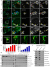
Fig 1
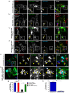
Fig 1. HOPS subunits are recruited to LAMP1-positive SCVs and SIFs during Salmonella infection.
a-d) Representative confocal micrographs of HeLa cells infected with DsRed-expressing Salmonella (red). At different time points post infection (p.i.), cells were fixed and stained for endogenous Vps41 (green) and LAMP1 (blue). Different panels represent a higher magnification of the boxed areas, showing recruitment of Vps41 on SCVs and SIFs (marked by yellow arrowheads). Red arrowheads indicate lysosomal localization of Vps41 in panels (a) and (b). Bars: (main) 10 μm; (insets) 5 μm. e) Time-lapse microscopy of HeLa cells co-transfected with plasmids encoding GFP-Vps41 and untagged-Arl8b, and infected with DsRed-expressing Salmonella (red). Time-lapse series were recorded 9 hr p.i., and still images shown here correspond to S2 Movie. Different panels represent a higher magnification of the boxed area indicating Vps41-positive SIFs emanating from the SCVs showing extension, retraction and bifurcation (white arrowheads). Red arrowheads indicate fusion of Vps41-positive vesicles with SIFs. Bars: (main) 10 μm; (insets) 5 μm. f and g) Quantification of endogenous (f) or HA-tagged Vps41 (g)-positive SCVs at different time points p.i. Data represent percentage of Vps41-postive SCVs scored for ~100 SCVs for each time point. The mean ± S.D. is shown for three independent experiments. h) SCVs were isolated from Salmonella-infected HeLa cells at 3 hr and 8 hr p.i. using sucrose density ultracentrifugation, followed by second round of ultracentrifugation of fractions 8–10 on a ficoll cushion (labeled as SCV). Different fractions were resolved on SDS-PAGE gel and immunoblotted using indicated antibodies. i) Salmonella-modified membranes were isolated from HeLa cells infected with sseF-deficient strain of Salmonella harboring an expression vector with a C-terminal epitope-tagged sseF and its cognate chaperone sscB (sseF/pSseF-HA) at 8 hr p.i. by differential centrifugation. The enriched fraction was further subjected to affinity immunoprecipitation (IP) using anti-HA antibody-conjugated agarose beads or anti-Myc antibody-conjugated agarose beads as a control. The eluted samples were analyzed for presence of effector protein (SseF) and host proteins by Western blotting as indicated.
Fig 2
Fig 2. Depletion of HOPS subunits impairs Salmonella replication.
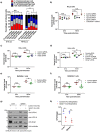
a) HeLa cells transfected with indicated siRNA were infected with Salmonella, fixed at the indicated time points, and immunostained with antibodies to Salmonella and LAMP1. Using confocal microscopy, the number of intracellular bacteria was enumerated in ~300 cells per experiment. These numbers were grouped according to the legend, and expressed as a percentage of the total infected cell population. The mean ± S.D. is shown for three independent experiments (n.s., not significant; *, P < 0.05; **, P < 0.01; Student’s t test). b-f) Intracellular replication assay. HeLa (b-d) and RAW264.7 (e and f) cells treated with indicated siRNA and infected with Salmonella were harvested at indicated times p.i. The number of colony forming units (CFU) per well are shown as dot plot and data represent mean ± S.D. (n.s., not significant; *, P < 0.05; **, P < 0.01; ***, P < 0.001; ****, P < 0.0001; Student’s t test). g and h) Mice were injected intravenously (i.v.) with control- or Vps41 specific-vivo-morpholinos (sample size of six for each treatment) for 2 days at an interval of 24 hr, followed by an i.v. challenge with 1.3 × 10³ CFU Salmonella. On day 3 p.i., mice were sacrificed and bacterial loads in spleen and liver were determined by plating serial dilutions of tissue homogenates on agar plates containing streptomycin (*, P < 0.05; **, P < 0.01; Student’s t test). The tissue homogenates were resolved on SDS-PAGE and immunoblotted with indicated antibodies by Western blotting.
Fig 3
Fig 3. Depletion of HOPS subunits delays but does not block SCV maturation.
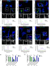
a-f) Representative confocal micrographs of control siRNA-, Vps41 siRNA- or Vps39 siRNA-treated HeLa cells infected with DsRed-expressing Salmonella (red). At different time points p.i., cells were fixed and stained for early endosomes marker, EEA1 (green), and LAMP1 (blue). Insets depict higher magnification of the boxed areas showing localization of different markers on the SCVs. Intensity line scan profile of EEA1/LAMP1 across the width of a single SCV (indicated by an arrow in the boxed region) is shown below the individual image. Bars: (main) 10 μm; (insets) 5 μm. g and h) Quantification of percentage of infected cells displaying EEA1/LAMP1-accumulation around SCVs at the indicated time point p.i. Data represent mean ± S.D. for ~50 SCVs from three independent experiments (n.s., not significant; *, P < 0.05; **, P < 0.01; ***, P < 0.001; ****, P < 0.0001; Student’s t test).
Fig 4
Fig 4. Depletion of HOPS subunits does not alter Rab7 recruitment to SCV.
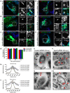
a-f) Representative confocal images of control-, Vps41-, or Vps39-siRNA treated HeLa cells, and subsequently transfected with GFP-Rab7 and infected with DsRed-expressing Salmonella (red). At different times p.i., cells were fixed and stained for LAMP1 (blue). Insets depict higher magnification of the boxed areas showing localization of Rab7 and LAMP1 around the SCVs. Bars: (main) 10 μm; (insets) 5 μm. g) Quantification of Rab7-positive SCVs in control-, Vps41- or Vps39-siRNA treated HeLa cells. Data represent mean ± S.D. over three independent experiments at the indicated time points where ~100 SCVs were counted in each experiment. h and i) Quantification of GFP-Rab7 intensity around the SCVs in control-, Vps41- or Vps39-siRNA treated cells over three independent experiments at the indicated time points p.i. where intensity profile of ~50 SCVs were quantified in each experiment. Data represent mean ± S.E.M. j-m) Representative immunogold EM images of control siRNA (j and k)- or Vps41 siRNA (l and m)-treated HeLa cells infected with Salmonella for 2 hr and transfected with HA-tagged Rab7 and fixed at 10 hr p.i. Cells were processed for immunogold labeling with anti-LAMP1 (10 nm) and anti-HA (15 nm) antibodies. Arrowheads indicate localization of Rab7 (red) and LAMP1 (black) around the SCVs. Bar: 500 nm.
Fig 5
Fig 5. Interaction of dextran-loaded lysosomes with SCVs is impaired upon Vps41 depletion.
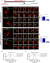
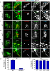
a) Schematic illustrating the protocol used for loading of lysosomes with Alexa-Fluor 647-conjugated dextran in HeLa and RAW264.7 cells, prior to infection with GFP-expressing Salmonella. b-e) HeLa cells treated with control siRNA (b) or Vps41 siRNA (c) or RAW264.7 cells transduced with control shRNA (d) or Vps41 shRNA (e) were pre-incubated with Alexa-Fluor 647-conjugated dextran (red) to label lysosomes, followed by infection with GFP-expressing Salmonella (green). Time-lapse series for Alexa-Fluor 647-conjugated dextran loaded and infected cells were recorded at 10 hr p.i., and still images from representative time lapse series are shown (S7–S10 Movies). Different panels represent a higher magnification of the boxed area and the white arrowheads indicate the SCVs. Bars: (main) 10 μm; (insets) 5 μm. f and g) Quantification of Alexa-Fluor 647-conjugated dextran-positive SCVs in control and Vps41 depleted HeLa and RAW264.7 cells fixed at 10 hr p.i. Data represent mean ± S.D. over three independent experiments where ~100 SCVs were counted in each experiment (****, P < 0.0001; Student’s t test). h and i) Quantification of Alexa-Fluor 647-conjugated dextran signal intensity around the SCVs in control and Vps41 depleted HeLa and RAW264.7 cells. Data represent mean ± S.E.M. of signal intensity from three independent experiments at 10 hr p.i. where ~50 SCVs were counted in each experiment.
Fig 6
Fig 6. Silencing of Vps41 abrogates docking of late endosomes and lysosomes at SCVs and impairs nutrient acquisition by auxotrophic strain of Salmonella.
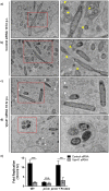
a-d) Representative TEM images of control (a and b) and Vps41 (c and d) siRNA treated HeLa cells infected with Salmonella for 10 hr. Higher magnification of multiple SCVs (marked by yellow arrowheads) interacting with late endosomes (containing MVBs) and lysosomes (containing lamellar membrane sheets) in control siRNA treated cells are shown in the panels on the right. In Vps41 siRNA treated cells, white arrowheads depict the MVBs containing late endosomal compartments. Bar: 500 nm. e) Intracellular replication of wild-type (WT) Salmonella and a mutant strain auxotrophic for proline (proC) was determined at indicated times p.i. in control siRNA- or Vps41 siRNA-transfected HeLa cells. The fold change in intracellular proliferation was calculated as the ratio of CFU at 16 hr p.i./CFU at 2 hr p.i. To complement the intracellular proliferation of proC Salmonella strain, cell culture medium was supplemented with proline (0.8 mM). Shown are the means ± S.D. from three independent experiments (n.s., not significant; ***, P < 0.001; ****, P < 0.0001; Student’s t test).
Fig 7
Fig 7. Deletion of Salmonella effector SifA abrogates Vps41 recruitment to SCV membranes.
a-f) HeLa cells were infected with either DsRed expressing-wild-type (WT) strain of Salmonella (NCTC 12023 in (a) and SL1344 in (c)) or sifA sseJ, sseF, sseG, and pipB2 strains, followed by transfection with HA tagged-Vps41. Cells were fixed at 10 hr p.i., and co-stained with anti-HA (green) and anti-LAMP1 (blue) antibodies. Different panels represent a higher magnification of the boxed areas. Bars: (main) 10 μm; (insets) 5 μm. g and h) Quantification of Vps41-positive SCVs in HeLa cells infected with different Salmonella strains (as labeled) and fixed at 10 hr p.i. Data represent mean ± S.D. from three independent experiments where ~100 SCVs were counted in each experiment (n.s., not significant; ****, P < 0.0001; Student’s t test).
Fig 8
Fig 8. Bacterial effector SifA localizes with HOPS subunit Vps39 in a SKIP-dependent manner.
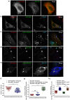
a-e) Representative confocal micrographs of HeLa cells co-transfected with HA-Vps39 (red) and Myc-SifA (green) (a), HA-Vps39 (red) and GFP-SKIP (green) (b), HA-Vps39 (red) and GFP-PLEKHM1 (green) (c), HA-Vps39 (red), GFP-SKIP (green) and Myc-SifA (blue) (d), or HA-Vps39 (red), GFP-PLEKHM1 (green) and Myc-SifA (blue) (e). Cells in (b) and (c) were also stained for lysosomes using with anti-LAMP1 (blue) antibodies. Bars: 10 μm. f-h) Pearson’s correlation coefficient was calculated for the indicated protein pairs in transfected cells as labeled. Data represent mean ± S.D. over three independent experiments where ~25–30 transfected cells were analyzed in each experiment (n.s., not significant; ****, P < 0.0001; Student’s t test).
Fig 9
Fig 9. SKIP is required for interaction between bacterial effector SifA and HOPS complex.
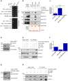
a) Yeast three-hybrid assay. Co-transformants were spotted on -Leu-Trp medium to check for viability, and on -Leu-Trp-His-Met media to test the interaction between SifA and Vps39 in the presence of SKIP (WT), SKIP (G828D) mutant or PLEKHM1. b) GST or GST tagged-SifA were immobilized on resin and incubated with lysates prepared from HEK293T cells transfected with HA-Vps39 alone or co-transfected with HA-Vps39 and FLAG-SKIP or FLAG-PLEKHM1. The precipitates were resolved by SDS-PAGE and immunoblotted with indicated antibodies. Ponceau S stain was done to visualize purified protein. c) Densitometric analysis of immunoblots of HA-Vps39 pulldown (normalized to input signal band intensity) by GST tagged-SifA in presence of FLAG-SKIP or FLAG-PLEKHM1. d) Control siRNA- or SKIP siRNA-treated HeLa cell lysates were resolved by SDS-PAGE, and immunoblotted with anti-SKIP antibody for assessing the knockdown efficiency and with anti-α-tubulin antibody as the loading control. e) Lysates from HEK293T cells treated with control- or SKIP-siRNA and transfected with indicated plasmids were immunoprecipitated using anti-Myc antibodies-conjugated resin. The cell lysates (inputs) and immunoprecipitates were resolved by SDS-PAGE and immunoblotted by Western blotting with the indicated antibodies. f) Densitometric analysis of immunoblots of HA-Vps39 co-immunoprecipitated (normalized to input signal band intensity) with Myc-SifA in control siRNA-, SKIP siRNA- or SKIP siRNA rescue construct-transfected HEK293T cell lysates. g) Lysates from control siRNA- or PLEKHM1 siRNA-treated HeLa cells were resolved by SDS-PAGE, and immunoblotted with anti-PLEKHM1 antibody for assessing the knockdown efficiency and with anti-α-tubulin antibody as the loading control. h) Lysates from HEK293T cells treated with control- or PLEKHM1-siRNA and expressing HA-Vps39 and Myc-SifA were immunoprecipitated with anti-HA antibodies-conjugated resin and precipitates were resolved on SDS-PAGE and immunoblotted with indicated antibodies.
Fig 10
Fig 10. Recruitment of HOPS subunit Vps41 to SCVs requires SifA-SKIP interaction.
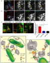
a and b) Representative confocal micrographs of control siRNA- or SKIP siRNA-treated HeLa cells infected with Salmonella, followed by co-transtection with plasmids expressing GFP-Vps41 (green) and untagged-Arl8b. Cells were fixed at 10 hr p.i., and immunostained with antibodies to Salmonella (blue) and LAMP1 (red). Arrowheads in the insets depict localization of Vps41 around SCV membranes (marked by LAMP1). c and d) Representative confocal micrographs of HeLa cells infected with Salmonella strains sifA/pSifA (WT)-2HA (c) or sifA/pSifA (L130D)-2HA (d), followed by co-transfection with plasmids expressing GFP-Vps41 (green) and untagged-Arl8b. Cells were fixed 10 hr p.i., and immunostained with antibodies to Salmonella (blue) and LAMP1 (red). Arrowheads in the insets depict localization of Vps41 around SCV membranes (marked by LAMP1). e and f) Representative confocal micrographs of HeLa cells treated with SKIP siRNA, infected with DsRed-expressing Salmonella, followed by transfection with plasmids expressing HA-Vps41, untagged-Arl8b and vector (e) or siRNA-resistant FLAG-SKIP (rescue construct) (f). Cells were fixed 10 hr p.i., and immunostained with antibodies to HA tag (green) and LAMP1 (blue). Different panels represent a higher magnification of the boxed areas, indicating Vps41 localization around SCV membranes (marked by arrowheads). Bars: (main) 10 μm; (insets) 5 μm. g) Quantification of HA-Vps41-postive SCVs at 10 hr p.i. in indicated siRNA treated HeLa cells. Data represent mean ± S.D. of ~50 SCVs scored in each experiment for three independent experiments (n.s., not significant; ***, P < 0.001; ****, P < 0.0001; Student’s t test). h) Quantification of HA-Vps41-positive SCVs at 10 hr p.i. in HeLa cells infected with indicated Salmonella strains. Data represent mean ± S.D. of ~100 SCVs scored in each experiment for three independent experiments (****, P < 0.0001; Student’s t test).
Fig 11
Fig 11. SifA-dependent lysosome clustering requires both SKIP and Vps39.
a-b) Representative confocal micrographs of HeLa cells co-transfected with Myc-SifA and GFP-LAMP1 plasmids. The cells were fixed and co-stained using anti-Myc (blue) and anti-Vps18 (a, red) or anti-Vps41 (b, red) antibodies. Different panels represent a higher magnification of the boxed areas and HOPS subunits enrichment on clustered lysosomes is indicated by the arrowheads. c-e) HeLa cells treated with indicated siRNA were transfected with Myc-SifA expressing plasmid. The cells were fixed and co-stained using anti-Myc (green) and anti-LAMP1 (red) antibodies. Insets show higher magnification of the boxed areas clustered lysosomes induced by SifA expression, which is dependent upon Vps39 and SKIP expression. Bars: (main) 10 μm; (insets) 5 μm. f) Average size of LAMP1-positive compartments calculated in cells treated with indicated siRNA and transfected with Myc-SifA plasmid. Data represent mean ± S.D. of ~25–30 transfected per experiment over three independent experiments (**, P < 0.01; Student’s t test). g) Schematic depicting the molecular machinery required for SCV fusion with late endosomes and lysosomes. Multisubunit tethering factor-HOPS complex is a target for Salmonella effector SifA, which associates with its known binding partner-SKIP to recruit HOPS complex to SCV membranes, thereby enabling SCV fusion with Arl8b-positive lysosomes.

Similar articles

Cited by

References

    1. LaRock DL, Chaudhary A, Miller SI (2015) Salmonellae interactions with host processes. Nat Rev Microbiol 13: 191–205. doi: 10.1038/nrmicro3420 - DOI - PMC - PubMed
    1. Liss V, Hensel M (2015) Take the tube: remodelling of the endosomal system by intracellular Salmonella enterica. Cell Microbiol 17: 639–647. doi: 10.1111/cmi.12441 - DOI - PubMed
    1. Figueira R, Holden DW (2012) Functions of the Salmonella pathogenicity island 2 (SPI-2) type III secretion system effectors. Microbiology 158: 1147–1161. doi: 10.1099/mic.0.058115-0 - DOI - PubMed
    1. Bujny MV, Ewels PA, Humphrey S, Attar N, Jepson MA, et al. (2008) Sorting nexin-1 defines an early phase of Salmonella-containing vacuole-remodeling during Salmonella infection. J Cell Sci 121: 2027–2036. doi: 10.1242/jcs.018432 - DOI - PubMed
    1. Steele-Mortimer O, Meresse S, Gorvel JP, Toh BH, Finlay BB (1999) Biogenesis of Salmonella typhimurium-containing vacuoles in epithelial cells involves interactions with the early endocytic pathway. Cell Microbiol 1: 33–49. - PubMed