. 2018 Mar 1;314(3):L388-L396.
doi: 10.1152/ajplung.00555.2016. Epub 2017 Oct 26.

Autophagy inhibitor 3-methyladenine protects against endothelial cell barrier dysfunction in acute lung injury

Affiliations

Autophagy inhibitor 3-methyladenine protects against endothelial cell barrier dysfunction in acute lung injury

Spencer A Slavin et al. Am J Physiol Lung Cell Mol Physiol. .

Abstract

Autophagy is an evolutionarily conserved cellular process that facilitates the continuous recycling of intracellular components (organelles and proteins) and provides an alternative source of energy when nutrients are scarce. Recent studies have implicated autophagy in many disorders, including pulmonary diseases. However, the role of autophagy in endothelial cell (EC) barrier dysfunction and its relevance in the context of acute lung injury (ALI) remain uncertain. Here, we provide evidence that autophagy is a critical component of EC barrier disruption in ALI. Using an aerosolized bacterial lipopolysaccharide (LPS) inhalation mouse model of ALI, we found that administration of the autophagy inhibitor 3-methyladenine (3-MA), either prophylactically or therapeutically, markedly reduced lung vascular leakage and tissue edema. 3-MA was also effective in reducing the levels of proinflammatory mediators and lung neutrophil sequestration induced by LPS. To test the possibility that autophagy in EC could contribute to lung vascular injury, we addressed its role in the mechanism of EC barrier disruption. Knockdown of ATG5, an essential regulator of autophagy, attenuated thrombin-induced EC barrier disruption, confirming the involvement of autophagy in the response. Similarly, exposure of cells to 3-MA, either before or after thrombin, protected against EC barrier dysfunction by inhibiting the cleavage and loss of vascular endothelial cadherin at adherens junctions, as well as formation of actin stress fibers. 3-MA also reversed LPS-induced EC barrier disruption. Together, these data imply a role of autophagy in lung vascular injury and reveal the protective and therapeutic utility of 3-MA against ALI.

Keywords: adherens junctions; autophagy; endothelial cells; lung vascular injury.


Figures
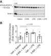
Fig. 1.
Fig. 1.
Effect of 3-methyladenine (3-MA) on lipopolysaccharide (LPS)-induced autophagy in the lung. Mice were treated with 3-MA (35 mg/kg ip) at 7 and 1 h before and 7 h after LPS challenge as illustrated in Fig. 2A. After 18 h of LPS inhalation, an antibody that detects ATG5 conjugated with ATG12 (ATG5-ATG12) was used to analyze ATG5 levels in lung homogenates. A and B: immunoblots and quantitative analysis of ATG5-ATG12 (~55 kDa) abundance in lungs from mice exposed to LPS in the presence of 3-MA or saline control. +ve, positive control (HEK 293 cell lysate); Veh, vehicle. Values are means ± SE (n = 5 for each condition) and expressed as fold increase over saline control.
Fig. 2.
Fig. 2.
Timeline of protective (Prot) and therapeutic (Ther) administration of 3-MA and its effects on LPS-induced lung pathology. A and B: mice were treated with 3-MA (35 mg/kg ip) at 7 and 1 h before and 7 h after LPS challenge (A) or 1 and 7 h after LPS challenge (B). C: at 18 h after LPS inhalation, lung sections were prepared, and alterations in lung histology were detected by hematoxylin-eosin staining. Images are representative of similar images from 4–5 mice exposed to LPS in the presence or absence of 3-MA.
Fig. 3.
Fig. 3.
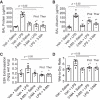
Protective and therapeutic effect of 3-MA on LPS-induced increase in proinflammatory mediators and neutrophil sequestration in the lung. Mice were treated with 3-MA and LPS as shown in Fig. 2, A and B. At 18 h after LPS challenge, lungs were analyzed for levels of proinflammatory mediators [vascular cell adhesion molecule-1 (VCAM-1), monocyte chemoattractant protein-1 (MCP-1), and interleukin (IL)-1β] by ELISA (AC) and neutrophil sequestration by tissue myeloperoxidase (MPO) activity (D). A460, absorbance at 460 nm. Values are means ± SE [n = 4–5 (saline-treated groups) and 4–6 (LPS-treated groups)].
Fig. 4.
Fig. 4.
Protective and therapeutic effect of 3-MA on LPS-induced lung vascular leak and tissue edema. Mice were treated with 3-MA and LPS as shown in Fig. 2, A and B. At 18 h after LPS challenge, lungs were analyzed for bronchoalveolar lavage (BAL) protein (A), BAL albumin (B), Evans blue albumin (EBA) extravasation (C), and wet-to-dry weight ratio (D). Values are means ± SE [n = 4–6 (saline-treated groups) and 5–7 (LPS-treated groups)].
Fig. 5.
Fig. 5.
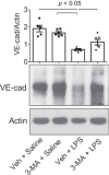
Protective effect of 3-MA on LPS-induced decrease in VE-cadherin (VE-Cad) levels. Mice were treated with 3-MA and LPS as shown in Fig. 2A. At 18 h after LPS challenge, VE-cadherin levels in lung homogenates were analyzed by immunoblotting. Actin levels were used to monitor loading. Effect of 3-MA on LPS-induced decrease in VE-cadherin levels was normalized to actin levels. Values are means ± SE (n = 5–6 for each condition).
Fig. 6.
Fig. 6.
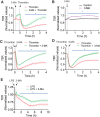
Effect of 3-MA on endothelial barrier function. A: confluent human pulmonary artery endothelial cells (HPAEC) grown on gold electrode plates were treated with 3-MA (2 mM) for ~35 min and then challenged with thrombin (2.5 U/ml). Changes in transendothelial electrical resistance (TER) were measured to monitor endothelial barrier function. Values are means ± SE (n = 4 for each condition). *P < 0.05, thrombin vs. 3-MA + thrombin (control). B: confluent HPAEC grown on gold electrode plates were treated with 3-MA (2 mM) or left untreated (control), and changes in TER were measured to monitor endothelial barrier integrity. Values are means ± SE (n = 3–4 for each condition). *P < 0.05, 3-MA vs. untreated. C and D: confluent HPAEC grown on coverslips were treated with 3-MA for 5 min (C) or 15 min (D) after thrombin challenge, and changes in TER were measured to monitor endothelial barrier function. Values are means ± SE (n = 3–5 for each condition). *P < 0.05, thrombin-treated vs. thrombin + 3-MA (control). E: confluent HPAEC grown on coverslips were treated with 3-MA at ~0.5 h after LPS challenge, and changes in TER were measured to monitor endothelial barrier function. Values are means ± SE (n = 4–8 for each condition). *P < 0.05, LPS vs. LPS + 3-MA (control).
Fig. 7.
Fig. 7.
Effect of the phosphoinositide 3-kinase (PI3K) inhibitor LY294002 on endothelial barrier function. A: confluent HPAEC grown on gold electrode plates were pretreated with LY294002 (LY, 30 μM) for ~30 min and then challenged with thrombin (2.5 U/ml). Changes in TER were measured to monitor endothelial barrier function. Values are means ± SE (n = 3 for LY alone; n = 6 for both thrombin and LY + thrombin). *P < 0.05, thrombin vs. LY + thrombin (control). B: confluent HPAEC grown on gold electrode plates were pretreated with LY294002 (LY, 30 μM) and 3-MA (2 mM) for ~55 and 15 min, respectively, and then challenged with thrombin (2.5 U/ml). Changes in TER were measured to monitor endothelial barrier function. Values are means ± SE (n = 5 for each condition). *P < 0.05, LY + thrombin vs. LY + 3-MA + thrombin-(control).
Fig. 8.
Fig. 8.
Effect of ATG5 knockdown on thrombin-induced endothelial barrier disruption. HPAEC were transfected with siRNA-control (si-Con) or siRNA-ATG5 (si-ATG5). A: after 24–36 h, total cell lysates were immunoblotted with an anti-ATG5 antibody. Total protein levels (bottom blot) were used to monitor loading. M, markers. B: quantitative analysis of ATG5 abundance in endothelial cells transfected with si-Con or si-ATG5. Values are means ± SE (n = 4 for each condition). C: after 24–36 h, cells were reseeded on gold electrode plates and allowed to reach confluency. Confluent monolayers were treated with thrombin, and changes in TER were measured to determine endothelial barrier function. Value are means ± SE (n = 4–6 for each condition). *P < 0.05, si-Con + thrombin vs. si-ATG5 + thrombin (control).
Fig. 9.
Fig. 9.
Effect of 3-MA on thrombin-induced actin stress fiber formation and loss of cell surface VE-cadherin. A: confluent HPAEC grown on coverslips were treated with 3-MA (2 mM) for 1 h and then challenged with thrombin (2.5 U/ml) for 15 min. Alexa 488-labeled phalloidin was used to visualize actin stress fibers by fluorescence microscopy. Images are representative of 3 experiments. B: confluent HPAEC monolayers grown on coverslips were treated with 3-MA (2 mM) for 1 h and then challenged with thrombin (2.5 U/ml) for 15 min. Immunofluorescence was performed using VE-cadherin antibody to visualize adherens junctions. Arrows indicate disruption of VE-cadherin staining. Images are representative of 3 experiments. C: confluent HPAEC monolayers were treated with thrombin (2.5 U/ml) for the indicated time periods. Total cell lysates were analyzed by immunoblotting to monitor cleavage of VE-cadherin (cVE-Cad, ~90 kDa). Actin levels were used to monitor loading. Top: quantitative analysis of thrombin-induced time-dependent generation of cVE-Cad. Values are means ± SE (n = 7 for each condition). Bottom: immunoblots. D: confluent HPAEC monolayers were treated with 3-MA (2 mM) for 1 h and then challenged with thrombin (2.5 U/ml) for 15 min. Total cell lysates were analyzed by immunoblotting to monitor cleavage of VE-cadherin (cVE-Cad, ~90 kDa). Actin levels were used to monitor loading. Top: quantitative analysis of thrombin-induced generation of cVE-Cad in the presence of 3-MA. Values are means ± SE (n = 3 for each condition). Bottom: immunoblots.

Similar articles

Cited by

References

    1. Bhattacharya J, Matthay MA. Regulation and repair of the alveolar-capillary barrier in acute lung injury. Annu Rev Physiol 75: 593–615, 2013. doi:10.1146/annurev-physiol-030212-183756. - DOI - PubMed
    1. Bijli KM, Fazal F, Slavin SA, Leonard A, Grose V, Alexander WB, Smrcka AV, Rahman A. Phospholipase C-ε signaling mediates endothelial cell inflammation and barrier disruption in acute lung injury. Am J Physiol Lung Cell Mol Physiol 311: L517–L524, 2016. doi:10.1152/ajplung.00069.2016. - DOI - PMC - PubMed
    1. Bijli KM, Kanter BG, Minhajuddin M, Leonard A, Xu L, Fazal F, Rahman A. Regulation of endothelial cell inflammation and lung polymorphonuclear lymphocyte infiltration by transglutaminase 2. Shock 42: 562–569, 2014. doi:10.1097/SHK.0000000000000242. - DOI - PMC - PubMed
    1. Birukova AA, Shah AS, Tian Y, Moldobaeva N, Birukov KG. Dual role of vinculin in barrier-disruptive and barrier-enhancing endothelial cell responses. Cell Signal 28: 541–551, 2016. doi:10.1016/j.cellsig.2016.02.015. - DOI - PMC - PubMed
    1. Brest P, Corcelle EA, Cesaro A, Chargui A, Belaïd A, Klionsky DJ, Vouret-Craviari V, Hebuterne X, Hofman P, Mograbi B. Autophagy and Crohn’s disease: at the crossroads of infection, inflammation, immunity, and cancer. Curr Mol Med 10: 486–502, 2010. doi:10.2174/156652410791608252. - DOI - PMC - PubMed

Publication types

MeSH terms