. 2017 Oct 24;21(4):1021-1035.
doi: 10.1016/j.celrep.2017.09.091.

Absence of Carbohydrate Response Element Binding Protein in Adipocytes Causes Systemic Insulin Resistance and Impairs Glucose Transport

Affiliations

Absence of Carbohydrate Response Element Binding Protein in Adipocytes Causes Systemic Insulin Resistance and Impairs Glucose Transport

Archana Vijayakumar et al. Cell Rep. .

Abstract

Lower adipose-ChREBP and de novo lipogenesis (DNL) are associated with insulin resistance in humans. Here, we generated adipose-specific ChREBP knockout (AdChREBP KO) mice with negligible sucrose-induced DNL in adipose tissue (AT). Chow-fed AdChREBP KO mice are insulin resistant with impaired insulin action in the liver, muscle, and AT and increased AT inflammation. HFD-fed AdChREBP KO mice are also more insulin resistant than controls. Surprisingly, adipocytes lacking ChREBP display a cell-autonomous reduction in insulin-stimulated glucose transport that is mediated by impaired Glut4 translocation and exocytosis, not lower Glut4 levels. AdChREBP KO mice have lower levels of palmitic acid esters of hydroxy stearic acids (PAHSAs) in serum, and AT. 9-PAHSA supplementation completely rescues their insulin resistance and AT inflammation. 9-PAHSA also normalizes impaired glucose transport and Glut4 exocytosis in ChREBP KO adipocytes. Thus, loss of adipose-ChREBP is sufficient to cause insulin resistance, potentially by regulating AT glucose transport and flux through specific lipogenic pathways.

Keywords: ChREBP; Glut4 trafficking; PAHSA; adipose tissue inflammation; adipose-carbohydrate response element binding protein; de novo lipogenesis; glucose transport; palmitic acid hydroxy stearic acid; systemic insulin resistance.


Figures

Figure 1
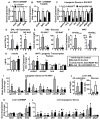
Figure 1. Deletion of ChREBP selectively in adipocytes abrogates sucrose-induced DNL in WAT but not liver
Expression of total ChREBP and its isoforms (A), ChREBP target genes (B), and lipogenic genes (C) in SQ WAT (n=7–8/group). DNL from all substrates (D) and glucose (E) in WAT in ad lib-fed (n=7–11/group) and sucrose re-fed (n=3–5/group) mice. (F) Expression of chrebptotal and chrebpβ in PG WAT. (G) Serum insulin levels. Expression of lipogenic transcription factors (H) and lipogenic genes (I) in PG WAT. n=3–5/group for F–I (J) DNL from all substrates (left) and glucose (right) in liver in ad lib-fed (n=7–11/group) and sucrose re-fed (n=3–5/group) mice. Hepatic expression of ChREBP and its isoforms (K) and lipogenic genes (L) (n=3–5/group). Data are mean±SEM. 16-week old female mice were used for all experiments. mRNA expression was normalized to tbp and shown as fold change over ChREBPfl/fl (A–C) or ad lib-fed controls (F, H–I, K–L). In D–G & J, F= ad lib-fed and R= sucrose re-fed. *p<0.05; $p<0.06 vs. ChREBPfl/fl, same feeding condition; #p<0.05 vs. ad lib-fed, same genotype. (also see Fig S1)
Figure 2
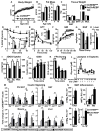
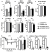
Figure 2. Loss of adipose-ChREBP causes insulin resistance and WAT inflammation in lean mice
Body weight (A), fat mass [per mouse (left) and relative to body weight (right)] (B), and wet tissue weight (C) (n=6–11/group). ITT (0.5U/kg, i.p.) (D) Inset: AAC (mg/dl*min*103) (left) and slope0–15 (mg/dl/min) (right). GTT (2g/kg, i.p.) area under the curve (AUC, mg/dl*min*103) (E) in 9–11 week-old mice n=7–8/group. GIR (F) and endogenous glucose production (EGP) (G) during hyperinsulinemic-euglycemic clamps. #-p<0.05 vs. basal, same genotype. (H) Liver TG levels 6h after food removal. (I) In vivo tissue 2DOG transport at the end of the clamp. n=7–8/group for F–I GSIS (J) and serum FFAs (K) in overnight fasted mice during GTT (n=19–23/group). †p<0.05 vs. T0, same genotype; ϕp<0.05 vs. T0 ChREBPfl/fl; #-p<0.05 vs. all, same genotype, t-test. (L) Glycerol secretion from PG WAT explants after 6h of food removal. #-p<0.05, $p<0.06 vs. no insulin, no 8-Br cAMP, same genotype; †p<0.05 vs. 8-BrcAMP, no insulin, same genotype (n=7/group). (M) Insulin signaling (1U/kg, 5min) (n=4–5/group). #p< 0.05 vs. saline. (N) Numbers of total, pro-inflammatory (M1) and anti-inflammatory (M2), TNFα+, and IL-1β+ adipose tissue macrophages (ATMs) in PG WAT of 8 week-old male mice measured by flow cytometry (n=9/group). Data are mean±SEM. A–M were performed in chow-fed female mice. *p< 0.05 vs. ChREBPfl/fl, same condition. (also see Fig S2–3)
Figure 3
Figure 3. Loss of adipose-ChREBP lowers WAT PAHSA levels
(A) Relative abundance of fatty acids in SQ WAT (left). Δ9 desaturation index and C16 elongation index (right) (n=7/group). Levels of total PAHSAs (B) and individual PAHSA isomers (C) in SQ WAT (n=3/group except n=2 for 9-PAHSA levels in ChREBPfl/fl mice). Data are mean±SEM. 16 week-old chow-fed female ChREBPfl/fl and AdChREBP KO mice were used. *p<0.05 vs. ChREBPfl/fl
Figure 4
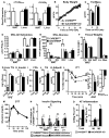
Figure 4. ChREBP deletion in adipocytes results in insulin resistance even in the obese state
Female mice were used for all experiments. (A) ChREBP expression in SQ WAT and liver of 16 week HFD-fed mice (n=5–6/group). Body weight (B) and fat mass (C) (n=26–27/group) with HFD. Ad lib-fed DNL from all substrates (D) and glucose (E) in HFD-fed mice (n=6/group). (F) Terminal wet tissue weight after 16 weeks of HFD (n=5–6/group). (G) Liver TG content after 8 weeks of HFD (n=5/group). Chow-fed mice (n=2) were used for comparison. #p<0.05 vs. chow, Ad lib-fed serum insulin (H), FFA (I), TG (J) and adiponectin (AdipoN) (K) levels after 16 week of HFD. ITT (0.5U/kg, i.p) (L) and GTT (2g/kg, i.p) (M) after 16 weeks of HFD. n=13–14/group for H–M (N) Insulin signaling (1U/kg, 15min) after 20 weeks of HFD. (n=3–7/group). #p< 0.05 (Two-Way ANOVA), @p< 0.05 (t-test) vs. saline. (O) Numbers of total, and pro-inflammatory (M1) and anti-inflammatory (M2) ATMs in PG WAT measured by flow cytometry after 16–20 weeks of HFD (n=5/group). Data are mean±SEM. *p<0.05 vs. ChREBPfl/fl, same condition; (also see Fig S4)
Figure 5
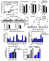
Figure 5. Absence of ChREBP in adipocytes impairs insulin-stimulated glucose transport cell autonomously
(A) Glucose transport in primary adipocytes isolated from 16 week-old chow-fed female mice (n=7–8/group). Adipocyte number (B) and size (C) in PG and SQ WAT of female mice (n=17–25/group). (D) Expression of glucose transporters (Gluts) in SQ WAT of chow-fed mice. (E) Glut4 protein content in total cellular membranes from PG WAT. (D–E) Data are fold change over ChREBPfl/fl (n=5–9/group). (F) Expression of chrebptot (n=6–7/group) (left) and chrebpβ (n=2–6/group) (right) during differentiation of SVF-derived adipocytes. Data are fold change over Day 0 control (ChREBPfl/fl). Expression of differentiation markers (G) and lipogenic genes (H) in Day 7 differentiated SVF-derived adipocytes. Data are fold change over preadipocytes (G) or control (H) (n=8/group). (I) Insulin-stimulated glucose transport in SVF-derived adipocytes. Data are normalized to protein concentration (n=3 experiments). (J) Insulin-stimulated glucose transport in 3T3L1 adipocytes with scrambled (control) or ChREBP shRNA (ChREBP KD) after pre-incubation with 9-PAHSA (20μM, overnight). Data are normalized to protein concentration (n=6/group). Data are mean±SEM. mRNA was normalized to tbp, and protein to p85. *p<0.05 vs. ChREBPfl/fl (A–E) or control (F–J), same condition; #p<0.05 vs. no insulin (I) or no 9-PAHSA (J), same genotype. (also see Fig S5)
Figure 6
Figure 6. Adipocytes lacking ChREBP have defects in insulin-stimulated Glut4 translocation
(A) Quantitation of western blot analysis for Glut4 in 3T3L1 adipocytes with scrambled (Con) or ChREBP shRNA (KD). Glut4 levels are normalized to p85 and expressed as fold change over Control (n=5–8/group). (B) Western blot analysis of insulin (10nM, 10min) signaling in control and ChREBP KO adipocytes (left). The ratio of phosphorylated/total protein is expressed as fold change over control/insulin (n=8–12/group). (C) Representative images of Glut4 translocation using the HA-Glut4-GFP reporter assay (left). Surface:Total HA-Glut4-GFP was quantified under basal and insulin (1nM)-stimulated conditions (right). Insulin (1nM)-stimulated Glut4 exocytosis (D), exocytosis rate constant (kex) (E), and effect of 9-PAHSA (9P) (20μM, 2h) on Glut4 exocytosis (F). Data represented as a fold over control (D, F). B–F were performed in SVF-derived adipocytes C–F n=>50 cells from 2–3 combined experiments (G) Insulin (1nM)-stimulated glucose transport in primary adipocytes isolated from chow-fed female mice. Cells were pre-incubated with DMSO or increasing concentrations of 9-PAHSA for 2.5h prior to insulin (1nM) stimulation (n=8–20/group). D=DMSO (F–G) Data are mean±SEM. *p<0.05 vs. ChREBPfl/fl(control) same condition (B–E), ChREBPfl/fl DMSO insulin (F–G). #p<0.05 vs. no insulin/basal (B, C), DMSO+insulin (F, G), same genotype. (also see Fig S6)
Figure 7
Figure 7. Restoring reduced PAHSA levels in AdChREBP KO mice reverses their insulin resistance
7–9 week old male mice were treated with vehicle (V) or 9-PAHSA (9P, 15mg/kg/d, oral gavage) for 26 days. Serum (A) and WAT (B) PAHSA levels (n=4–5/group). Body weight (C), fat mass (D), and ad lib-fed serum insulin (E), and TG (F) levels after 26 days of treatment. (G) ITT (0.5U/kg, i.p.) after 14 days of treatment. (C–G) n=7–10/group (H) Numbers of total, TNFα+ and IL-1β+ ATMs in PG WAT of AdChREBP KO mice after 26 days of treatment (n=5/group). Data are mean±SEM. *p< 0.05 vs. ChREBPfl/fl, same treatment; #p< 0.05 vs. vehicle. (also see Fig S7)

Similar articles

Cited by

References

    1. Abel ED, Peroni O, Kim JK, Kim YB, Boss O, Hadro E, Minnemann T, Shulman GI, Kahn BB. Adipose-selective targeting of the GLUT4 gene impairs insulin action in muscle and liver. Nature. 2001;409:729–733. - PubMed
    1. Baraille F, Planchais J, Dentin R, Guilmeau S, Postic C. Integration of ChREBP-Mediated Glucose Sensing into Whole Body Metabolism. Physiology (Bethesda) 2015;30:428–437. - PubMed
    1. Benhamed F, Denechaud PD, Lemoine M, Robichon C, Moldes M, Bertrand-Michel J, Ratziu V, Serfaty L, Housset C, Capeau J, et al. The lipogenic transcription factor ChREBP dissociates hepatic steatosis from insulin resistance in mice and humans. J Clin Invest. 2012;122:2176–2194. - PMC - PubMed
    1. Blot V, McGraw TE. Use of quantitative immunofluorescence microscopy to study intracellular trafficking: studies of the GLUT4 glucose transporter. Methods Mol Biol. 2008;457:347–366. - PubMed
    1. Bruss MD, Khambatta CF, Ruby MA, Aggarwal I, Hellerstein MK. Calorie restriction increases fatty acid synthesis and whole body fat oxidation rates. Am J Physiol Endocrinol Metab. 2010;298:E108–116. - PMC - PubMed

MeSH terms

LinkOut - more resources