. 2017 Oct 17;21(3):612-627.
doi: 10.1016/j.celrep.2017.09.072.

USP7 Is a Tumor-Specific WNT Activator for APC-Mutated Colorectal Cancer by Mediating β-Catenin Deubiquitination

Affiliations

USP7 Is a Tumor-Specific WNT Activator for APC-Mutated Colorectal Cancer by Mediating β-Catenin Deubiquitination

Laura Novellasdemunt et al. Cell Rep. .

Abstract

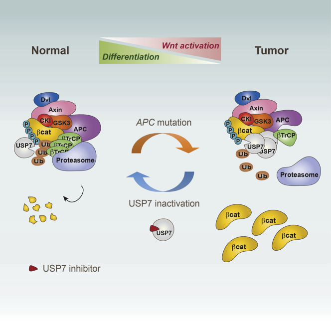
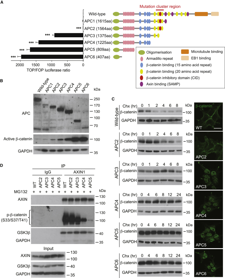
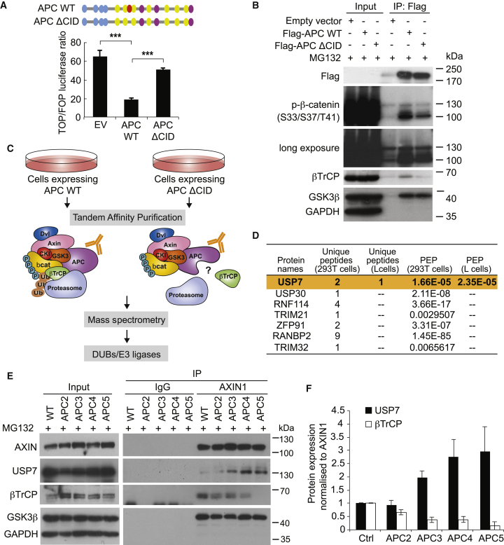
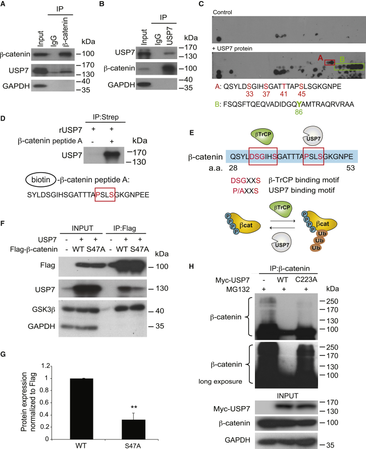
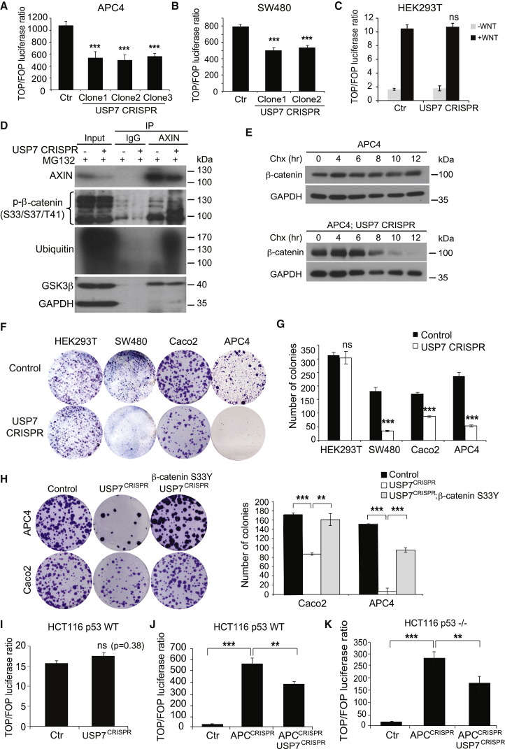
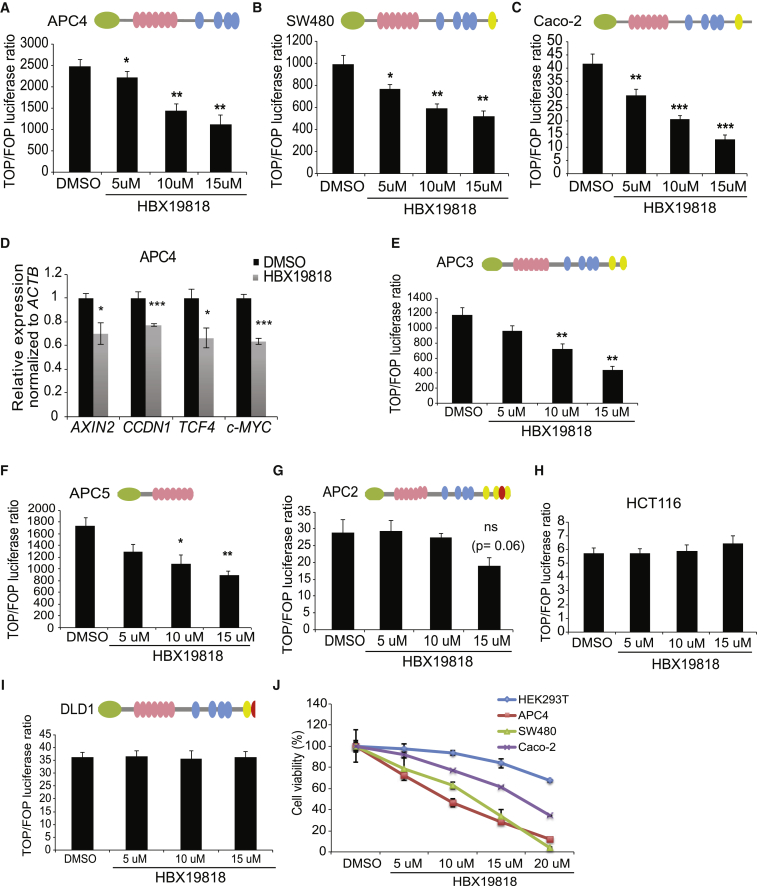
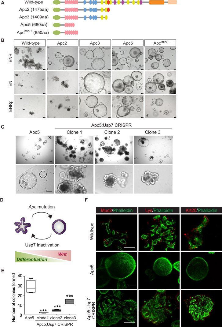
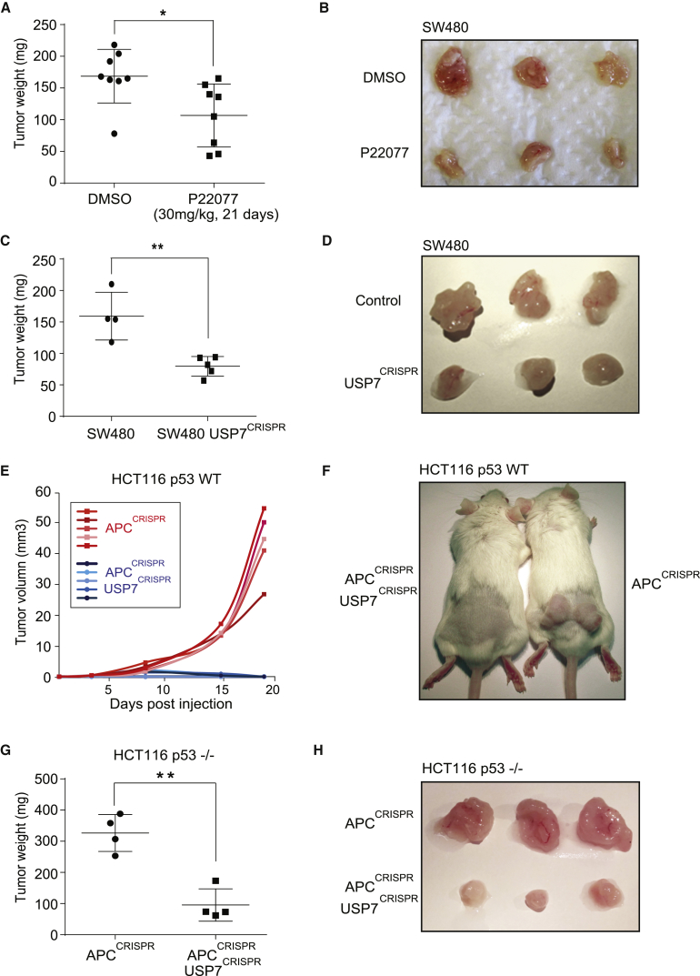
The tumor suppressor gene adenomatous polyposis coli (APC) is mutated in most colorectal cancers (CRCs), resulting in constitutive Wnt activation. To understand the Wnt-activating mechanism of the APC mutation, we applied CRISPR/Cas9 technology to engineer various APC-truncated isogenic lines. We find that the β-catenin inhibitory domain (CID) in APC represents the threshold for pathological levels of Wnt activation and tumor transformation. Mechanistically, CID-deleted APC truncation promotes β-catenin deubiquitination through reverse binding of β-TrCP and USP7 to the destruction complex. USP7 depletion in APC-mutated CRC inhibits Wnt activation by restoring β-catenin ubiquitination, drives differentiation, and suppresses xenograft tumor growth. Finally, the Wnt-activating role of USP7 is specific to APC mutations; thus, it can be used as a tumor-specific therapeutic target for most CRCs.

Keywords: APC; USP7; Wnt signaling; colorectal cancer; ubiquitination; β-catenin.


Figures

None
Graphical abstract
Figure 1
Figure 1
CID Is the Threshold for the Pathological Level of Wnt Activation (A) Schematic representations of human WT APC protein and the corresponding truncating mutants generated by the CRISPR-Cas9 technique. Relative TOP/FOP luciferase activities were measured in different lines. Error bars represent ± SE from at least three independent experiments (∗∗∗p < 0.001). (B) Cell lysates of HEK293T WT and APC truncated cell lines were analyzed by western blot using the indicated antibodies. GAPDH protein levels were used as loading control. (C) Cells were treated with cycloheximide (Chx) (50 μg/mL) and collected at different time points as indicated. Stimulated lysates were subjected to western blot analysis using the indicated antibodies. Immunostaining of β-catenin (green) was performed in the indicated cell lines using phospho-insensitive β-catenin antibody (BD Biosciences). Scale bar, 20 μm. (D) Different cell lysates were immunoprecipitated with AXIN1 antibody followed by western blot analysis using the indicated antibodies. IgG, immunoglobulin G. See also Figure S1.
Figure 2
Figure 2
Reciprocal Binding of β-TrCP and USP7 to the β-Catenin-Destruction Complex in APC-Truncated Cells (A) Schematic representation of the central region (1,265–2,060 aas) of APC (APC WT) and the CID-deleted mutant (APC ΔCID). Relative TOP/FOP activities of the APC truncated cell line SW480 transfected with empty vector (EV), APC WT, or APC ΔCID plasmids. Error bars represent SE from at least three independent experiments (∗∗∗p < 0.001). (B) HEK293T cells were transfected with EV, FLAG-tagged APC WT, or APC ΔCID. Lysates were subjected to anti-FLAG IP followed by western blotting using the indicated antibodies. (C) Schematic representation of the experimental procedure used for mass spectrometry analysis. (D) DUBs and E3 ligases identified from mass spectrometry in HEK293T and L cells that showed differential bindings to WT versus ΔCID complex. PEP, posterior error probability (p value for protein identification). (E) Cell lysates were immunoprecipitated with AXIN1 antibody followed by western blotting using the indicated antibodies. (F) Quantitation of USP7 and β-TrCP protein binding normalized to AXIN1 protein pulled down in (E). See also Figure S2.
Figure 3
Figure 3
USP7 Directly Interacts with the N Terminus of β-Catenin to Mediate β-Catenin Ubiquitination (A and B) IP of endogenous β-catenin (A) or USP7 (B) in HEK293T cells followed by western blotting using the indicated antibodies. (C) A β-catenin 26-mer peptide array was probed with vehicle control or USP7 recombinant protein. Two USP7-specific binding regions were identified. Reported modification sites were indicated. (D) Biotinylated β-catenin peptide A, including the putative USP7 binding motif (highlighted in the red box), was incubated with recombinant USP7 protein (rUSP7) followed by streptavidin-pull-down assays. (E) Top: presence of β-TrCP and USP7 binding motif in the β-catenin peptide sequence aas 28–53. Conserved sequences are highlighted in red. Bottom: schematic representation of the regulation of β-catenin ubiquitination by β-TrCP and USP7. Phosphorylation sites indicated represent GSK3-mediated (Ser33, Ser37, and Thr41) and CK1-mediated Ser45 phosphorylation. (F) USP7 showed reduced binding to the β-catenin S47A mutant IP complex compared to the WT. (G) Quantitation of the amount of USP7 in the FLAG-β-catenin complex after transfecting β-catenin WT and comparing with β-catenin S47A. Error bars represent SD from three independent experiments. p < 0.05. (H) Empty vector, MYC-USP7 WT, or MYC-USP7-C223A plasmids were transfected followed by endogenous β-catenin IP and immunoblotting using the indicated antibodies. See also Figure S3.
Figure 4
Figure 4
USP7 Depletion in APC-Truncated CRC Suppresses Wnt Activation by Restoring β-Catenin Ubiquitination (A–C) Relative TOP/FOP activities of the APC4 (A), SW480 (B), and HEK293T (C) cells with control or USP7 CRISPR targeting. (D) AXIN1 complexes were immunoprecipitated in APC4 cells with or without USP7 targeting followed by western blotting using the indicated antibodies. (E) Cells were treated with cycloheximide (Chx) (50 μg/μL), and lysates were collected at different time points as indicated for immunoblotting of ABC (active β-catenin) and control GAPDH. (F) Colony formation assay in parental HEK293T, SW480, Caco2, and APC4 cells and the corresponding USP7 CRISPR-deleted cells. (G) Quantitation of number of colonies in (F). Experiments were performed in triplicates. (H) Colony formation assay of APC4 and Caco2 cells upon transient transfection of USP7 CRISPR and/or β-catenin S33Y mutant plasmids. The corresponding quantitation is indicated on the right. (I–K) Relative TOP/FOP activities of the HCT116 p53 WT (I and J) and HCT116 p53−/− (K) cells with indicated CRISPR targeting. Error bars represent SE from at least three independent experiments (∗∗p < 0.01; ∗∗∗p < 0.001; ns, not significant). See also Figure S4.
Figure 5
Figure 5
USP7 Inhibitor Treatment Suppresses Wnt Activation in CID-Deleted APC Mutant CRCs (A–C) Relative TOP/FOP activities of APC4 (A), SW480 (B), and Caco-2 (C) cells treated with HBX19818 at the indicated concentrations or DMSO as control. (D) mRNA expression of the indicated Wnt target genes was analyzed by qRT-PCR in APC4 cells. Data are presented as fold change normalized to β-actin control in triplicate and are representative of at least three independent experiments. (E–I) Relative TOP/FOP activities of APC3 (E), APC5 (F), APC2 (G), HCT116 (H), and DLD1 (I) cells treated with DMSO or HBX19818 at the indicated concentrations. (J) MTT assay in the indicated cell lines treated with DMSO or HBX19818 at the indicated concentrations for 24 hr. Error bars represent ± SE from at least three independent experiments (p < 0.05; ∗∗p < 0.01; ∗∗∗p < 0.001; ns, not significant). See also Figure S5.
Figure 6
Figure 6
Usp7 Deletion in Apc-Mutated Intestinal Organoids Induces Differentiation and Suppresses Growth (A) Schematic representation of the mouse WT Apc protein and the corresponding truncating mutants generated by the CRISPR-Cas9 technique. (B) Morphological changes of organoids cultured in the indicated conditions. E, Egf; N, Noggin, R, R-spondin; p, porcupine inhibitor (IWP2). Scale bars, 100 μm. (C) Representative images of Apc5 organoids with or without Usp7 CRISPR targeting. Scale bars, 100 μm. (D) Schematic representation of the changes on the morphology of intestinal organoids upon Apc loss and Usp7 inactivation. (E) Clonogenic assay of Apc5 organoids with or without Usp7 CRISPR targeting after 7 days in culture. Error bars represent SE from at least three independent experiments (∗∗∗p < 0.001). Scale bar, 100 μm. (F) Immunofluorescent staining of WT, Apc5, and Apc5;Usp7 CRISPR-targeted organoids using the indicated antibodies. Red signal shows markers of differentiation, including Mucin2 (Muc2), Lysozyme (Lys), and Keratin20 (Krt20). Scale bars, 100 μm. See also Figure S6.
Figure 7
Figure 7
USP7 Inactivation Suppresses APC-Mutated Colorectal Cancer Tumor Growth In Vivo (A) SW480-derived tumor weights between DMSO control group and P22077 treatment group (30 mg/kg) at the end of treatment (21 days) (p < 0.05) (n = 8 per condition). (B) Representative photos of SW480-derived tumors at the end of treatment. (C) Comparison of weights between parental SW480- and SW480 USP7 CRISPR-derived tumors at the end of treatment (25 days) (∗∗p < 0.01) (n = 4 and n = 5, respectively). (D) Representative photos of SW480 and SW480 USP7 CRISPR-derived tumors at the end of treatment. (E) Tumor volumes derived from HCT116 APCCRISPR compared to HCT116 APCCRISPR USP7CRISPR throughout the experiment (19 days) (n = 5 per condition). (F) Representative photos of the mice injected with HCT116 APCCRISPR cells compared to HCT116 APCCRISPR USP7CRISPR cells at the end of the experiment. (G) Weight comparison between HCT116 p53−/− APCCRISPR- and HCT116 p53−/− APCCRISPR USP7CRISPR-derived tumors at the end of the experiment (13 days) (n = 4 per condition; ∗∗p < 0.01). (H) Representative photos of the tumors derived from HCT116 p53−/− APCCRISPR and HCT116 p53−/− APCCRISPR USP7CRISPR cells. See also Figure S7.

Similar articles

Cited by

References

    1. Aberle H., Bauer A., Stappert J., Kispert A., Kemler R. beta-catenin is a target for the ubiquitin-proteasome pathway. EMBO J. 1997;16:3797–3804. - PMC - PubMed
    1. The Cancer Genome Atlas Network Comprehensive molecular characterization of human colon and rectal cancer. Nature. 2012;487:330–337. - PMC - PubMed
    1. Chen B., Dodge M.E., Tang W., Lu J., Ma Z., Fan C.W., Wei S., Hao W., Kilgore J., Williams N.S. Small molecule-mediated disruption of Wnt-dependent signaling in tissue regeneration and cancer. Nat. Chem. Biol. 2009;5:100–107. - PMC - PubMed
    1. Choi S.H., Estarás C., Moresco J.J., Yates J.R., 3rd, Jones K.A. α-Catenin interacts with APC to regulate β-catenin proteolysis and transcriptional repression of Wnt target genes. Genes Dev. 2013;27:2473–2488. - PMC - PubMed
    1. Clevers H., Nusse R. Wnt/β-catenin signaling and disease. Cell. 2012;149:1192–1205. - PubMed

MeSH terms

Substances