. 2017 Oct 17;7(1):13393.
doi: 10.1038/s41598-017-13750-0.

Expression and function of Abcg4 in the mouse blood-brain barrier: role in restricting the brain entry of amyloid-β peptide

Affiliations

Expression and function of Abcg4 in the mouse blood-brain barrier: role in restricting the brain entry of amyloid-β peptide

Agnès Dodacki et al. Sci Rep. .

Abstract

ABCG4 is an ATP-binding cassette transmembrane protein which has been shown, in vitro, to participate in the cellular efflux of desmosterol and amyloid-β peptide (Aβ). ABCG4 is highly expressed in the brain, but its localization and function at the blood-brain barrier (BBB) level remain unknown. We demonstrate by qRT-PCR and confocal imaging that mouse Abcg4 is expressed in the brain capillary endothelial cells. Modelling studies of the Abcg4 dimer suggested that desmosterol showed thermodynamically favorable binding at the putative sterol-binding site, and this was greater than for cholesterol. Additionally, unbiased docking also showed Aβ binding at this site. Using a novel Abcg4-deficient mouse model, we show that Abcg4 was able to export Aβ and desmosterol at the BBB level and these processes could be inhibited by probucol and L-thyroxine. Our assay also showed that desmosterol antagonized the export of Aβ, presumably as both bind at the sterol-binding site on Abcg4. We show for the first time that Abcg4 may function in vivo to export Aβ at the BBB, in a process that can be antagonized by its putative natural ligand, desmosterol (and possibly cholesterol).


Conflict of interest statement

The authors declare that they have no competing interests.

Figures

Figure 1
Figure 1
Immunodetection of Abcg4 in wild-type (WT) mice. Brain capillaries are identified by endothelial cell-specific P-gp labelling (magenta, middle panel) and nuclei are counterstained by Topro3 (cyan). Abcg4 staining (yellow, left hand panel) is localized to parenchymal cells (arrows) and endothelial cells of the capillaries (full arrowheads). The overlay image of all three (right hand panel) indicates co-localization of Abcg4 with P-gp in the brain capillaries. Scale bar: 30 µm.
Figure 2
Figure 2
Quantification of transcripts of several ABC transporters in wild-type (WT) and Abcg4-KO mice BBB. The mRNA levels of each gene were determined by qRT-PCR and normalized to those of β-actin as described in Methods. Data are means ± S.D. (n = 3 experiments). *p < 0.05 (Student’s unpaired t test).
Figure 3
Figure 3
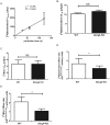
Abcg4 function at the mouse BBB. Panel A shows the time course of [3H]desmosterol uptake by the right hemisphere of C57BL/6 mice, expressed as apparent volume of distribution (Vbrain, µl/g brain), determined by in situ brain perfusion technique, r2 = 0.944 for regression analysis of individual data. Data are means ± S.D. of 3 animals per data point. Panels B shows the brain uptake clearance (Clup, µL/g/s) of [3H]desmosterol and Panel C for [3H]Aβ1–40 (C) in WT and Abcg4-KO mice, measured by in situ brain perfusion technique. Panel D shows the efflux ratio (ER) measured in WT and Abcg4-KO mice: ratio for [3H]desmosterol and panel E that for [3H]Aβ1–40, Clup obtained in the presence or absence of probucol (10 µM) (ERProbucol/Ctrl). Panels B-E: Data are means ± S.D. of 8–9 mice. *p < 0.05, NS non-significant (two-way ANOVA analysis followed by a Bonferroni post-test).
Figure 4
Figure 4
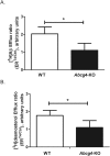
L-thyroxine (T4) inhibition of Abcg4-mediated efflux of [3H]Aβ1–40 and [3H]desmosterol at the mouse BBB level. Efflux ratio of [3H]desmosterol (Panel A) or [3H]Aβ1–40 (Panel B) obtained by dividing the brain uptake clearance (Clup, µL/g/s) measured in the presence of T4 (10 µM) over the Clup measured in the absence (Ctrl) of T4 (ERT4/ctrl) in WT and Abcg4-KO mice. Data are means ± S.D. of 5–8 mice. *p < 0.05 (two-way ANOVA analysis followed by a Bonferroni post-test).
Figure 5
Figure 5
In vivo interaction between [3H]Aβ1–40 and desmosterol at the mouse BBB level. Efflux ratio of [3H]Aβ1–40 obtained by dividing the brain uptake clearance (Clup, µL/g/s) measured in the presence of desmosterol (20 µM) over the Clup measured in the absence (Ctrl) of desmosterol (ERdesmosterol/ctrl) in WT and Abcg4-KO mice. Data are means ± SD of 5–7 mice. *p < 0.05 (two-way ANOVA analysis followed by a Bonferroni post-test).
Figure 6
Figure 6
Computed structural and binding features of the Abcg4 dimer model. Mouse Abcg4 sequence was submitted to the ModBase modeling server which constructed an Abcg4 model (blue) after selecting the x-ray crystal structure of ABCG5/ABCG8 (red cartoon) as the template in an unbiased search (Panel A). Panel B shows docking of desmosterol across the entire transmembrane surface identified two putative desmosterol binding sites (red dots represent the placement of desmosterol after each docking run). The overwhelming majority of final binding modes for desmosterol on Abcg4 were located in the homologous sterol binding cleft found in ABCG5/ABCG8 suggesting conserved sterol binding domain is found in both dimers. Best predicted binding mode of desmosterol (pink and white spheres) in the cleft formed by Abcg4 dimers (gray and blue, Panel C). Gray monomer rotated 90° away from the viewer along the axis of symmetry reveals competitive binding between desmosterol and Aβ (green mesh, panel D).
Figure 7
Figure 7
Dual Aβ binding sites in Abcg4. The Abcg4 dimer is comprised of two monomers (gray surface) that form two nearly identical binding sites for Aβ (green mesh, panel A). This view of the membrane-spanning domain from the intracellular aspect shows two Aβ molecules (green mesh) occupying the binding clefts. The membrane-spanning domain viewed from the side, panel B, shows the competitive binding mode of desmosterol (yellow surface) with Aβ (red mesh) while another Aβ molecule is present in the opposite binding cleft (green mesh). This configuration explains the observed preference for Aβ (two sites) and inhibitory effect of sterols on Aβ efflux.
Figure 8
Figure 8
Abcg4-knockout mice line establishment. Panel A depicts a cartoon of the Abcg4 knockout targeting vector used for homologous recombination. The gene structure is not drawn to scale. Targeting was engineered to result in expression of a fusion protein encoding partial Abcg4 fused to eGFP prior to the transmembrane domains. The neomycin gene cassette is indicated by Neo. The homologous targeting removes a significant portion of coding sequences in exon 7 and all of exon 8. The Bgl I sites are as shown. Panel B shows the validation of the homologous recombination by Southern blotting. Mouse DNA was digested using Bgl I and detected using a genomic fragment spanning exons 3–5 shown in panel A. The targeted fragment (6.4 kb) is larger than the WT fragment (4.5 kb). The molecular weight marker positions were estimated from the ethidium-stained gel. Panel C shows Northern blot from WT and knockout (KO) tissues with a cDNA probe (Li: liver, Lu: lung, Sp: spleen, H: heart, E: eye, Br: brain, Te: testes, Ki: kidney). In RNA from Abcg4-knockout mice (‘KO’), multiple shorter transcripts were detected, compared to WT. Additionally, expression was more wide-spread, with signals detected in lung, spleen and testes, compared to WT mice. RT-PCR analyses of RNA from brains of WT (panel D, tracks 1–3) or KO (panel D, tracks 4–6) using primers in exon 4–10 (tracks 1 and 4), exon 5–10 (tracks 2 and 5) and exon 6–10 (tracks 3 and 6) led to the bona fide sized products in wild-type RNA, but these were smaller for samples from KO brain. All of the largest products, as well as one of the smaller products (arrow) were sequenced. For wild-type RNA, there was contiguous sequential exon splicing, but for Abcg4-KO, exon 6 was spliced to exon 10 in the largest PCR fragments and in the smaller fragment, exon 5 was spliced to exon 10 (see text for discussion).

Similar articles

Cited by

References

    1. Abbott NJ, Patabendige AAK, Dolman DEM, Yusof SR, Begley DJ. Structure and function of the blood-brain barrier. Neurobiol. Dis. 2010;37:13–25. doi: 10.1016/j.nbd.2009.07.030. - DOI - PubMed
    1. Bernacki J, Dobrowolska A, Nierwińska K, Małecki A. Physiology and pharmacological role of the blood-brain barrier. Pharmacol Rep. 2008;60:600–622. - PubMed
    1. Cecchelli R, et al. Modelling of the blood-brain barrier in drug discovery and development. Nat Rev Drug Discov. 2007;6:650–661. doi: 10.1038/nrd2368. - DOI - PubMed
    1. Quaegebeur A, Lange C, Carmeliet P. The neurovascular link in health and disease: molecular mechanisms and therapeutic implications. Neuron. 2011;71:406–424. doi: 10.1016/j.neuron.2011.07.013. - DOI - PubMed
    1. Jones PM, George AM. The ABC transporter structure and mechanism: perspectives on recent research. Cell. Mol. Life Sci. 2004;61:682–699. doi: 10.1007/s00018-003-3336-9. - DOI - PMC - PubMed

MeSH terms